0922
Correcting Eddy Current Induced Geometric Distortion for High Resolution Multi-Band Diffusion Weighted SE-EPI with Magnetic Field Monitoring at 7T1Center for Magnetic Resonance Research, University of Minnesota, Minneapolis, MN, United States, 2Electrical and Computer Engineering, University of Minnesota, Minneapolis, MN, United States
Synopsis
DW-EPI suffers from geometric distortion induced by eddy current from strong diffusion gradients. Magnetic field monitoring using 19F probes followed by algebraic reconstruction incorporating higher order field perturbation has been proposed to address such issue and was applied for single-band acquisition at 3T. In this study, this approach was implemented for high resolution multiband in vivo brain DW-EPI acquisition at 7T, taking into account of scanner imposed global phase modulation for eddy-current correction. Preliminary results showed successful correction of the geometric distortion across all diffusion gradients. Further investigation needs to be made to increase the SNR of DW images.
Introduction:
Multiband-accelerated-diffusion-weighted-echo-planer-imaging (MB-DW-EPI) at ultra-high field enables high-resolution brain structural mapping1. One challenge of DW-EPI is its susceptibility to image distortion induced by eddy-current. Magnetic field monitoring followed by higher than 0th and 1st order field correction using conjugate-gradient SENSE (CG-SENSE)2 has been proposed to successfully address such issue for single-band-DW-EPI at 3T3. In this work, we implemented this method for high-resolution MB-DW-EPI acquisition at 7T to correct the eddy-current induced geometric distortion.Methods:
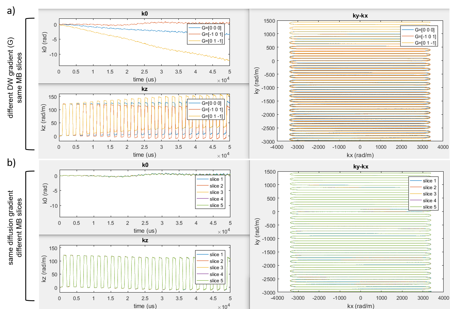
MR data were acquired in one subject on a whole body 7T scanner (Siemens,Erlangen,Germany), with a 32-channel head coil (Nova Medical,Wilmington,MA,USA). A 2D single-shot monopolar DW-Spin-Echo-EPI sequence was used with following parameters: diffusion encoding (bvalue=1000s/mm2) applied in six standard orientations, plus one reference with bvalue=0, TR/TE=7000/71.2ms, resolution=1.05x1.05mm2, slice-thickness=1.05mm, 10 slices, in-plane GRAPPAx3, blipped-CAIPI4 MBx2 with a gap of 21mm.
The field monitoring was conducted in a separate acquisition. A clip-on magnetic field camera (Skope,Zurich,CH) with 16(19F) probes was used. The phase coefficients of up to 2nd order spherical harmonics were fitted by the field camera system.
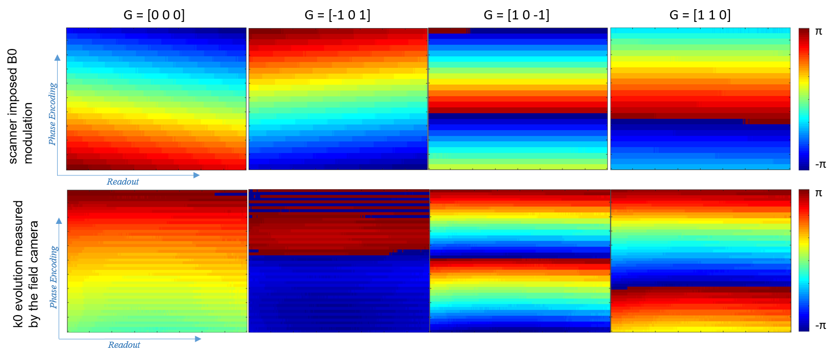
Eddy-current induced 0th order B0 modulation are compensated by the scanner by real time RF synthesizer frequency modulation (B0-RF). We measured these RF modulations by running the exact same diffusion sequence and protocol while feeding one receive-channel with a sinusoid RF at Larmor frequency using an external RF synthesizer synchronized to the 10MHz clock of the scanner, and subsequently reading afterwards the data written by the console after B0-RF modulation.
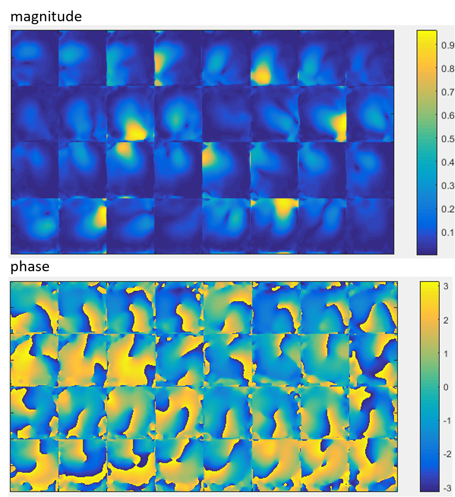
The image reconstruction scheme was implemented in MATLAB 2016a (Mathworks,USA), using CG-SENSE (10 iterations) with: 1) the scanner imposed B0-RF modulation term removed by multiplying the raw k-space data with the conjugate of the raw data written during B0-RF measurement; 2) coil sensitivity maps generated using ESPIRiT algorithm5.
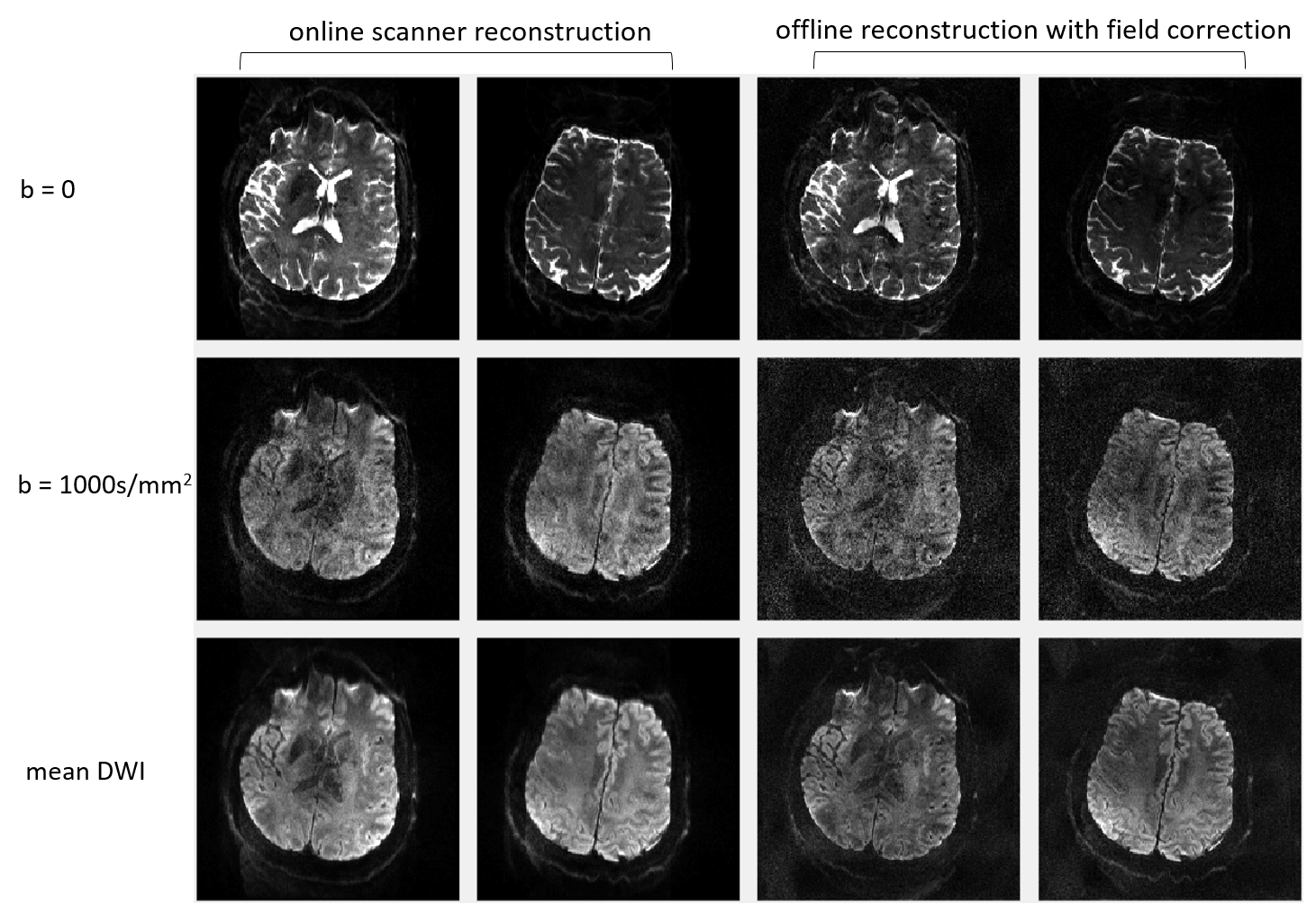
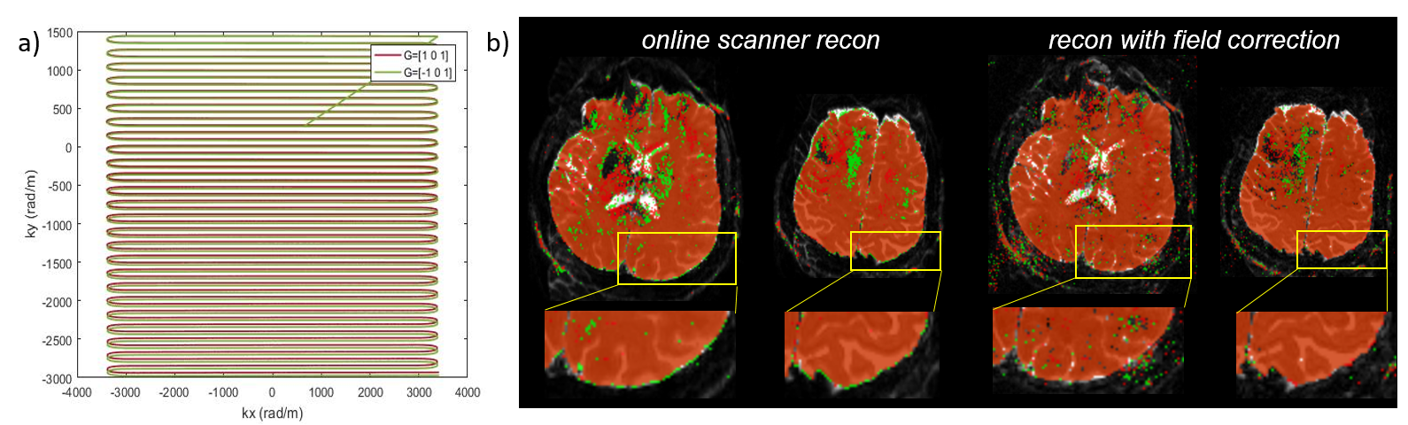
The effect of utilizing field evolution measured by the field camera was examined by the following two approaches: 1) As previously described3, the six DW images with different diffusion orientations were averaged separately for images reconstructed on the Siemens console (dicom) (i.e. no field correction) and reconstructed offline with field correction. The sharpness of the edge of the brain was compared visually. 2) A 2D binary mask of the brain was created from each DW image with and without field correction using the multithresh function in MATLAB. Each mask was first overlaid on the DW image that it was derived from to verify the accuracy of brain edge detection. The masks obtained with different DW directions were compared between each other either with or without field correction to evaluate the corresponding brain edge alteration.
Results:
Field monitoring: As illustrated in Fig.1a, much larger deviations of 0th and 1st order terms were observed when DW gradients were added. As expected, no significant differences were observed when monitoring the field during different multi-slice excitations (Fig.1b), indicating that eddy-currents were in stable steady-state for a given DW direction for different slabs.
Removal of B0-RF: As seen in Fig.2, noticeable differences were observed between the console driven B0-RF modulation and the actual k0 term measured by the field camera, both in the absence (first column) and in the presence (2nd-4th column) of DW gradients. This highlights the need for removing from the raw data the system induced B0-RF modulation from the raw data and replacing this correction with the measured k0 excursion.
Image reconstruction: The coil sensitivity maps from one slice are displayed in Fig.3. Two simultaneously acquired slices obtained from the scanner and reconstructed with field correction are shown in Fig.4. The two reconstruction strategies (sGRAPPA4 and CG-SENSE) showed comparable image quality for the bvalue=0 images. However, for DW images with lower SNR, a higher residual noise level was observed for images reconstructed with CG-SENSE. Nevertheless, the averaged DW images showed sharper brain edges after correction for field deviations. The correction of geometric distortions resulting from integrating the monitored field variation in the reconstruction can be visualized in Fig.5, with a clear improvement of brain mask matching between images obtained with different DW directions.
Discussion and conclusion:
Using the monitored field evolution, successful correction of eddy-current induced geometric distortion was obtained at 7T with a high-resolution MB-DW-EPI in vivo acquisition. A higher level of residual noise was observed in our offline reconstruction, regardless of whether field correction was used or not, suggesting the need for an improved implementation of the offline CG-SENSE reconstruction. In the current protocol, the measured field variations did not affect the two simultaneously excited slices differently. However, this may be different if the gap between slices are much larger than 21mm as in the present case.Acknowledgements
The authors would like to give acknowledgement to Bertram Wilms for the insightful discussions and Paul Weavers for his support with the monitoring camera system.
Supporting grants: P41 EB015894 ‘Biotechnology Research Center’; P30 NS076408 ‘Institutional Center Cores for Advanced Neuroimaging’;
References
1. Vu AT, Auerbach E. et al. High resolution whole brain diffusion imaging at 7T for the Human Connectome Project. Neuroimage. 2015;122:318-331
2. Wilm B, Barmet C, Pavan M et al. Higher order reconstruction for MRI in the presence of spatiotemporal field perturbations. Magn Reson Med. 2011;65:1690-1701
3. Wilm B, Nagy Z, Barmet C et al. Diffusion MRI with concurrent magnetic field monitoring. Magn Reson Med. 2015;74:925-933
4. Setsompop K, Gagoski B et al. Blipped-controlled aliasing in parallel imaging for simultaneous multislice echo planar imaging with reduced g-factor penalty. Magn Reson Med. 2012;67:1210-1224.
5. Uecker M, Lai P, Murphy M et al. ESPIRiT-An eigenvalue approach to autocalibrating parallel MRI: Where SENSE meets GRAPPA. Magn Reson Med. 2014;71:990-1001.
Figures