0917
An automated deep neural network for denoising task-based fMRI dataZhengshi Yang1, Xiaowei Zhuang1, Karthik Sreenivasan1, Virendra Mishra1, and Dietmar Cordes1,2
1Cleveland Clinic Lou Ruvo Center for Brain Health, Las Vegas, NV, United States, 2University of Colorado, Boulder, CO, United States
Synopsis
Deep neural networks (DNN) recently have gained increasing interest in neuroimaging research for different applications. However, it remains to be an open question whether and how artificial neural networks can be used for denoising neuroimaging data. In this study, we have designed a DNN network for denoising task-based fMRI data. The result showed that DNN can efficiently reduce physiological fluctuation and achieve more homogeneous fMRI activation maps.
Introduction
Deep neural networks (DNN) recently have gained increasing interest in neuroimaging research for different applications, such as automatic tissue/tumor segmentation, group classification and age prediction [1]. However, it remains to be an open question whether and how artificial neural networks can be used for denoising neuroimaging data. The blood-oxygen-level dependent (BOLD) signal captured in fMRI data is an indirect measure of neuronal activity and is contaminated by a large proportion of non-neural fluctuations [2]. The non-neural fluctuation introduced by these noise sources could considerably affect the result and interpretation of any task-based fMRI experiments. In this study, we proposed a DNN network to denoise task-based functional magnetic resonance imaging (fMRI) data without explicitly modeling noise.Methods
Subjects: The structural and functional MRI data used in this study were obtained from Human Connectome Project (HCP) database (https://ida.loni.usc.edu/login.jsp). The working memory task fMRI data were acquired from 88 healthy subjects (males, age 26-30 years old). The minimally preprocessed fMRI data [2] with additional detrending step were treated as raw fMRI data in our analysis. DNN architecture: The DNN network consists of four layers in a sequential order, namely a 1-dimensional convolutional layer with V nodes, a long short-term memory (LSTM) layer with L nodes, a time-distributed fully-connected layer with K nodes and a selection layer with a single node, as shown in Fig.1. Each voxel is treated as a sample and each time point is treated as a feature. The 1-dimensional convolutional layer, unlike a fixed high-pass or low-pass filter, adaptively filters fMRI data. The LSTM layer takes the information from previous time points to inform the current time points, this property makes LSTM particularly useful for sequential data, such as fMRI time series. The time-distributed fully-connected layer weights the output of the LSTM layer and the selection layer determines the output denoised fMRI time series. In the network, gray matter (GM) voxels and non-GM (including white matter and ventricle) voxels are treated as two input datasets but share exactly the same network. The conventional cost function of DNN networks requires known true values or classes, however, the true BOLD signal in fMRI data is not available. Instead, a customized cost function is defined as the correlation difference between the denoised GM and non-GM time series with task design matrix to optimize model parameters. The correlation between time series and design matrix is calculated by applying the general linear model. Analysis: Multiple techniques were used to process fMRI data, including the proposed DNN method, the ICA-based denoising technique FIX [3] with nuisance regression included and 0.01-0.1 Hz temporal filtering (TF). GLM is applied to calculate the correlation map between different denoised data and task design matrix for further comparison.Results
With the hypothesis that the voxels having lower correlation with task design matrix are less likely to be active in the task, the correlation difference between 20% high-correlation voxels and 20% low-correlation voxels is calculated to evaluate how well a method distinguishes active from inactive voxels. The median values of correlation difference were 0.168, 0.137, 0.245, 0.344, and 0.374 for raw, FIX, FIX+TF, DNN, and FIX+DNN, respectively (see Fig.2a). The subjects are expected to have similar brain correlation maps corresponding to the working memory task. The Jaccard similarity coefficient indicates that the similarity is considerably increased by DNN denoising and further improved with additional FIX denoising. With the assistance of externally recorded respiratory and cardiac signals, we have calculated the remaining physiological variance after denoising. Physiological fluctuation accounts for the variance in the denoised time series with median percentage as 7.7%, 4.8%, 3.6%, 4.4% and 4.0% for raw, FIX, FIX+TF, DNN and FIX+DNN respectively. Compared to raw data, all denoising techniques have significantly reduced physiological variance in the time series with p<10-4. We have also attempted to evaluate motion-related artifacts in the dataset, however, motion-FC correlation barely has association with inter-node distance even in the raw data (see Fig.2d) and thus no further steps are applied. In the activation count maps [4] (Fig.3), DNN and FIX+DNN processed datasets have more robust activation than the other three datasets in terms of cluster size and magnitude.Discussion and Conclusion
In this study, a subject-level artificial neural network is designed to denoise task-based fMRI data without assuming any explicit noise models. The result showed that DNN can efficiently reduce physiological fluctuation and achieve more homogeneous fMRI activation maps. To the best of our knowledge, this is the first study using a deep learning algorithm for denoising task fMRI data.Acknowledgements
This research project was supported by the NIH (grant 1R01EB014284 and COBRE grant 5P20GM109025) and a private grant from Peter and Angela Dal Pezzo. Data collection and sharing for this project was provided by the Human Connectome Project (HCP; Principal Investigators: Bruce Rosen, M.D., Ph.D., Arthur W. Toga, Ph.D., Van J. Weeden, MD).References
[1]. Shen, D., Wu, G., Suk, H., 2017. Deep learning in medical image analysis. Annual Review of Biomedical Engineering Vol.19:221-248. [2]. Bianciardi, M., Fukunaga, M., van Gelderen, P., Horovitz, S.G., de Zwart, J.A., Shmueli, K., Duyn, J.H., 2009. Sources of functional magnetic resonance imaging signal fluctuations in the human brain at rest: a 7 T study. Magnetic resonance imaging 27, 1019-1029. [3]. Glasser, M.F., Sotiropoulos, S.N., Wilson, J.A., Coalson, T.S., Fischl, B., Andersson, J.L., Xu, J., Jbabdi, S., Webster, M., Polimeni, J.R., 2013. The minimal preprocessing pipelines for the Human Connectome Project. Neuroimage 80, 105-124. [4]. Griffanti, L., Salimi-Khorshidi, G., Beckmann, C.F., Auerbach, E.J., Douaud, G., Sexton, C.E., Zsoldos, E., Ebmeier, K.P., Filippini, N., Mackay, C.E., 2014. ICA-based artefact removal and accelerated fMRI acquisition for improved resting state network imaging. Neuroimage 95, 232-247. [5]. Barch, D.M., Burgess, G.C., Harms, M.P., Petersen, S.E., Schlaggar, B.L., Corbetta, M., Glasser, M.F., Curtiss, S., Dixit, S., Feldt, C., 2013. Function in the human connectome: task-fMRI and individual differences in behavior. Neuroimage 80, 169-189.Figures
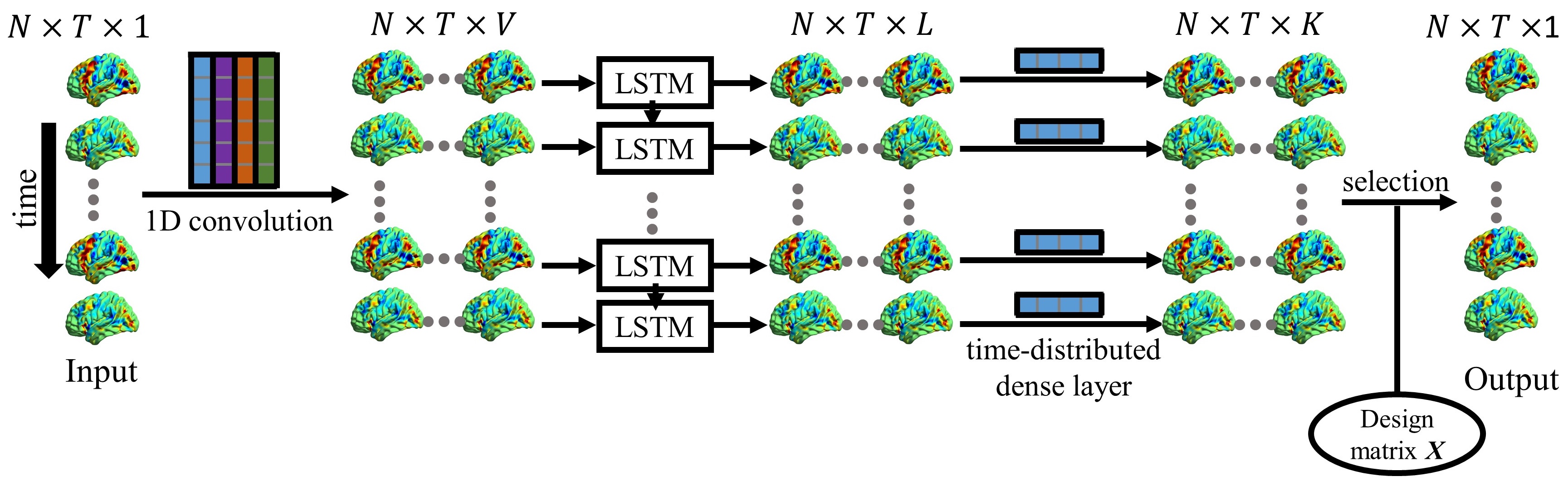
Figure 1. The architecture of the DNN network.
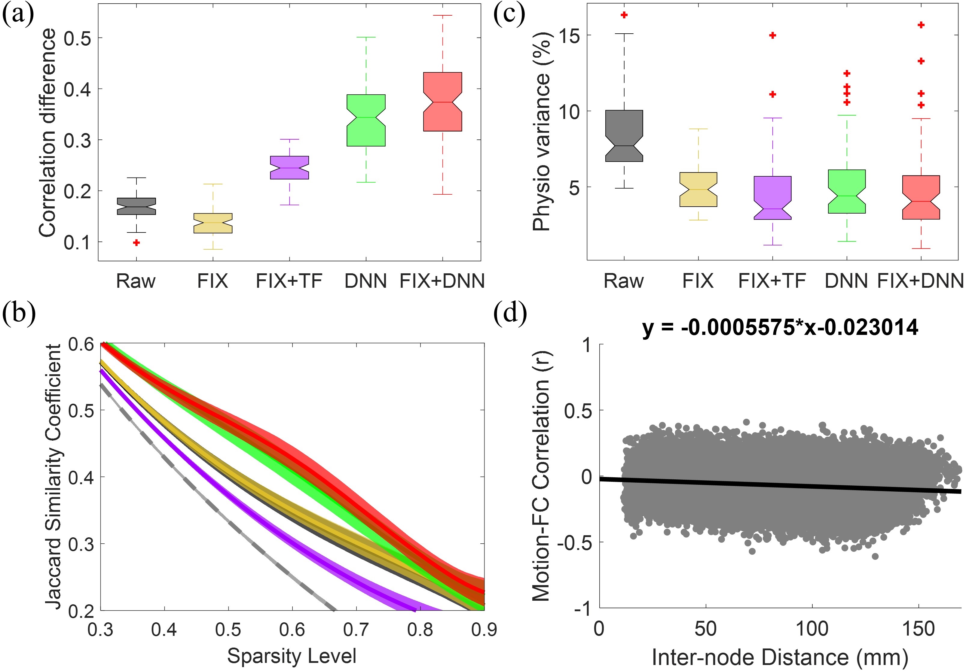
Figure 2. Results from working memory task fMRI data. (a) Boxplot of
correlation difference between top and bottom 20% voxels within gray matter
mask. (b) Similarity between subjects’ correlation maps at varying sparsity
levels. (c) Variance of physiological noise accounted for fMRI data. (d) Plot
of the correlation between pairwise connectivity from raw data (34,716 unique
connections) and mean framewise displacement across subject versus inter-node
distance.
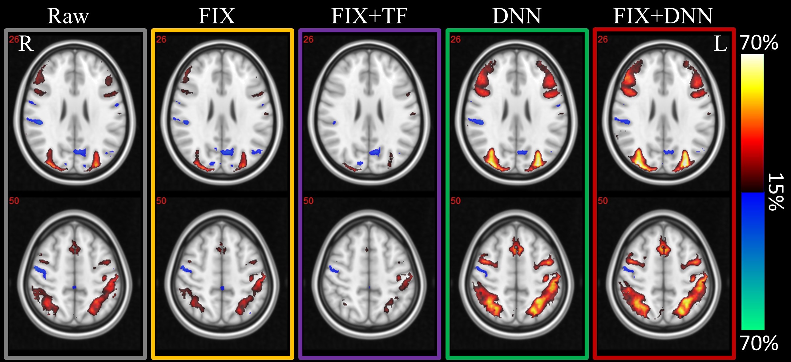
Figure
3. Activation count maps from different denoised fMRI data. The activation
count maps were generated to demonstrate the proportion of subjects that showed
activation or deactivation at a z-threshold of 1.96 (uncorrected two-tailed p
value 0.05) for the target contrast.