0908
Principal gradient of resting-state functional connectivity represents a principal propagating direction of spontaneous brain activity1Biomedical Engineering, Pennsylvania State University, State College, PA, United States, 2Institute for CyberScience, Pennsylvania State University, State College, PA, United States
Synopsis
Decomposing functional connectome to low-dimensional spaces revealed a principal gradient cross brain hierarchy from the primary sensorimotor areas to the default mode network. However, the physiological meaning underlying this principal gradient remains unclear. Here, we showed significant propagating activities of resting-state fMRI along this gradient direction. We further used simulation to demonstrate that the propagating activity can result in a gradient in its propagating direction with the use of the low-dimensional embedding method. Overall, the findings suggest that the principal connectivity gradient actually represents the major propagating direction of spontaneous brain activity, which is likely across brain hierarchies.
Introduction
Decomposing the human functional connectome using a low-dimensional embedding method has revealed a principal gradient showing a gradual transition from the primary sensorimotor regions to the higher-order default mode network (DMN) [1]. This principal gradient suggests a hierarchical organization of the human neocortex, similar to those mapped with myelination contents [2], semantic perceptions [3], and general brain functions [4]. However, the physiological meaning of the principal connectivity gradient remains largely unknown due to the data-driven nature of the applied approach. Specifically, it is unclear what aspect of functional connectivity leads to this spatial gradient of obvious functional relevance. In this study, we examined the relationship between the principal connectivity gradient and propagating structures of resting-state fMRI (rsfMRI) signals by using experimental data and simulation.Methods
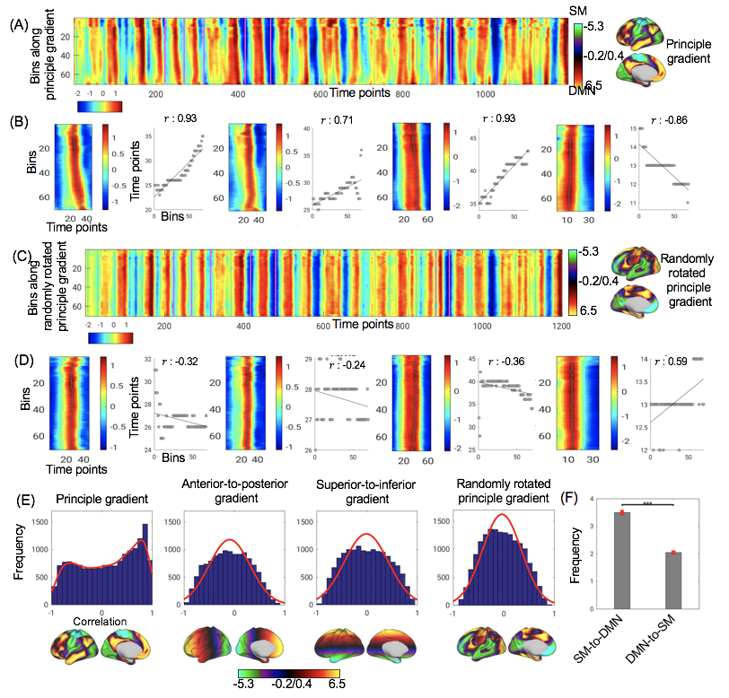
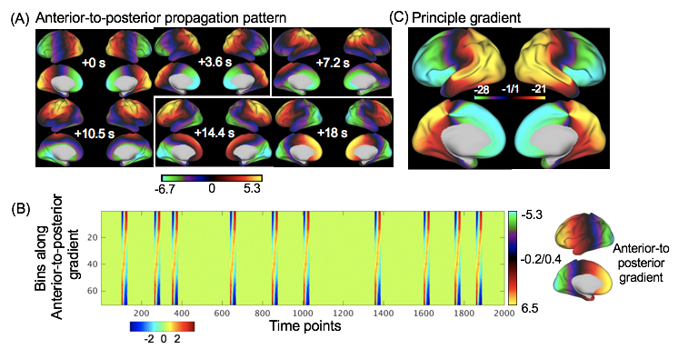
We used rsfMRI data from the 900-subject release of the human connectome project (HCP). We quantified the propagating activity along a given direction, e.g., the principal gradient direction, in the following way. First, we reduced the spatial dimension by dividing 59412 cortical surface vertices into 70 bins along the given direction and then averaged fMRI signals within each bin. Secondly, we cut fMRI signals into temporal segments according to troughs of the global mean signal. For each segment, we located signal peaks of the 70 bins and then examine the correlation between the relative timing of these peaks (time) and their relative position (space) along the given direction. A significant space-time relationship would suggest propagation of these peaks along the given direction for a specific segment. Using this strategy, we summarized and compared propagating activities along the principal gradient and the three other directions generated artificially. We also simulated fMRI signal containing the propagating activity along the anterior-to-posterior direction and then applied the diffusion embedding mapping, which was used to derive the principal gradient, to obtain the associated spatial gradient.Results
Projecting rsfMRI signals along a given direction results in a corresponding space-time plot (Fig. 1A and 1C). Visual inspection of the space-time plot for the direction of the principal connectivity gradient identified instances suggestive of propagating activities between the two ends of this axis: signal peaks move continuously across space over time. Relative timing of these signal peaks is significantly correlated with their relative location along the principal gradient direction, confirming the existence of a space-time relationship. These propagations are mostly from the primary sensorimotor regions towards the DMN, but instances traveling on the opposite direction also exist (Fig. 1B). In contrast, the space-time plots for the randomly rotated principal gradient, do not show similar propagating structures (Fig. 1D). To quantify the above qualitative observations, we examined the space-time relationship, i.e., the correlation between the relative timing of signal peaks and their spatial position along the given direction, for fMRI segments delineated by troughs of the global mean signal. Compared with the other directions, the space-time correlations for the principal gradient direction show a distinct bimodal distribution with a much higher peak on the positive side, suggesting a dominant propagation from the sensory regions to DMN (Fig. 1E). The diffusion embedding method for deriving the principal gradient takes the input of functional connectome, which intuitively does not contain any time-varying information. However, the space-time relationship of propagation could be embedded in the connectome data through the relative strength of functional connectivity. To test this hypothesis, we simulated fMRI data with apparent propagating structure along the anterior-to-posterior direction (Fig. 2A and 2B). The application of the diffusion embedding on the functional connectome constructed on this dataset resulted in a spatial pattern showing this anterior-to-posterior gradient (Fig. 2C).Discussion
Here we showed that the principal gradient of resting-state functional connectivity may actually represent a direction characterized by major propagating activities. Given the correspondence between the principal connectivity gradient and the hierarchical organization of the brain, this also suggests a relationship between the brain hierarchy and spontaneous propagating activity. Propagating structures have been found in rsfMRI data as quasi-periodic patterns [5] and lag threads [6], which might at least partially originate from the propagating instances observed here. We further showed that these propagating activities are largely along a direction defining the brain hierarchy. The finding is consistent with previous observations in rodents with optical imaging that spontaneous activities initiated at different unimodal sensory regions converge to transmodal parietal association area [7]. The functional relevance of these across-hierarchy propagation could be an important topic for future research.Conclusion
The principal gradient of connectivity may originate from spontaneous activity propagating across the brain hierarchy, mostly from the primary sensorimotor regions to the DMN.Acknowledgements
This research was supported by the NIH Pathway to Independence Award (K99/R00) 4R00NS092996-02.References
[1] D. S. Margulies et al., “Situating the default-mode network along a principal gradient of macroscale cortical organization,” Proc. Natl. Acad. Sci., vol. 113, no. 44, pp. 12574–12579, 2016.
[2] J. M. Huntenburg, P. L. Bazin, A. Goulas, C. L. Tardif, A. Villringer, and D. S. Margulies, “A Systematic Relationship Between Functional Connectivity and Intracortical Myelin in the Human Cerebral Cortex,” Cereb. Cortex, vol. 27, no. 2, pp. 981–997, 2017.
[3] A. G. Huth, W. A. De Heer, T. L. Griffiths, F. E. Theunissen, and J. L. Gallant, “Natural speech reveals the semantic maps that tile human cerebral cortex,” Nature, vol. 532, no. 7600, pp. 453–458, 2016.
[4] C. Baldassano, J. Chen, A. Zadbood, J. W. Pillow, U. Hasson, and K. A. Norman, “Discovering Event Structure in Continuous Narrative Perception and Memory,” Neuron, vol. 95, no. 3, p. 709–721.e5, 2017.
[5] G. J. Thompson, W. J. Pan, M. E. Magnuson, D. Jaeger, and S. D. Keilholz, “Quasi-periodic patterns (QPP): Large-scale dynamics in resting state fMRI that correlate with local infraslow electrical activity,” Neuroimage, vol. 84, pp. 1018–1031, 2014.
[6] A. Mitra, A. Z. Snyder, T. Blazey, and E. Marcus, “Correction for Mitra et al., Lag threads organize the brain’s intrinsic activity,” Proc. Natl. Acad. Sci., vol. 112, no. 52, pp. E7307–E7307, 2015.
[7] M. H. Mohajerani et al., “Spontaneous cortical activity alternates between motifs defined by regional axonal projections,” Nat. Neurosci., vol. 16, no. 10, pp. 1426–1435, 2013.
Figures