0898
Brain Tumor Simulator: Creating Ground Truth for Evaluation of Complex MR Acquisition and Reconstruction Methodologies1Department of Biomedical Engineering, University of Wisconsin-Madison, Madison, WI, United States, 2Department of Radiology, University of Wisconsin-Madison, Madison, WI, United States, 3Department of Medical Physics, University of Wisconsin-Madison, Madison, WI, United States, 4Department of Neurological Surgery, University of Wisconsin-Madison, Madison, WI, United States
Synopsis
New dynamic MRI methods promise to better characterize and monitor the vexing problem of brain cancer. Assessing these methods and the assumptions they rely upon is difficult as 1) no known gold standard is available for brain tumors and 2) the time window of availability for brain cancer volunteers is narrow. We present a simulator that generates realistic, heterogeneous, tumor models overlaid over normal brain tissue with user-specified permeability parameters. The simulator generates raw data for arbitrary k-space acquisition strategies. Using this simulator, new methodologies for various tumor types and sizes can be assessed before the first volunteer is ever recruited.
INTRODUCTION
New dynamic contrast enhanced (DCE) MRI strategies hold promise to better detect, characterize, and monitor new and recurrent brain tumors and their margins 1. These strategies utilize combinations of compressed sensing, parallel imaging, and machine learning and thus their performance relies upon several assumptions. Testing their performance and robustness for delivering spatial and temporal resolution is difficult as (1) no gold standard is available for brain cancer imaging and (2) the availability of brain cancer volunteers is limited. Therefore, we modified a previously built breast cancer simulator 2 to generate realistic tumor models of varying sizes and shapes that are overlaid over normal brain tissue. The simulator generates raw data for tumors with user-specified permeability parameters for arbitrary user-provided, k-space acquisition strategies. This platform allows the simulation of raw data that can be reconstructed by new research schemes to later be compared to the digital truth generated from clinically infeasible yet fully sampled acquisitions.METHODS
Tumor Generation:
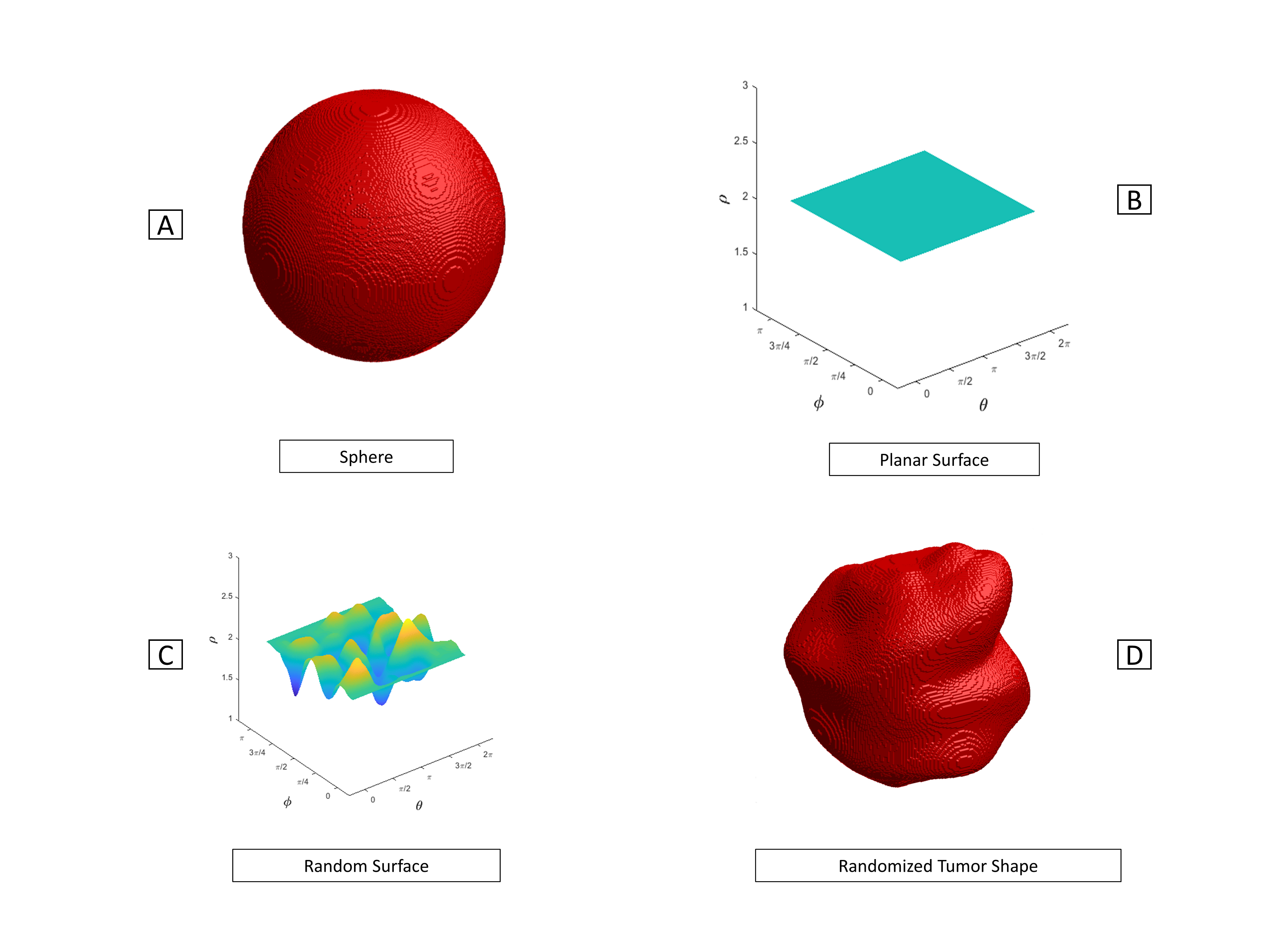
To represent the asymmetry of a typical brain tumor, we randomized the surface of a round sphere to create a randomized yet spherical tumor shape. This process is illustrated in Fig.1.Once the tumor shape is generated, another spherical surface with smaller size and a different randomized pattern is generated and placed inside the previous one. The space enclosed between the smaller surface and the outer surface becomes a tumor rim whose width varies spatially, as shown in Fig. 2A.
Normal tissue layers:
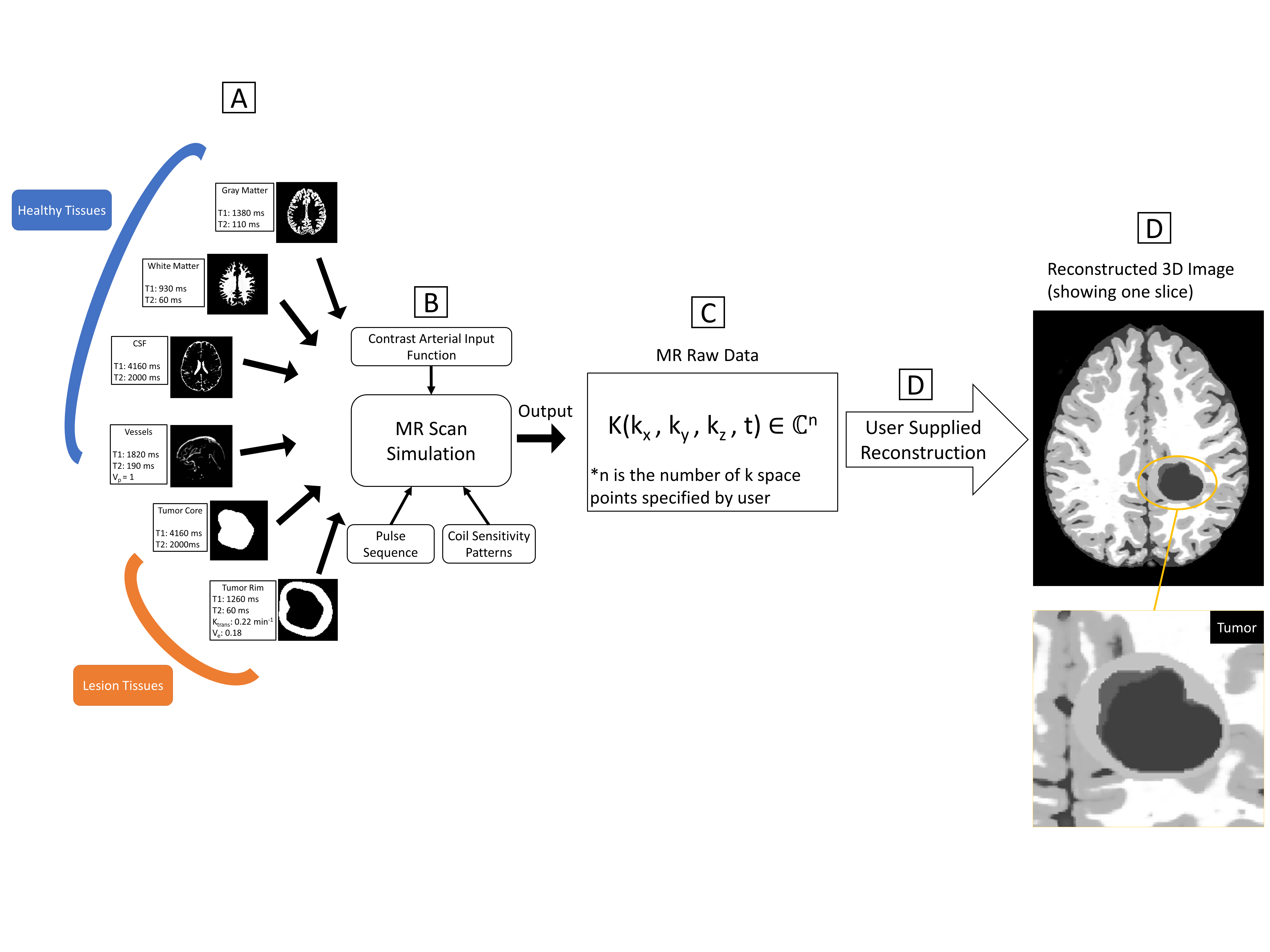
We obtained segmented brain tissue mappings from BrainWeb 3-7. To reduce the computational load, we limited our initial model to the four most prevalent tissues in the brain: white matter, gray matter, cerebral spinal fluid (CSF) and vessels, as shown in Fig. 2A. The tumor generated in Fig. 1 is then superimposed over the normal tissue at a user-specified 3D center coordinate, as shown in Fig. 2A.
Signal generation:
The base simulator package allows MR properties (T1, T2) and pharmacokinetic parameters (volume transfer coefficient (Ktrans), extravascular extracellular volume fraction (Ve), blood plasma volume (Vp)) to be specified for each tissue layer. The simulator uses these to calculate a Gadolinium contrast agent concentration C(t) in tissue layers based on a contrast arterial input function, which then modifies the signal provided by the inherent tissue relaxation properties for each tissue layer (Fig. 2B). Coil sensitivity patterns and the k-space sampling pattern, K (kx,ky,kz,t), from a pulse sequence under test need to be provided by the user and inputted at the stage shown in Fig. 2B. The raw MR data vector for each coil is output by the MR simulation algorithm in Fig. 2C and then cascaded to the reconstruction method under test.
Gold standard generation:
A ground truth can be easily generated by simply providing a fully sampled, 3D mapping of points in Fig. 2 at any desired snapshot point in time and generating a reconstruction. Gold standards were created for a commonly performed clinical, low resolution Cartesian reconstruction [1] and a recently published 3D radial method utilizing compressed sensing and local low rank iterative reconstruction processing, termed STELLR 8
RESULTS
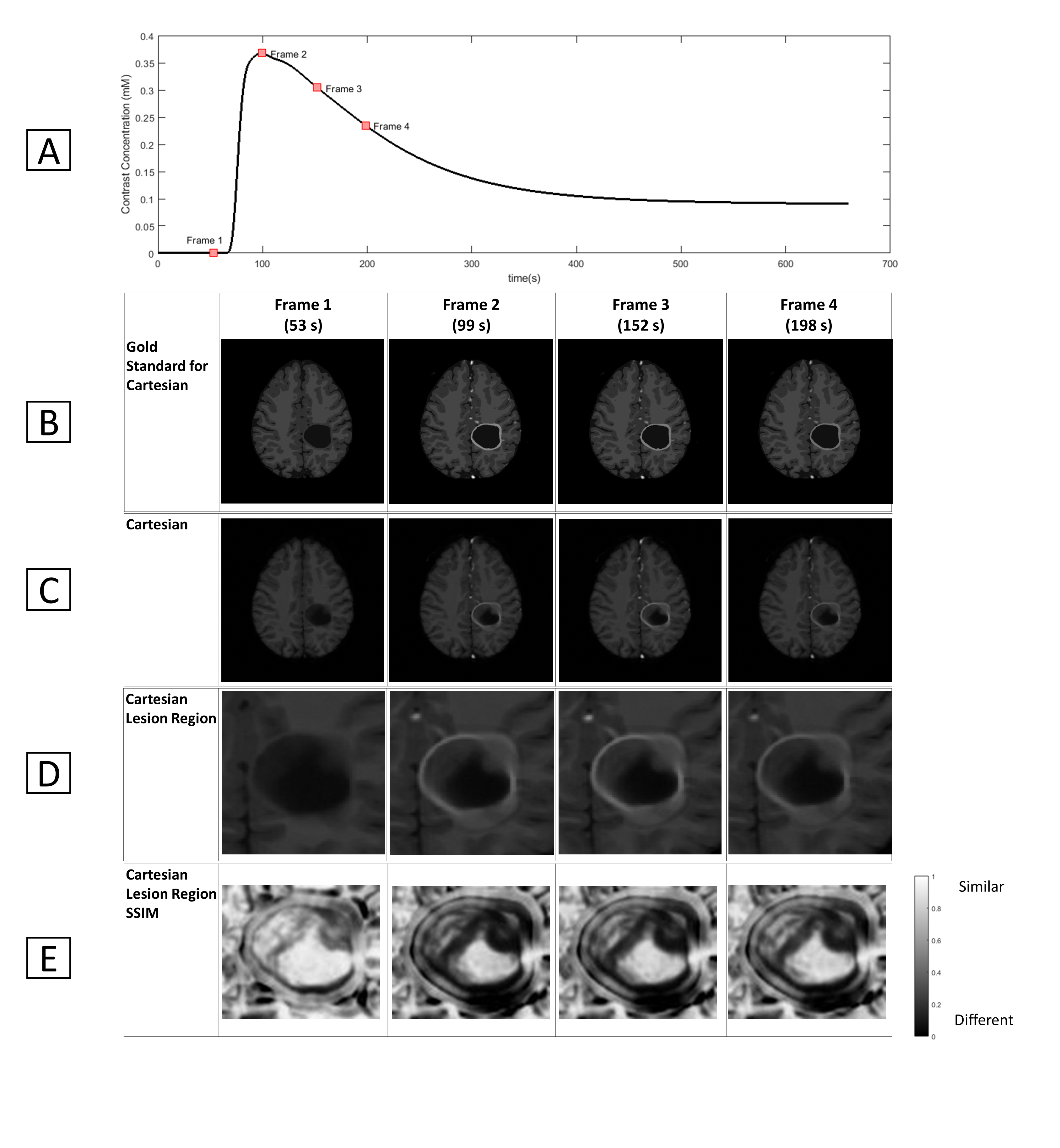
We first created a tumor with a mean radius of 25mm with a necrotic core and a rim with mean width of 2.5mm as shown in Figure 2D. We then simulated a conventional clinical Cartesian DCE acquisition sequence used previously in a brain cancer trial [1] with 0.93*1.3*7mm3 spatial and 6.6s temporal resolution. Structural similarity (SSIM) tests 9 were performed between the conventional Cartesian reconstructed imaged and the gold standards. As shown in Figure3, the relatively thick axial slab obscures the enhancing rim structure due to partial voluming from other tissues.
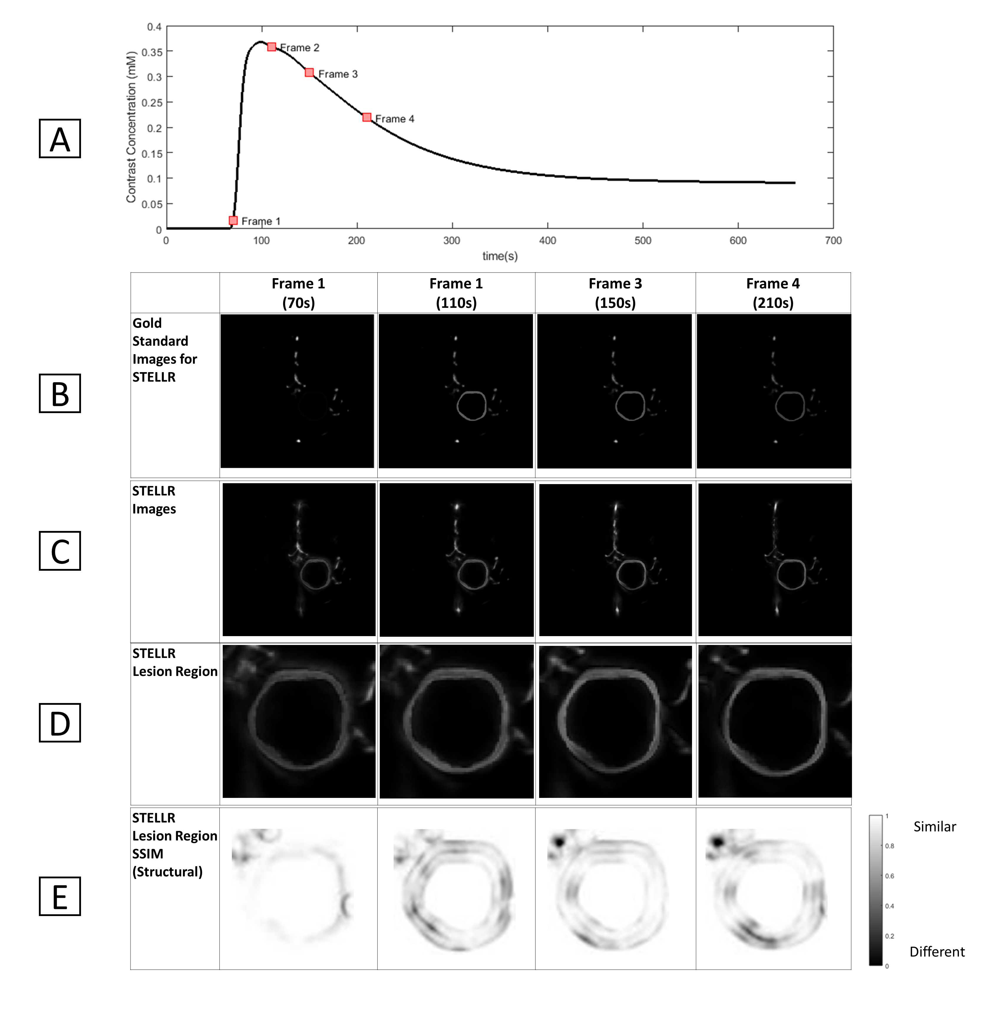
Simulation and evaluation of the STELLR reconstruction algorithm with mask subtraction was also performed on the same lesioned brain to create 0.8mm3 isotropic spatial and 20s temporal resolution (Fig.4). The simulation shows that STELLR performs well at capturing fine details of the enhancing rim but also creates some ringing artifacts around the rim.
DISCUSSION
The two acquisition and reconstruction methods were shown to capture some tumor features correctly while some features were erroneously depicted. We did not include computation of coil sensitivity because doing so was computationally expensive in this preliminary work. Additionally, we simply replaced a normal region of tissue with the brain lesion instead of displacing the normal tissues, as would happen in an actual tumor.CONCLUSION
Given the significant efforts and uncertainty in assessing new imaging methods for brain cancer, the proposed simulator allows one to test and predict performance on a variety of tumor sizes and types before ever recruiting a brain tumor patient. It also provides a much needed capability for objectively comparing the performance of competing methods.Acknowledgements
We acknowledge the support from Vilas associate professor research funding from University of Wisconsin-MadisonReferences
1. Y. Guo et al., High-resolution whole-brain DCE-MRI using constrained reconstruction: Prospective clinical evaluation in brain tumor patients, (in eng), Med Phys, vol. 43, no. 5, p. 2013, May 2016.
2. Henze Bancroft LC et al., An Anthropomorphic Digital Breast Phantom for Simulation and Analysis of MRI Techniques: Implementation for dynamic contrast-enhanced MRI, Proceedings of Joint Annual Meeting ISMRM-ESMRMB 2010, Stockholm, Sweden.
3. BrainWeb: Simulated Brain Database (Accessed Nov. 5). Available: http://brainweb.bic.mni.mcgill.ca/brainweb/
4. C. A. Cocosco, V. Kollokian, R. K.-S. Kwan, and A. C. Evans, BrainWeb: Online Interface to a 3D MRI Simulated Brain Database, vol. 5, ed. NeuroImage, 1997.
5. R. K. Kwan, A. C. Evans, and G. B. Pike, MRI simulation-based evaluation of image-processing and classification methods, (in eng), IEEE Trans Med Imaging, vol. 18, no. 11, pp. 1085-97, Nov 1999.
6. D. L. Collins et al., Design and construction of a realistic digital brain phantom, (in eng), IEEE Trans Med Imaging, vol. 17, no. 3, pp. 463-8, Jun 1998.
7. R. K. Kwan, A. C. Evans, and G. B. Pike, An Extensible MRI Simulator for Post-Processing Evaluation, vol. 1131, ed. Lecture Notes in Computer Science: Springer-Verlag, 1996, pp. 135-140.
8. J. E. Jimenez, R. M. Strigel, K. M. Johnson, L. C. Henze Bancroft, S. B. Reeder, and W. F. Block, Feasibility of high spatiotemporal resolution for an abbreviated 3D radial breast MRI protocol, (in eng), Magn Reson Med, vol. 80, no. 4, pp. 1452-1466, Oct 2018.
9. W. Zhou, A. C. Bovik, H. R. Sheikh, and E. P. Simoncelli, Image Qualifty Assessment: From Error Visibility to Structural Similarity, vol. 13, ed. IEEE Transactions on Image Processing, 2004, pp. 600-612.
Figures