0893
Structural Connectivity abnormality in children treated for Medulloblastoma1The Hospital for Sick Children, Toronto, ON, Canada
Synopsis
Curative treatments for medulloblastoma impart significant toxicity on the developing brain. Though changes to white matter have been described, there remains a limited understanding of the effects of treatment on the structural connectome, which is thought to subserve complex and dynamic behaviors. Identifying compromise within the connectome may elucidate mechanisms of toxicity among survivors. We analyzed connectomic differences between survivors and age-matched controls. We identified two networks situated posteriorly with reduced white matter microstructure in survivors. Our findings complement studies showing long-term effects of treatment on brain structure, and localize these effects to areas around the site of the tumor.
Background
Curative treatments for medulloblastoma impart significant toxicity on the developing brain and lead to prominent neuropsychological deficits [1]. Though diffusion Tensor Imaging (DTI) has been used to describe treatment-related changes to white matter connections, there remains a limited understanding of the effect of treatment on the structural connectome. The structural connectome refers to the dense, integrative network of white matter connections present in the brain [2]. Networks within the connectome are thought to sub-serve complex and dynamic behaviors [3], and changes within these networks may help to explain treatment-related deficits in medulloblastoma survivors. Recent investigations have utilized a network analysis approach to characterize structural network changes in attention deficit hyperactivity disorder [4] and acute mild traumatic brain injury [5]. The present study aims to describe global connectomic changes in children treated for medulloblastoma. Identifying areas of compromise within the structural connectome long after treatment may help elucidate some of the mechanisms of treatment-related toxicity that is common among pediatric brain tumor survivors.Methods
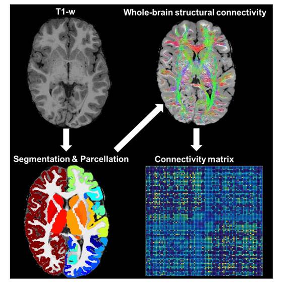
21 children treated for medulloblastoma (mean age = 14.27±2.86 years, 24% female) and 21 typically developing children (mean age = 14.25±2.46 years, 38% female) children were scanned at the Hospital for Sick Children in Toronto. The average time since treatment for patients was 7.03±4.09 years. MR images were acquired using a Siemens 3-Tesla scanner with a twelve-channel head coil according to the following protocol: 3D-T1 MPRAGE Grappa 2 protocol (TE/TR = 3.91/2300 ms, 160 contiguous axial slices, flip angle = 90o, 256 × 224 matrix, FOV = 256 × 224 mm, voxel size = 1 mm ISO) and a diffusion-weighted sequence using a single shot spin echo (TE/TR = 90/9000 ms, 70 contiguous axial slices, flip angle = 90o, 122 × 122 matrix interpolated to 244 × 244, FOV = 244 × 244 mm, b0 value = 1000 s/mm2, 30 directions, voxel size = 2mm ISO). Surface reconstruction, volumetric segmentation, and anatomic parcellation of MR images were completed using FreeSurfer (v.6.0) [6]. DTI map calculations and probabilistic streamline generation were completed using MRtrix3 (v0.3.16) [7] software. Connectivity matrices were derived by estimating the mean index (FA/MD/AD/RD) across streamlines connecting any two nodes; all resulting connections were extracted as a connectivity matrix (Fig. 1). Mass univariate comparisons of connections were carried out using the Network Based Statistic (NBS) Toolbox (5000 permutations at α = 0.05). DTI indices across significant networks were compared between patients and controls, with sex, handedness, and intracranial volume included as covariates.Results
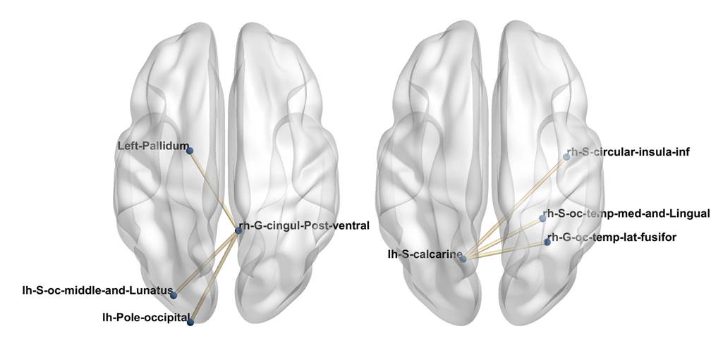
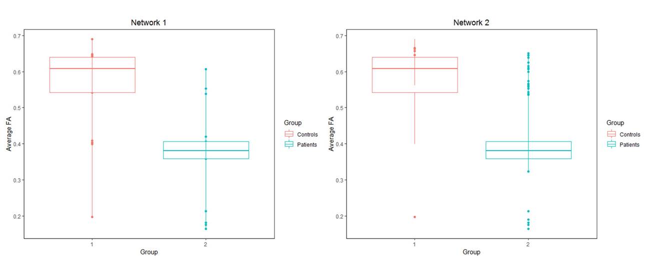
We identified two networks with significantly lower mean FA in patients (Fig. 2 & 3). Network comparisons for MD, AD, and RD were not significant. For the first significant network, group comparisons revealed significantly lower FA in patients (t=-5.25, p<0.0001) and no significant effect of sex (t= 1.03, p>0.05) or intracranial volume (t=-1.27, p>0.05). For the second significant network, group comparisons revealed significantly lower FA in patients (t=-2.79, p<0.01) and no significant effect of sex (t=1.70, p>0.05) or intracranial volume (t=-1.21, p>0.05).Discussion
Our results suggest reduced white matter microstructure in posteriorly situated structural networks long-term after treatment for medulloblastoma. Reductions in FA in patients may reflect treatment-related injury to these networks and/or altered white matter development subsequent to treatment.Conclusion
Treatment for medulloblastoma may restrict subsequent development of white matter within specific networks. Our findings complement prior studies showing the long-term effects of treatment on brain structure, and potentially localize these effects to networks located close to the site of the tumor. Future work will relate these findings to relevant cognitive and behavioral data in order to explore the potential impact of structural deficits within these networks.Acknowledgements
The authors thank Dr. S. Bells for support with data analysisReferences
1. Mabbott, DJ. et al (2009) Diffusion tensor imaging of white matter after cranial radiation in children for medulloblastoma: Correlation with IQ. Neuro-Oncology, 8(3), 244-252.
2. Sporns, O., Chialvo, DR., Kaiser, M., Hilgetag, CC. (2004) Organization, development and function of complex brain networks. Trends in cognitive sciences 8(9), 418-425.
3. Menon, V. (2011). Large-scale brain networks and psychopathology: a unifying triple network model. Trends in cognitive sciences, 15(10), 483-506.
4. Cao, Q. et al (2013) Probabilistic diffusion tractography and graph theory analysis reveal abnormal white matter structural connectivity networks in drug-naive boys with attention deficit/hyperactivity disorder. The Journal of Neuroscience 33(26), 10676-10687.
5. Yuan. W., Wade, SL., & Babcock, L. (2015) Structural connectivity abnormality in children with acute mild traumatic brain injury using graph theoretical analysis. Human brain mapping 36(2), 779-792.
6. Fischl, B. (2012). FreeSurfer. Neuroimage, 62(2), 774-781.
7. Tournier, J., Calamante, F., & Connelly, A. (2012). MRtrix: diffusion tractography in crossing fiber regions. International Journal of Imaging Systems and Technology, 22(1), 53-66.
Figures