0890
A functional connectome-based interspecies model boosts classification in neuropsychiatric disorders1Institute of Neuroscience, CAS Center for Excellence in Brain Science and Intelligence Technology, State Key laboratory of Neuroscience, CAS Key Laboratory of Primate Neurobiology, Chinese Academy of Sciences, Shanghai, China, 2University of Chinese Academy of Sciences, Beijing, China, 3Institute of Automation, CAS Center for Excellence in Brain Science and Intelligence Technology, Chinese Academy of Sciences, Beijing, China, 4Kunming Institute of Zoology, Chinese Academy of Sciences, Kunming 650223, China, Kunming, China
Synopsis
Cross-species comparative connectomices based on resting-state functional MRI is a promising method to investigate large-scale brain organization. Here we leveraged a transgenic monkey model overexpressing MECP2 and developed a novel connectome-based interspecies machine learning algorithm for clinical diagnosis of individuals with neuropsychiatric disorders. This fully cross-validated algorithm based on cross-species mapping of regional features significantly boosts the diagnostic performance of ASD and OCD, but not for ADHD, in independent human cohorts, which paves a new avenue to establish a translational path to dissect the neural circuit mechanisms underlying complexity and heterogeneity of human mental disorders.
Introduction
Growing evidence supports the feasibility of constructing genetically engineered non-human primates to study mental illness and develop more effective therapeutics for patients1, 2. The translational potential of non-human primate models for examining human psychiatric disorders stands to benefit greatly from resting state functional MRI methods3. Despite this potential, there are no clear roadmaps for how to establish cross-species translational mapping under disease conditions. Here, we propose a novel cross-species translational model based on a transgenic monkey with methyl-CpG binding protein 2 (MECP2) duplication exhibiting autism-like behavior2, to apply for the diagnostic classification of human subjects with neuropsychiatric disorders.Method
Our interspecies machine learning model was tested in a cohort of 144 monkey datasets obtained from 5 transgenic (TG) and 11 wild-type (WT) monkeys (TG, 45 datasets; WT, 99 datasets), and four human cohorts including two ASD (ABIDE-I: ASDs = 133, HCs = 203; ABIDE-II: ASDs = 60, HCs = 89)4, 5, one ADHD (ADHDs = 102, HCs = 173)6 agglomerative data from a public repository, and one cohort of OCD (OCDs = 92, HCs = 79)7. The connectivity network was then constructed with a total of 94 parcellated brain regions, including 80 cortical areas based on the Regional Map template8, and 14 subcortical areas based on the INIA19 and Freesurfer templates for monkeys9 and humans10, respectively.
We adopted the group lasso method11 to identify a subset of active brain regions in the MECP2 monkey and then projected to the human counterpart for constructing a classifier (monkey-based classifier). A leave-one-participant-out cross validation (LOOCV) procedure employing sparse logistic regression (SLR)12 was implemented to distinguish patients from healthy controls. The features (functional connections) were determined by using LASSO with nested 10-fold cross-validation in a training set.
We evaluated the performance of the human-based classifier (active regions identified from human dataset) in comparison with the monkey-based classifier in the same human cohorts using the same procedure. To assess the probability of obtaining specificity and sensitivity values higher than those obtained by chance, we randomly selected 9 out of 94 regions with 5,000 repetitions.
Results
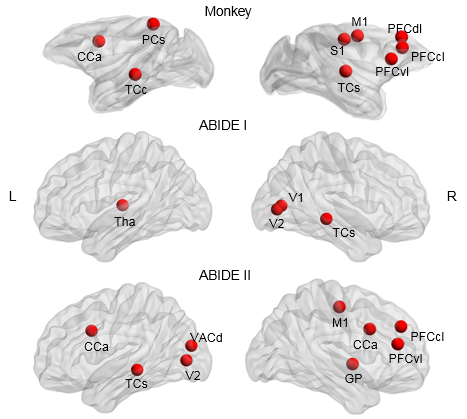
The algorithm automatically and objectively identified 9 active regions out of 94 nodes in the monkey cohort, which included the left TCc, right TCs, right PFCdl, right S1 and M1, left CCa, right PFCcl, left PCs, right PFCvl (Figure 1).
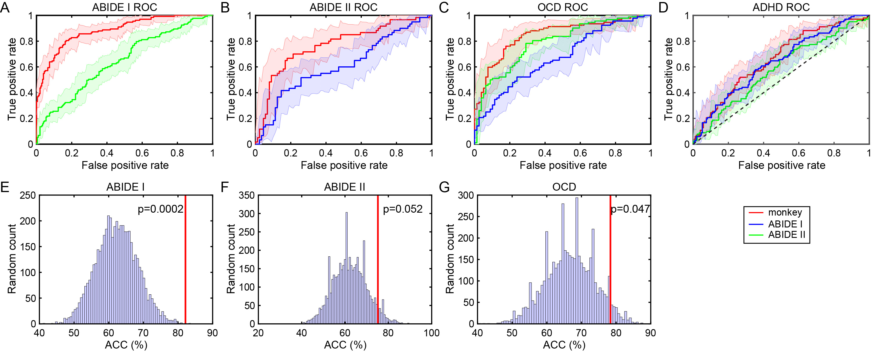
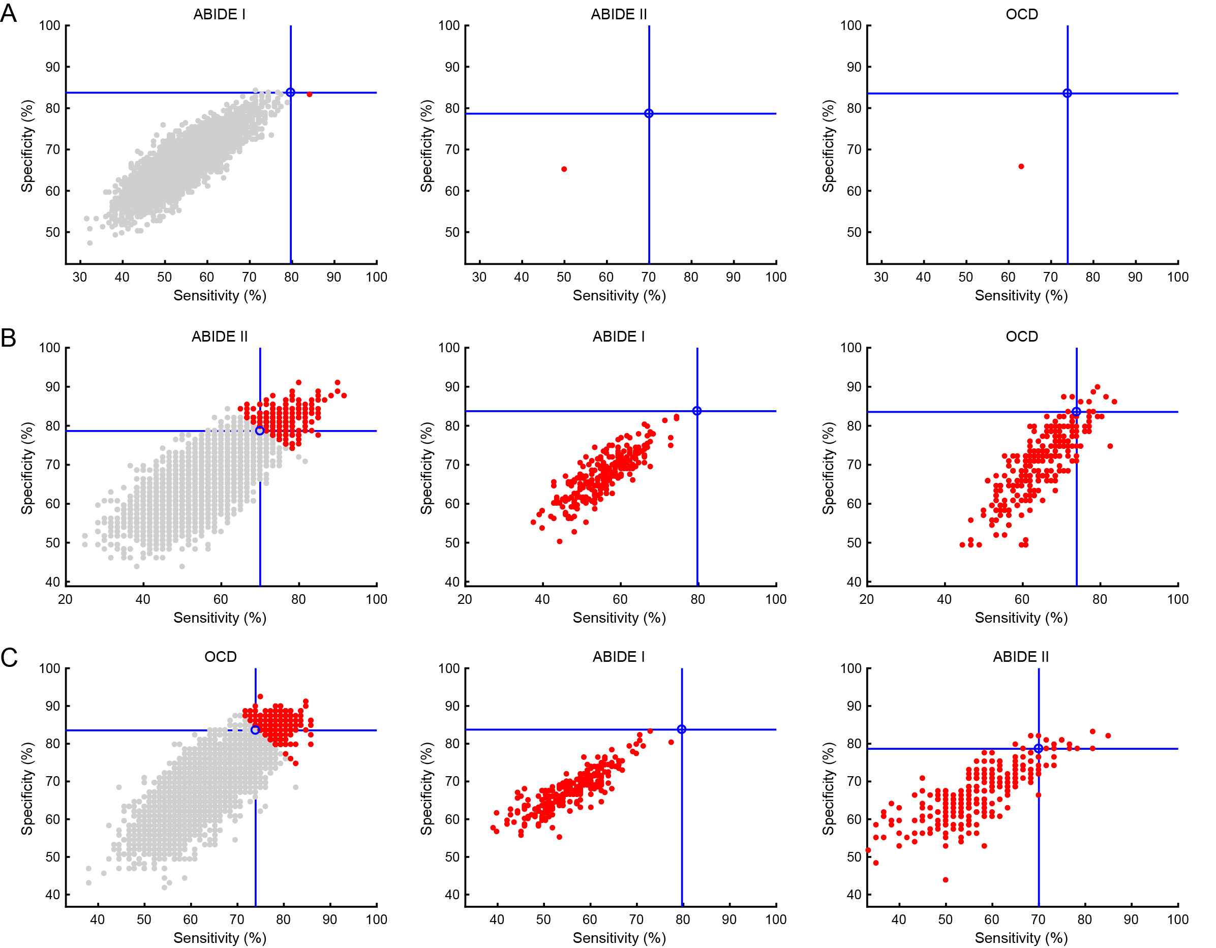
Our interspecies model boosted classification for ABIDE-I ASD (monkey-based accuracy = 82.14%, ABIDE-II based accuracy = 61.31%), validated in ABIDE-II ASD (monkey-based accuracy = 75.17%, ABIDE-I based accuracy = 60.40%). The same model applied in the OCD cohort also achieved high performance (monkey-based accuracy = 78.36%, ABIDE-I based accuracy = 69.59%, ABIDE-II based accuracy = 60.23%). By contrast, the monkey-based classifier performed poorly in the ADHD cohort with an accuracy of 64.73%, which yielded no significant difference relative to the human-based classifier (Figure 2 upper row). In addition, the performance of the monkey-based classifier was significantly better than expected by chance (Figure 2 bottom row). It is worthy to mention that the performance of the monkey-based classifier was not necessarily best for all the scenarios whereas it may hold better generalizability to other disease conditions compared to the random choice (Figure 3).
Discussion
The mapping of key nodes in the pathological circuits of transgenic monkeys to the homolog in the human conditions indicates a conserved role of these regions in the functional specialization of large-scale networks in the human and nonhuman primate brain. Knowledge about the active regions gleaned from the transgenic monkey model was useful to improve diagnostic classification of ASD and OCD cohorts, but not in ADHD, suggesting that these 9 regions may serve as key elements of a circuit endophenotype which has both marked generalizability and selectivity. Our findings help to strengthen the current understanding of the prevailing functional dysconnectivity hypothesis in ASD and OCD.Conclusion
We present a novel MRI-derived cross-species translational model for boosting the diagnostic classification performance of patients with ASD and OCD from healthy controls. The present cross-species modeling paves a new avenue to establish the mapping of distinct symptom clusters to neural circuits, thereby elucidating the neural circuit mechanisms underlying complexity and heterogeneity of mental disorders.Acknowledgements
No acknowledgement found.References
1. Jennings, C.G., R. Landman, Y. Zhou, et al., Opportunities and challenges in modeling human brain disorders in transgenic primates. Nat Neurosci, 2016. 19(9): p. 1123-30.
2. Liu, Z., X. Li, J.T. Zhang, et al., Autism-like behaviours and germline transmission in transgenic monkeys overexpressing MeCP2. Nature, 2016. 530(7588): p. 98-102.
3. Miranda-Dominguez, O., B.D. Mills, D. Grayson, et al., Bridging the gap between the human and macaque connectome: a quantitative comparison of global interspecies structure-function relationships and network topology. J Neurosci, 2014. 34(16): p. 5552-63.
4. Di Martino, A., D. O'Connor, B. Chen, et al., Enhancing studies of the connectome in autism using the autism brain imaging data exchange II. Sci Data, 2017. 4: p. 170010.
5. Di Martino, A., C.G. Yan, Q. Li, et al., The autism brain imaging data exchange: towards a large-scale evaluation of the intrinsic brain architecture in autism. Mol Psychiatry, 2014. 19(6): p. 659-67. 6. Consortium, H.D., The ADHD-200 Consortium: A Model to Advance the Translational Potential of Neuroimaging in Clinical Neuroscience. Front Syst Neurosci, 2012. 6: p. 62.
7. Yin, D., C. Zhang, Q. Lv, et al., Dissociable Frontostriatal Connectivity: Mechanism and Predictor of the Clinical Efficacy of Capsulotomy in Obsessive-Compulsive Disorder. Biol Psychiatry, 2018.
8. Bezgin, G., V.A. Vakorin, A.J. van Opstal, et al., Hundreds of brain maps in one atlas: registering coordinate-independent primate neuro-anatomical data to a standard brain. Neuroimage, 2012. 62(1): p. 67-76.
9. Rohlfing, T., C.D. Kroenke, E.V. Sullivan, et al., The INIA19 Template and NeuroMaps Atlas for Primate Brain Image Parcellation and Spatial Normalization. Front Neuroinform, 2012. 6: p. 27.
10. Fischl, B., D.H. Salat, E. Busa, et al., Whole brain segmentation: automated labeling of neuroanatomical structures in the human brain. Neuron, 2002. 33(3): p. 341-55.
11. Yuan, M. and Y. Lin, Model selection and estimation in regression with grouped variables. Journal of the Royal Statistical Society: Series B (Statistical Methodology), 2006. 68(1): p. 49-67.
12. Yamashita, O., M.A. Sato, T. Yoshioka, et al., Sparse estimation automatically selects voxels relevant for the decoding of fMRI activity patterns. Neuroimage, 2008. 42(4): p. 1414-29.
Figures