0885
Functional Connectivity Mediates the Relationship between Regional Excitation-Inhibition Balance and Default-Mode Network Deactivation1Neuroimaging Research Branch, National Institue on Drug Abuse, Baltimore, MD, United States, 2Department of Psychology, Zhejiang University, Hangzhou, China, 3Harvard Medical School, Boston, MA, United States, 4Beijing Normal University, Beijing, China
Synopsis
Aberrant default mode network (DMN) activity has been related to aging and various neuropsychiatric illnesses. To better understand the neural mechanisms underlying the DMN deactivation, we investigated the triple-relationship among the task-induced deactivation, regional excitation-inhibition balance, and the interregional functional connectivity strength associated with the DMN. Using mediation analysis, we found that the network interaction between DMN and the salience network partially mediated the association between the regional excitation-inhibition balance and the DMN deactivation. This finding bridges DMN-deactivation related findings from various neuroimaging modalities and may provide new insights into the neural mechanisms of the DMN deactivation.
Introduction
The default-mode network (DMN) features increased activity during self-referential thoughts and decreased activity during task performance that request external attention1,2. It has been demonstrated that task-elicited DMN deactivation, regional neurotransmitter concentrations and DMN functional connectivity are related with each other3-6. In this study, we took a further step to integrate these observations via a mediation model. We first examined the pairwise relationship between local resting-state glutamate/GABA concentrations, task-induced deactivation, and interregional functional connectivity (FC) associated with DMN when subjects performed an n-back working memory (WM) task. We then tested our hypothesis that functional connectivity mediates the relationship between the neurotransmitter concentrations and the task-induced deactivation at the posterior cingulate cortex/precuneus (PCC/PCu).Methods
Acquisition. Sixty-five participants (36 males, age range 17-49 years) pooled from two fMRI studies (Cohort 1: 35 subjects5; Cohort 2: 30 subjects6) gave informed consent approved by the NIDA-IRB. High-resolution T1w images and WM task BOLD fMRI data were collected on all subjects. GABA and glutamate were quantified on Cohort 2 subjects at the PCC/PCu, using a MEGA-PRESS and a PRESS sequence6. The n-back task was presented in a block-design pattern, during which, participants were asked to press a button for letter ‘D’ (0-back) or whenever a presented letter is the same as the one presented n trials before (n=1,2,3).
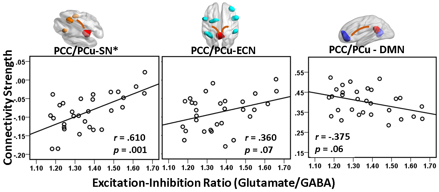
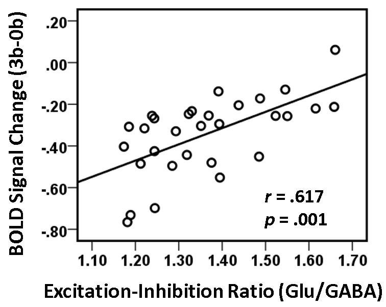
MRS Quantification. MRS data was quantified by fitting the MR spectra with a linear combination of basis sets using LCModel7. Spectra were excluded from further analysis if CRLB for GABA or glutamate was higher than 20%. Ratio of glutamate over GABA concentration level (denoted as Glu/GABA ratio) was computed as a measure of local excitation-inhibition balance.
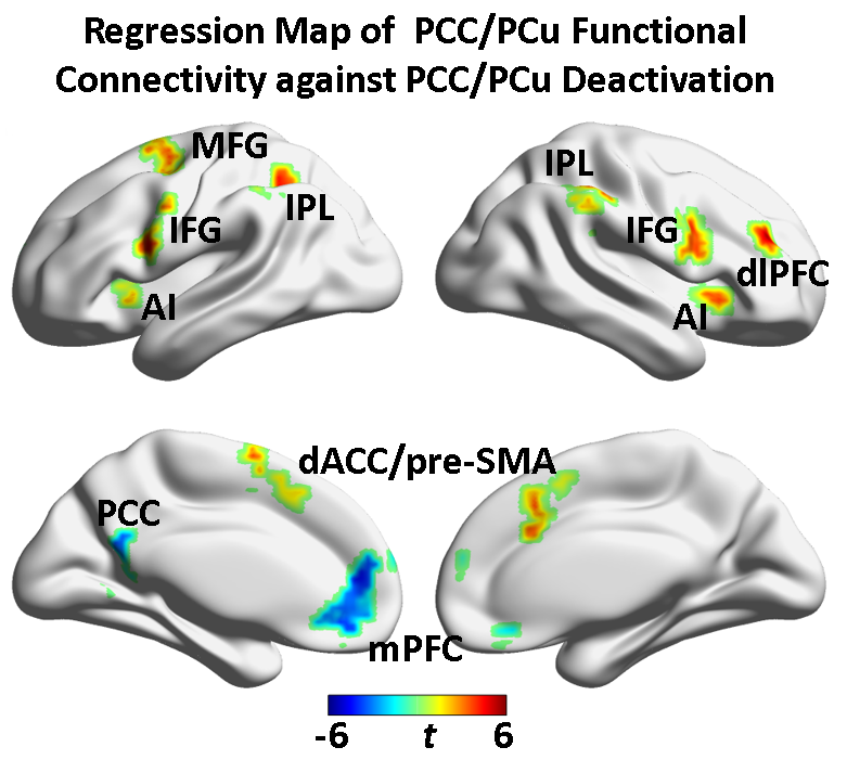
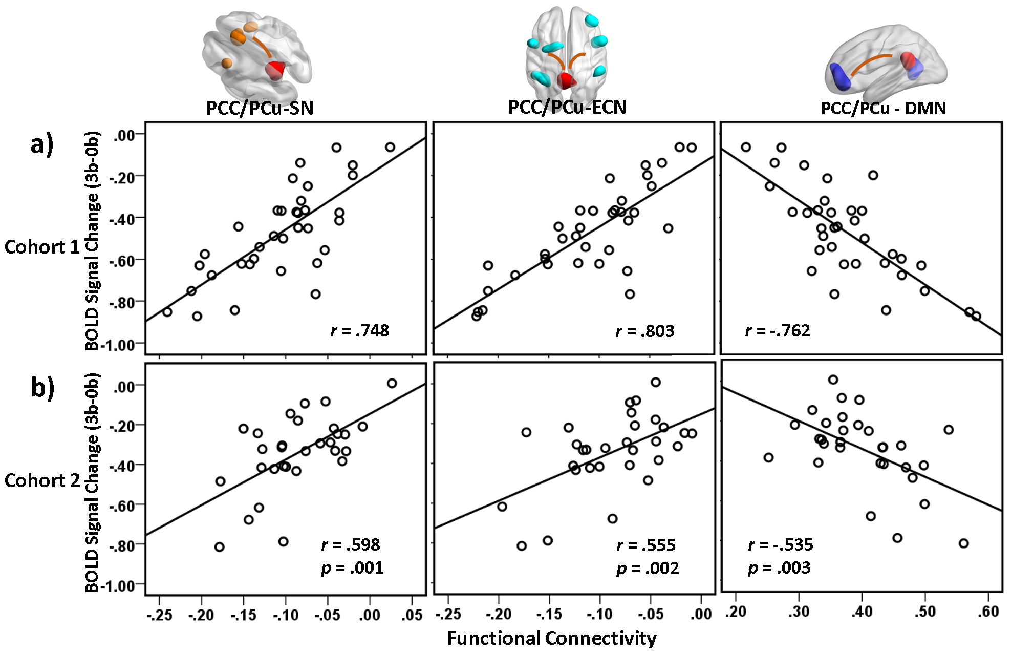
Preprocessing and Analysis. All fMRI data were preprocessed with a standard pipeline6. In task activation analysis, first-level individual activation maps under the 1-back, 2-back, and 3-back load versus the 0-back were obtained using GLM analysis. A second-level whole-brain repeated-measure ANOVA analysis was conducted on the 35 n-back activation maps from Cohort 1 to define the task-deactivated PCC/PCu region, which was used in the ensuing FC analysis as seed ROI. FC strength between PCC/PCu and other brain regions in the task context was assessed on the n-back task-removed residual time courses. To investigate which PCC/PCu related brain circuits correlate with the deactivation levels, a voxel-wise regression analysis was performed on the 35 PCC/PCu FC maps against the individual mean PCC/PCu BOLD signal changes at 3-back. Depending on their locations, the identified influential regions was grouped into three networks as SN (salience network), ECN (executive-control network), and DMN. The mean FC between PCC/PCu and the three networks was extracted from the 30 PCC/PCu FC maps of Cohort 2 to test whether this deactivation-predictive model derived from Cohort 1 can be generalized to independent datasets. Meanwhile, the relationship of the Glu/GABA ratio at rest with the DMN-SN/ECN inter-network / intra-DMN FC and with the task-induced deactivation was examined. The DMN inter-network (PCC/PCu-SN and PCC/PCu-ECN) and intra-network (PCC/PCu – DMN) circuits, whose FC correlated with both task-induced deactivation and the Glu/GABA ratio, will enter the ensuing mediation analysis as a mediator.
Results
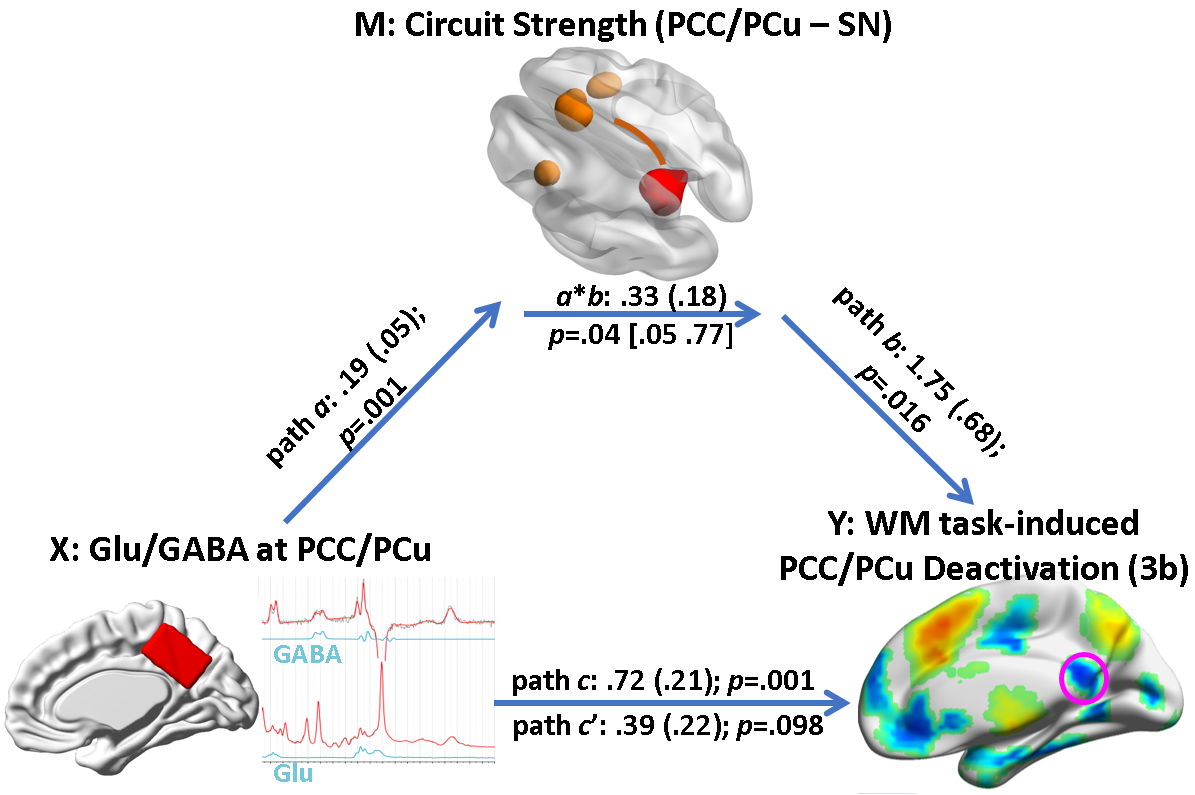
The voxel-wise regression analysis on Cohort 1 datasets revealed that the PCC/PCu deactivation was positively correlated with the DMN-SN and DMN-ECN coupling, and negatively correlated with the intra-DMN FC (Fig. 1). These relationships were replicated on Cohort 2 datasets (Fig.2). In addition, lower Glu/GABA ratio was associated with higher DMN-SN FC strength, but not with the DMN-ECN FC, nor with the intra-DMN coupling after Bonferroni correction (Fig.3). Consistent with our previous study, lower Glu/GABA ratio was associated with greater PCC/PCu deactivation (Fig.4). The mediation analysis revealed that the combined DMN–SN inter-network FC strength partially mediated the relationship between the local Glu/GABA ratio and the PCC/PCu deactivation at 3-back (Fig. 5).Discussion and Conclusion
Based on our model, the origin of deficits in DMN
deactivation during task performance may be rooted in an imbalance between
glutamate and GABA neurotransmission and/or disrupted DMN–SN functional
interaction. Identifying the nature of DMN dysfunction is important for the
development of effective interventional strategies. If the imbalance of
excitatory and inhibitory neurotransmission is the major cause, a pharmacologic
strategy to restore the balance may be more effective. If the deficit results
from impaired inputs from remote SN nodes that mediate DMN activity through FC,
a neuromodulation strategy to restore the long-range inputs by targeting such
regions as anterior insula may be more effective. Collectively, an integrated
application of pharmacological and neuromodulation strategies may achieve the
maximum outcome in rescuing DMN deactivation deficits.Acknowledgements
This work was supported by the Intramural Research Program of National Institute on Drug Abuse at the National Institutes of Health.References
1. Andrews-Hanna JR, Smallwood J, Spreng RN. The default network and self-generated thought: component processes, dynamic control, and clinical relevance. Ann NY Acad Sci. 2014;1316:29-52. 2. Buckner RL, Andrews-Hanna JR, Schacter DL. The brain’s default network: anatomy, function, and relevance to disease. Ann NY Acad Sci. 2008;1124:1-38. 3. Northoff G, Walter M, Schulte RF, et al. GABA concentrations in the human anterior cingulate cortex predict negative BOLD responses in fMRI. Nat Neurosci. 2007;10:1515-17. 4. Kapogiannis D, Reiter DA, Willette AA, et al. Posteromedial cortex glutamate and GABA predict intrinsic functional connectivity of the default mode network. Neuroimage. 2013;64:112-9. 5. Zou Q, Ross TJ, Gu H, et al. Intrinsic resting-state activity predicts working memory brain activation and behavioral performance. Hum Brain Mapp. 2013;34:3204-15. 6. Hu Y, Chen X, Gu H et al. Resting-state glutamate and GABA concentrations predict task-induced deactivation in the default mode network. J Neurosci. 2013;33:18566-73. 7. Provencher SW. Automatic quantitation of localized in vivo 1H spectra with LCModel. NMR Biomed. 2001;14:260-4.
Figures