0882
Aberrant Temporal Flexibility of Visual Network in Schizophrenia: a Resting State fMRI Study1UC Riverside, RIVERSIDE, CA, United States
Synopsis
Schizophrenia has been associated with large-scale hypo-connectivity between brain networks. Since functional connectivity (FC) is dynamic, characterizing the temporal dynamics of FC may provide valuable insights regarding the underlying FC aberrance of patients. We present a framework to investigate the aberrant functional interaction between brain networks in schizophrenia using sliding window correlation and modularity analysis and found aberrant temporal flexibility in ROIs for visual object recognition in patients. Examination of their temporal co-activation profiles revealed higher co-activation of
INTRODUCTION
Schizophrenia has been associated with disrupted neural connectivity between brain networks1. Traditional static functional connectivity (FC) analysis using resting-state fMRI data (rfMRI) has demonstrated large-scale hypo-connectivity between brain networks in schizophrenia2. Since FC is dynamic and non-stationary3, characterizing the temporal dynamics of FC may provide valuable insights regarding the underlying FC aberrance of patients. While prior studies of schizophrenia FC dynamics focused on changes of FC between networks4, how a functional region or ROI interacts with others remains unknown. We present a framework to investigate the aberrant functional interaction between brain networks in schizophrenia.METHODS
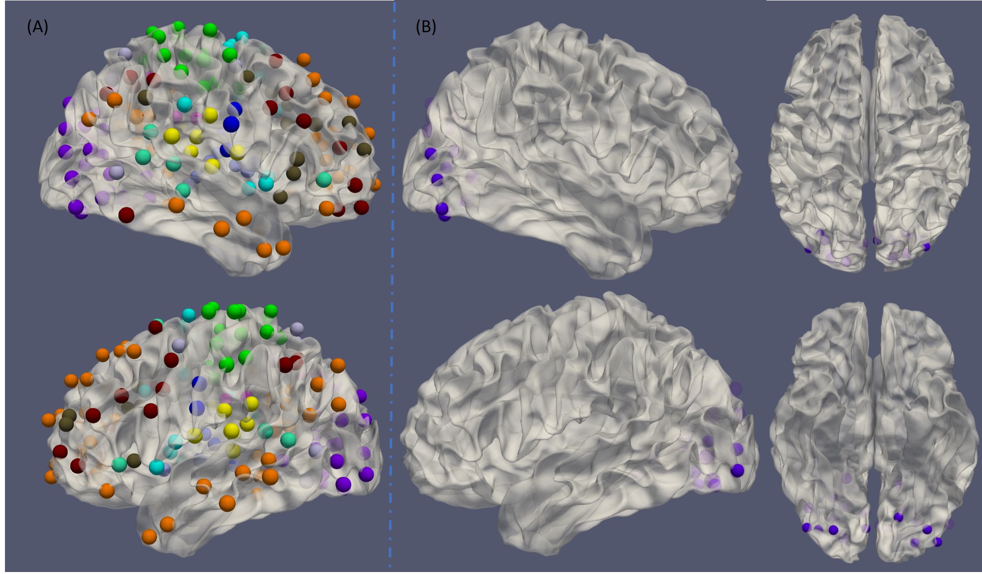
Fifty normal control (NC) subjects and 50 age- and sex-matched schizophrenia (SZ) patients from the UCLA Consortium for Neuropsychiatric Phenomics LA5c Study were included in the analysis. Standard prepossessing was performed on the rfMRI data using an in-house pipeline based on Freesurfer and AFNI. Nuisances including motion parameters, three principal components from BOLD signals of ventricles and local white matter signals were regressed out. Weighted sliding window correlation5 based on 236 ROIs6 (shown in Figure 1A) was then conducted on the residual signals, with a window size of 30 and weight parameter theta of 10, leading to 122 connectivity matrices for each subject. Modularity analysis was subsequently performed to ascertain the modules of each resultant matrix, revealing the functional co-activation of brain networks within short periods of time. A series of such analysis provided information on brain network interactions across the scan. We defined the temporal flexibility of an ROI similarly as Chen et al7. A key difference is we adopted predefined modules based on meta-analysis rather than the static modularity analysis to avoid biasing modules when different groups of subjects were involved. The temporal flexibility was statistically compared between NC and SZ subjects, and FDR was used to correct the statistical results. If a significant difference was detected, a post-hoc ANOVA test was used to examine differences in the temporal co-activation profiles.RESULTS
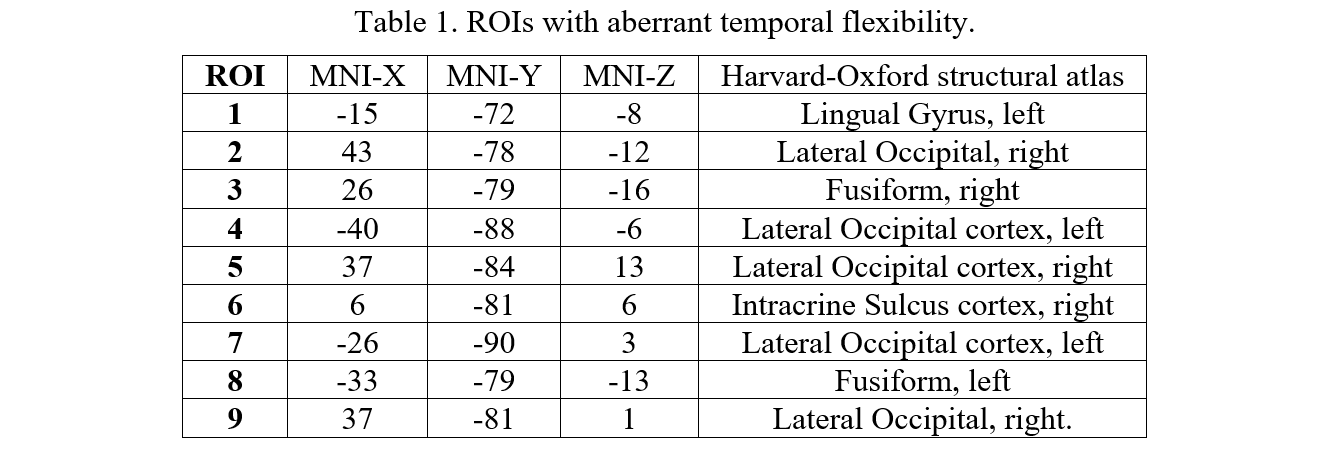
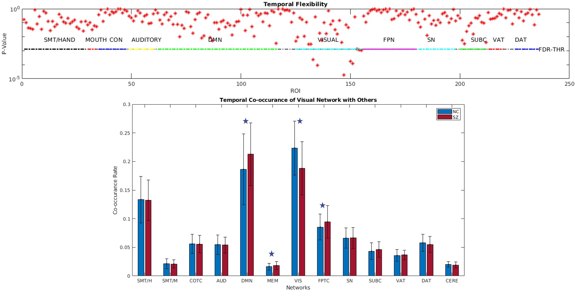
After FDR correction, significantly higher temporal flexibility in the SZ group were found in nine ROIs, all of which reside in the visual cortex (shown in Figure 1B). The specific locations in the standard MNI space and their probable anatomic structures in the Harvard-Oxford atlas are listed in Table 1. The average temporal flexibility of these ROIs for both groups are depicted in Figure 2. Post-hoc ANOVA analysis revealed that significant difference in co-occurrence pattern with visual network existed in DMN, MEM, VIS and FPTC networks. Other networks had no difference in temporal co-occurrence/flexibility.DISCUSSION AND CONCLUSION
Rather than changes in FC, the present study examined the temporal co-activation of a functional ROI with other networks using sliding window correlation and modularity analysis. Our experiment showed the existence of aberrant temporal flexibility in ROIs of visual cortex, most of which were located at high-level ventral visual pathway, regions for visual object recognition. This might be related to the hallucination symptoms that are commonly seen in schizophrenia. Examination of their temporal co-activation profiles revealed higher co-activation of visual network with DMN, MEM and FPTC in schizophrenia, indicating the patients might have active functional interaction between these systems under resting-state condition. This study indicates temporal co-occurrence may provide new insights regarding the functional organization of schizophrenia. Our future work will examine the aberrances with the symptoms of patients.Acknowledgements
We would like to thank the UCLA Consortium for Neuropsychiatric Phenomics LA5c study for sharing the data.References
1. Friston, K. J. The disconnection hypothesis. Schizophr Res 30, 115–125 (1998).
2. Dong, D., Wang, Y., Chang, X., Luo, C. & Yao, D. Dysfunction of Large-Scale Brain Networks in Schizophrenia: A Meta-analysis of Resting-State Functional Connectivity. Schizophrenia Bull (2017).
3. Hutchison, M. R. et al. Dynamic functional connectivity: Promise, issues, and interpretations. NeuroImage 80, 360–378 (2013).
4. Damaraju, E. et al. Dynamic functional connectivity analysis reveals transient states of dysconnectivity in schizophrenia. NeuroImage. Clinical 5, 298–308 (2014).
5. Thompson, W., Brantefors, P. & Fransson, P. From static to temporal network theory applications to functional brain connectivity. Netw Neurosci 1, 1–56 (2017).
6. Chen, M. & Hu, X. P. Individual Identification Using Functional Brain Fingerprint Detected by Recurrent Neural Network. Brain Connectivity (2018).
7. Chen, T., Cai, W., kanth Ryali, Supekar, K. & Menon, V. Distinct Global Brain Dynamics and Spatiotemporal Organization of the Salience Network. PLoS biology 14, e1002469 (2016).
Figures