0878
Super-resolution convolutional neural networks applied to functional lung MRI at 1.5T1Division of Radiological Physics, Department of Radiology, University of Basel, Basel, Switzerland, 2Department of Biomedical Engineering, University of Basel, Basel, Switzerland, 3High Field Magnetic Resonance, Max Planck Institute for Biological Cybernetics, Tübingen, Germany, 4Division of Pediatric Respiratory Medicine, Department of Pediatrics, Inselspital, Bern University Hospital, Bern, Switzerland, 5Department of Radiology, Inselspital, Bern University Hospital, Bern, Switzerland
Synopsis
High-resolution images are needed in many MR applications to enhance the diagnostic information at early stages of the disease. Often, the achievable resolution is limited by acquisition time constraints, in particular in moving organs such as the lung, where rapid imaging is a necessity. The low proton density in the lung parenchyma further constrains the resolution as sufficiently high signal-to-noise ratio (SNR) requires large voxel size. In this work, the concept of super-resolution is investigated to increase the spatial resolution and potentially shorten the acquisition time for functional assessment in the lung without SNR penalty.
INTRODUCTION
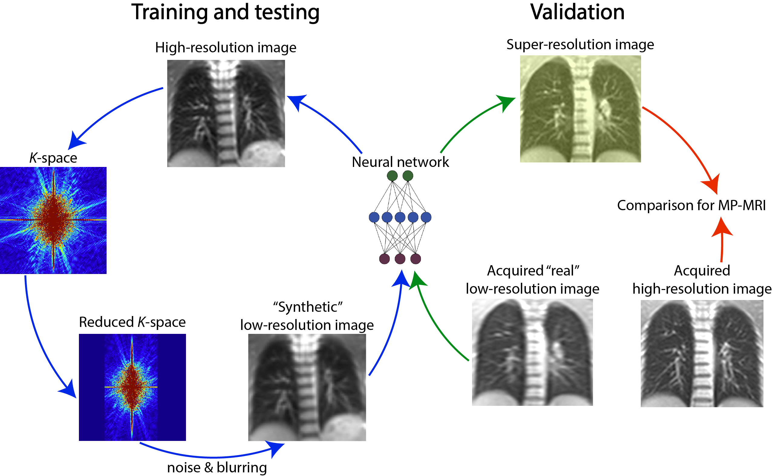
Lung MRI is commonly restricted to acquisitions at rather low resolution since the proton density in the parenchyma is low and rapid acquisitions are required to reduce motion sensitivity. Increasing the resolution by using standard interpolation methods may come at the cost of blurring and loss of contrast without adding new information. In contrast, super-resolution techniques have the potential to improve the resolution while ensuring that the increased information content of higher-resolution images is added with a priori learned knowledge1. From another point of view, imaging with super-resolution might benefit of a larger voxel sixe, shortened scanning time and an increased signal-to-noise ratio (SNR). Here, this concept is applied to acquisitions at 50% phase resolution with ultrafast steady-state free precession (ufSSFP)2,3 imaging for functional ventilation and perfusion mapping of the lung using matrix pencil (MP)4 decomposition, a technique derived from Fourier decomposition MRI.METHODS
MR Data
Fifty-five children (5-18 years old) with cystic fibrosis (CF) and 12 healthy (7-12 years old) underwent MR examinations at 1.5T during multiple visits for functional lung assessment with MP5,6. The data were acquired using time-resolved 2D ufSSFP pulse sequence2,3. Relevant imaging parameters were: matrix=128x128, field-of-view=450x450 mm2, phase resolution 100%, TE/TR = 0.67/1.46 ms; acquisition time for one image 120ms, followed by a 180 ms waiting time for longitudinal magnetization recovery, yielding 3.33 images per second. For MP MRI, 160 images were acquired during 48 seconds of free breathing and at multiple (8-10) slice locations (total scan time ~8min). From these time series, specific post-processing including deformable image registration7, lung segmentation8,9, and MP4 signal analysis allowed computing ventilation- and perfusion-weighted maps.
Super-Resolution Training Data
Out of the 67
subjects, 57 were randomly selected for training a fast super-resolution convolutional
neural network (FSRCNN)10, while the remaining 10 subjects were used as a
testing cohort. From every subject, 10 images per slice were used in the
atlases, totaling to about 8000 coronal images. From the acquired
high-resolution base images, “synthetic” images with phase resolution 50% were
generated by removing the outer ½ k-space and by adding random Gaussian noise
and blurring during the training. The network was trained for 12 hours on a GPU
to resolve high-resolution images from the low resolution one.
In the testing cohort,
time series of synthetic low-resolution images processed to super-resolution by
the trained network were evaluated using the peak signal-to-noise ratio index
and for MP ventilation and perfusion assessment.
Validation Data
An adult healthy subject was imaged with ufSSFP using the aforementioned acquisition parameters, as well as using a phase resolution lowered to 50% and yielding an acquisition time shortened to 80 ms. The “real” low-resolution images were processed with the trained network to recover high-resolution features, and subsequently investigated for MP decomposition. Figure 1 schematically summarizes the method.
RESULTS AND DISCUSSION
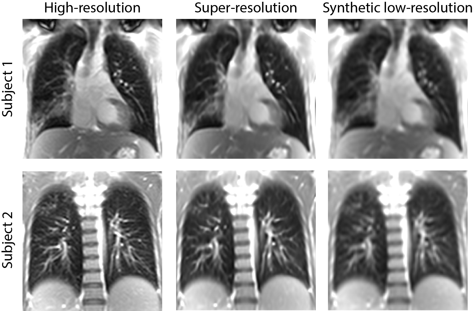
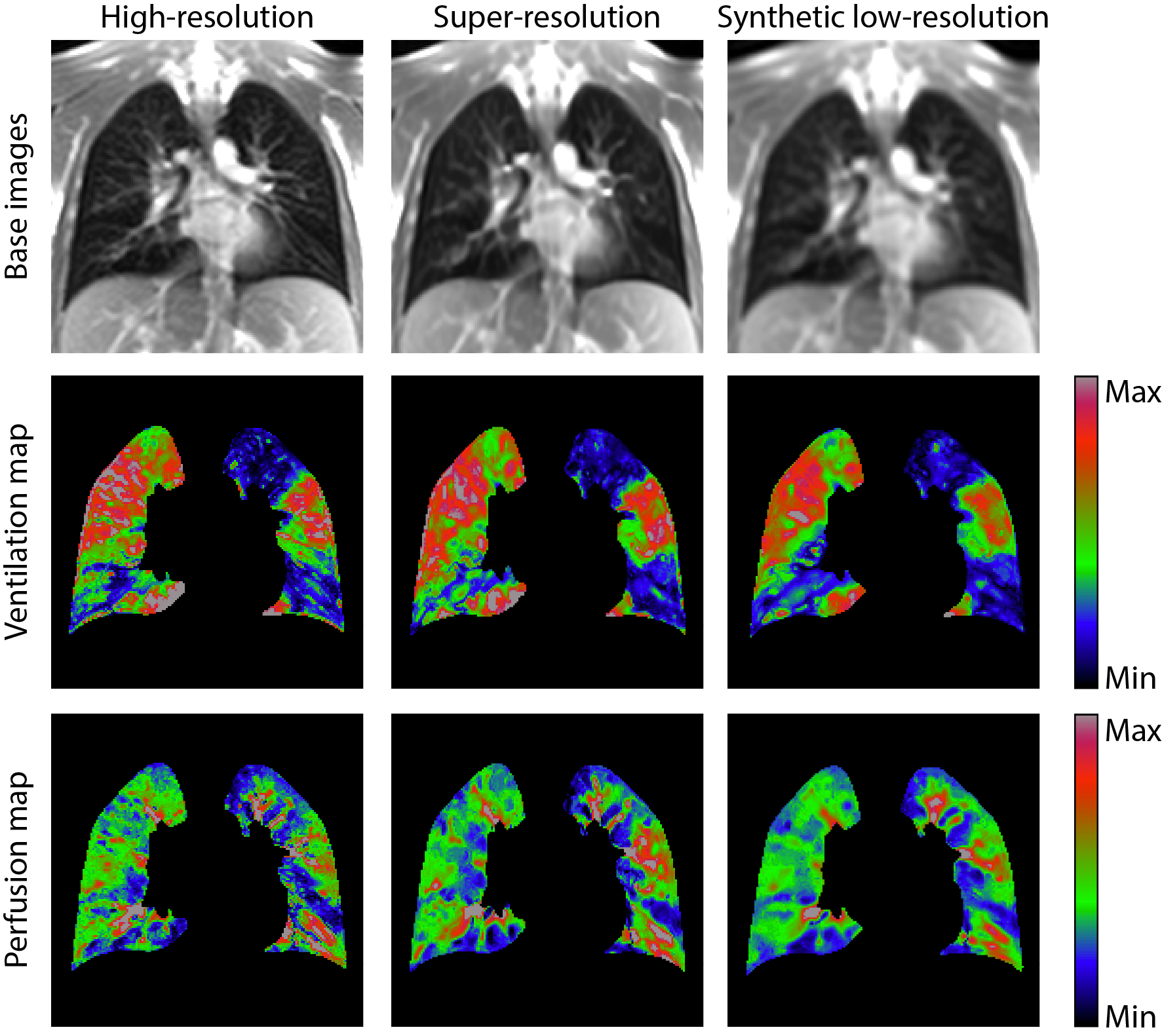
The super-resolution network processing time for one image was only ~0.2 s. Representative high-resolution, synthetic low-resolution and the resolved super-resolution images are shown in Figure 2. On the testing datasets, the network resolved hyperintense structures with a peak signal-to-noise ratio of 41.5 [dB] between high-resolution and super-resolution, and 37.7 [dB] between high-resolution and standard bicubic image interpolation.Figure 3 shows ventilation and perfusion imaging in a child with CF. The super-resolution approach did not alter the functional quantitative information derived with MP as compared to high-resolution imaging.
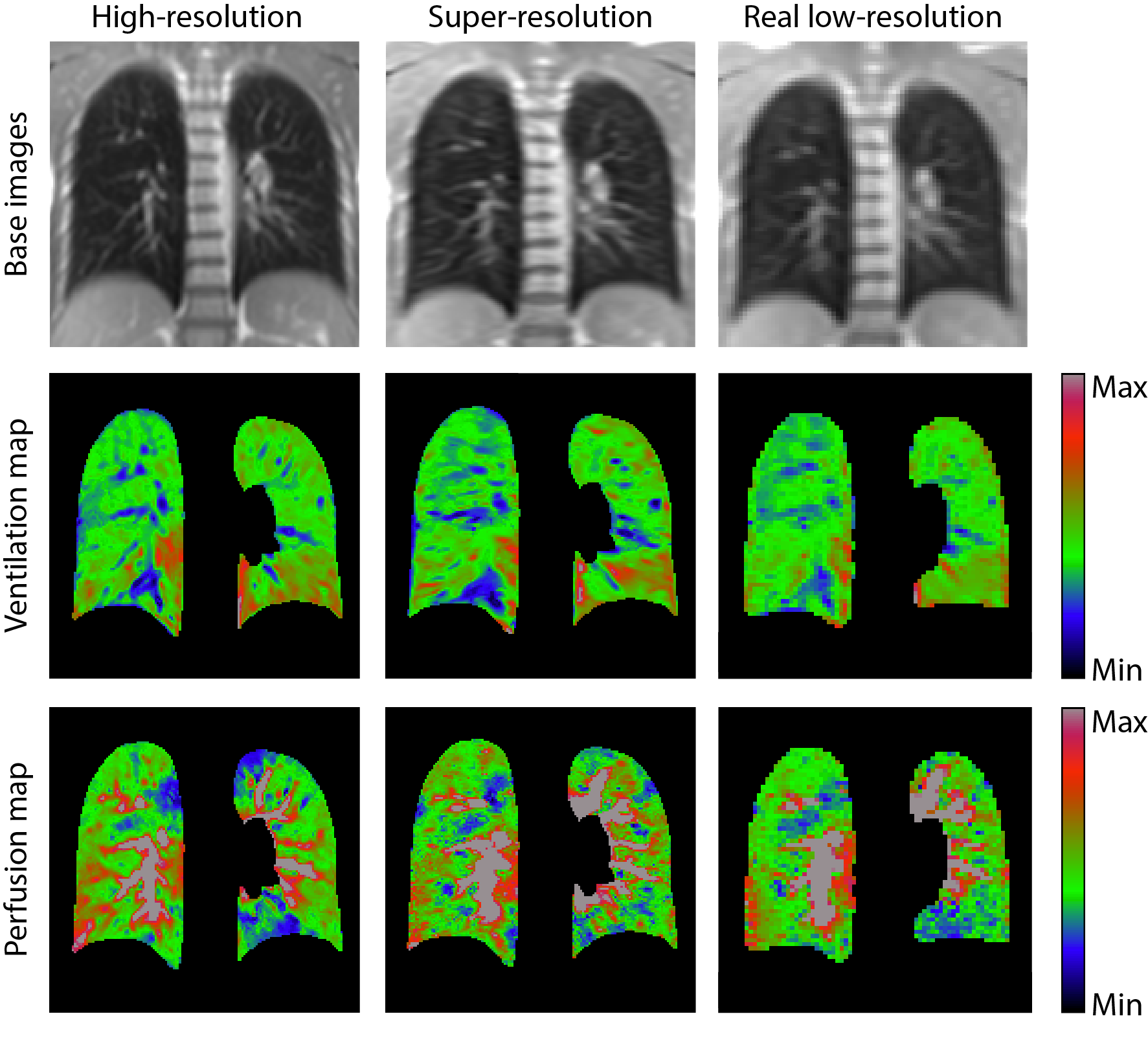
The validation of the network in a healthy subject is presented in Figure 4. The super-resolution network is able to enhance structures from the acquired low-resolution images (phase resolution 50%), but sub-pixel features not present on base images are not yet resolved. Also on the validation datasets, the super resolution approach alters only marginally perfusion and ventilation signal amplitudes.
CONCLUSIONS
Super-resolution neural networks show good potential for rapid MP imaging at low resolution while simultaneously recovering high-resolution imaging and functional image content. On the other hand, the increased SNR offered by lower resolution imaging might be beneficial, and moreover, the reduced acquisition time offered by super-resolution may be invested to increase the temporal resolution of MP imaging or to prolong the recovery period. An increased recovery period allows for regrowth of longitudinal magnetization for an even higher detectable signal and reduction of specific absorption rate.Acknowledgements
This work was supported by the Swiss National Science Foundation (SNF grant No. 320030_149576).References
1. Yang W, Zhang X, Titan Y et al. Deep Learning for Single Image Super-Resolution: A Brief Review. arXiv eprint, arXiv:1808.03344, 2018.
2. Bieri O. Ultra-fast steady state free precession and its application to in vivo 1H morphological and functional lung imaging at 1.5 tesla. Magn. Reson. Med. 2013;70:657–663.
3. Bauman G, Pusterla O, Bieri O. Ultra-fast Steady-State Free Precession Pulse Sequence for Fourier Decomposition Pulmonary MRI. Magn. Reson. Med. 2015:75:1647-53.
4. Bauman G, Bieri O. Matrix pencil decomposition of time-resolved proton MRI for robust and improved assessment of pulmonary ventilation and perfusion. Magn. Reson. Med. 2017;77:336–342.
5. Nyilas S, Bauman G, Sommer Get al. Novel Magnetic Resonance Technique for Functional Imaging Of Cystic Fibrosis Lung Disease. Eur Respir J 2017, 50 (6) 1701464.
6. Nyilas S, Bauman G, Pusterla O et al. Ventilation and perfusion assessed by functional MRI in children with CF: reproducibility in comparison to lung function. J Cyst Fibros, 2018.
7. Sandkühler R, Jud C, Pezold S, et al. Adaptive Graph Diffusion Regularisation for Discontinuity Preserving Image Registration - Biomedical Image Registration. Springer International Publishing, 2018.
8. Pusterla O, Andermatt S, Bauman G, et al. Deep Learning Lung Segmentation in Paediatric Patients. Proceedings of the 26th annual meeting of the ISMRM, 2018.
9. Andermatt S, Pezold S, Cattin P. Multi-dimensional Gated Recurrent Units for the Segmentation of Biomedical 3D-Data - Deep Learning and Data Labeling for Medical Applications. MICCAI 2016, Springer International Publishing, 2016.
10. Dong C, Loy C, He K, et al. Image super-resolution using deep convolutional networks,Transactions on Pattern Analysis and Machine Intelligence. IEEE, 2015.
Figures