0863
Changes in ACL T2* over the course of a menstrual cycle: a new biomarker for ACL injury risk?Erin C Argentieri1, Tatum W Braun1, Ryan E Breighner1, Alissa J Burge1, Joseph T Nguyen2, Matthew F Koff1, Ellen K Casey1, and Hollis G Potter1
1Radiology and Imaging, Hospital for Special Surgery, New York, NY, United States, 2Biostatistics, Hospital for Special Surgery, New York, NY, United States
Synopsis
This study assessed changes in ACL T2* metrics throughout the menstrual cycle. In the pre-ovulatory phase, ovulatory case subjects exhibited significant shortening of T2*S and PS in comparison to visit #4 (post-ovulatory phase). Non-ovulatory control subjects displayed no significant changes over time. Results of the current study suggest that there is a shift in bound water (T2*S) within the ACL from pre- to post-ovulatory phases. Shifts in tissue water content have been associated with altered mechanical properties and changes in ligament stiffness may alter proprioceptive sense and contribute to increases in laxity and risk of ACL-injury within the pre-ovulatory phase.
INTRODUCTION
Regardless of surgical or non-surgical treatment, anterior cruciate ligament (ACL) disruption is associated with the development of post-traumatic osteoarthritis (PTOA) within 5-10 years.1-3 Recent studies have focused on identifying risk factors associated with ACL-injury with the goal of reducing overall incidence. As a result, multiple demographic and morphologic features have been identified as risk factors for ACL-injury, and numerous studies have established that ACL-injury risk in females is more than double that of their male counterparts.4,7 Females also exhibit increased knee joint laxity and ACL-injury risk during the pre-ovulatory phase of their menstrual cycle.5,6 These findings suggest that sex hormones may directly impact the ACL structure and biomechanical integrity. Ongoing advances in MRI have led to the development of ultra-short echo (UTE) sequences which can capture the short T2* decay within ligaments, and allow for quantitative evaluation of tissue microstructure.8-11 The objective of this study was to determine if significant changes in ACL T2* metrics exist over the course of a menstrual cycle. We hypothesized that pre-menopausal females would exhibit significant changes in ACL T2* metrics over the course of a menstrual cycle, while no such differences would exist within non-ovulatory control subjects.METHODS
This IRB approved pilot study included 10 females with no history of knee injury. Six pre-menopausal females with normal menstrual cycles and no history of hormonal contraceptive use (>1 year) were included as ovulatory case subjects. Subjects within the non-ovulatory control group included 2 post-menopausal females, and 2 pre-menopausal subjects taking oral contraceptives with normal menses. All subjects participated in 4 study visits evenly spaced over the course of 1-month (1 per week). Study visit #1 for all pre-menopausal females coincided with onset of menses. Subjects were provided with commercially available ovulation predictor kits (ClearBlue Digital Ovulation Tests [Accuracy 99%]) to determine date of ovulation in case subjects, and to confirm anovulatory status in control subjects. MRI Acquisition: At each study visit, bilateral MRI examinations were obtained on a 3-Tesla clinical scanner (GE Healthcare) using an 8-channel phased array knee coil (Invivo). Three-dimensional, coronal-oblique, UTE sequences were acquired for the evaluation of T2* metrics (Voxel: 0.50x0.50x1.5mm3, TEs: 11 echoes between 0.03-25ms, TR: 166ms, RBW: ±83.3kHz, Flip-Angle: 16o). Imaging Analysis: Bi- exponential fits of SI to corresponding echo time were used to calculate ACL T2* metrics12: SI(TE) = A(-TE/T2*S) + B(-TE/T2*L)+noise, where T2*S and T2*L are respective short and long T2* components, A and B are corresponding short and long apparent proton densities, and PS is calculated as A/(A+B). Statistical Analysis: Generalized estimating equation modeling was used to cluster data points contributed from each leg of each patient, and longitudinal analyses were completed for each T2* metric (T2*S, T2*L, PS) within and between groups. Maximum likelihood estimates were used to establish parameter estimates for each study visit. Post-hoc pairwise evaluations with Bonferroni-adjustment for multiple comparisons were used to identify differences between visits.RESULTS
Significant differences were found within and between study groups for all T2* metrics (Tables 1 and 2). Specifically, at visit #1 (pre-ovulatory phase) ovulatory case subjects displayed significantly decreased T2*S and PS metrics in comparison to control subjects (mean difference: T2*S = −1.56ms, PS = −2.74%; p ≤ 0.01). Case subjects also exhibited significant changes over time in all T2* metrics, while control subjects displayed no such differences. Within case subjects, T2*S and PS were significantly shorter at study visit #1 (pre-ovulatory phase) in comparison to study visit #4 (mean difference: T2*S = −1.12ms, p = 0.04; PS = −1.23%, p = 0.01).DISCUSSION
ACLs of healthy pre-menopausal case subjects displayed significant changes in T2* metrics over the course of a menstrual cycle, while non-ovulatory control subjects displayed no such differences. At visit #1, when case subjects were in the pre-ovulatory phase, T2*S and PS were significantly shorter in comparison to visit #4 (post-ovulatory phase). These findings suggest that there is an increase in bound water (T2*S) within the ACL during the pre-ovulatory phase. Shifts in tissue water content have been associated with altered mechanical properties, and decreased T2* has been demonstrated in preclinical models of cyclic loading prior to gross collagen disruption.10,11 Subsequent changes in ligament stiffness over the course of the menstrual cycle may alter proprioceptive sense and contribute to increases in laxity and risk of ACL-injury within the pre-ovulatory phase.CONCLUSION
This is the first study to evaluate changes in ACL T2* metrics throughout the menstrual cycle. These data suggest that significant differences exist between pre- and post-ovulatory phases of the menstrual cycle and may be indicative of a new imaging biomarker for ACL-injury risk.Acknowledgements
HSS has an institutional research agreement with GE Healthcare. The authors would like to thank Kelly Zochowski and Erica Hooper, as well as all of the HSS MRI staff and technologists for their assistance with this studyReferences
(1) Oiestad 2010; (2) Potter 2012; (3) Lohmander 2007; (4) Beynnon 2014; (5) Beynnon 2006; (6) Shultz 2005; (7) Beynnon 2015; (8) Pauli 2012; (9) Diaz 2012; (10) Jerban 2017; (11) Koff 2014; (12) Juras 2013Figures
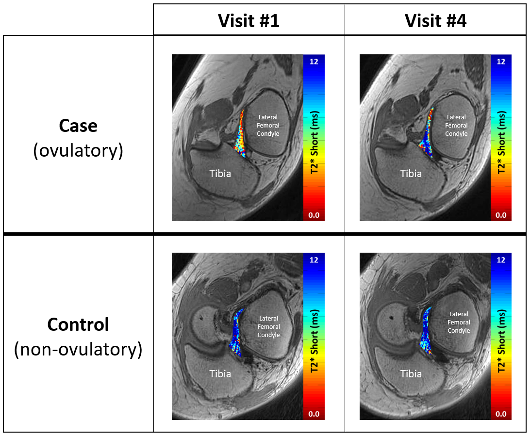
Figure 1: Multi-echo, coronal-oblique UTE-MRI
series. Example of
changes in T2*S within one case and one control subject from study visit #1
(pre-ovulatory) to study visit #4 (post-ovulatory). Significant changes were
found within case subjects only.
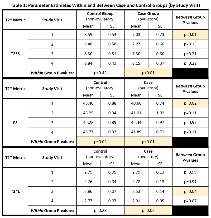
Table 1:Significant differences in T2* metrics were present within and between case and control groups
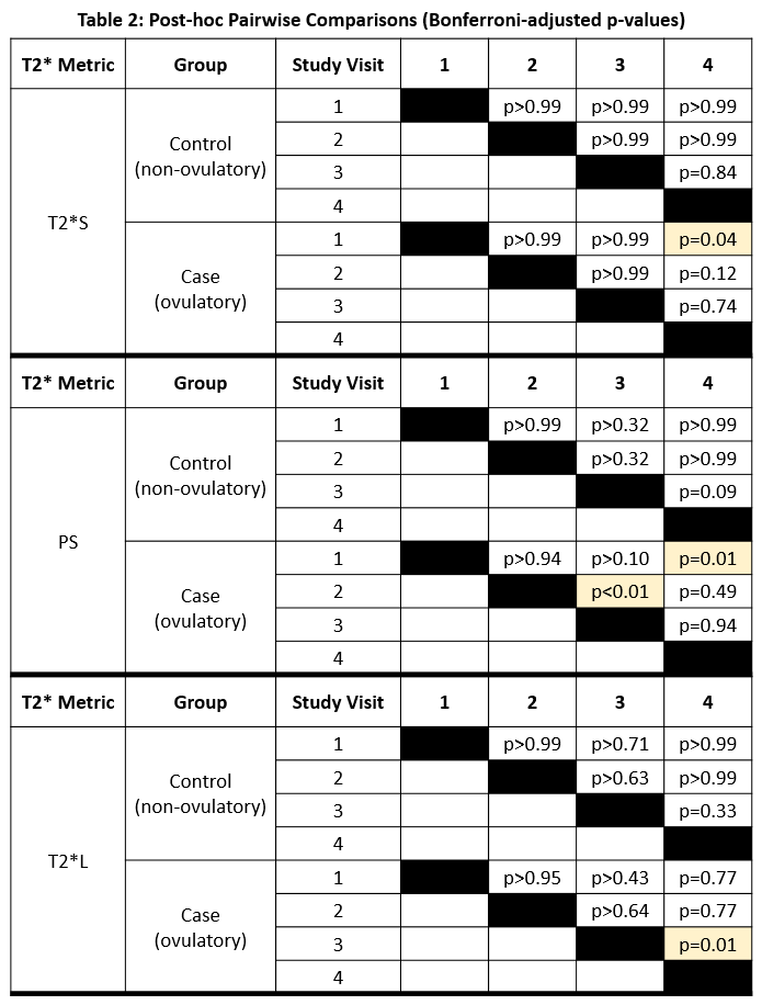
Table 2: Post-hoc pairwise comparisons adjusted for multiple comparisons (Bonferroni adjusted p-values). Ovulatory case subjects exhibited significant changes over the course of their menstrual cycle, while non-ovulatory control subjects exhibited no such differences.