0858
Hemispheric Specification of Remote Effect of Cerebral Glioma on White Matter Connectivity1Shenzhen Institutes of Advanced Technology,Chinese Academy of Sciences, Shenzhen, China, 2University of Chinese Academy of Sciences, Beijing, China, 3Department of Neurosurgery, Huashan Hospital of Fudan Univesity, Shanghai, China, 4Beijing Tian Tan hospital,Capital Medical University, Beijing, China
Synopsis
Biological aggressiveness of glioma extends beyond the radiological territory. Based on the atlas based method and diffusion metrics derived from diffusion tensor imaging, the remote effect of cerebral gliomas on the white matter connectivity was analyzed at the global level. LGGs and HGGs showed difference in the diffusion metrics in the contralesional brain regions with hemispheric and anatomical specifications, which possibly underlie the functional neuroplasticity confounding the aggressiveness assessment and management of glioma.
Introduction
The biological aggressiveness of glioma extends beyond the radiological territory.1,2 In this study, we aim to explore the remote effect (RE) of low grade glioma (LGG) and high grade glioma (HGG) on the white matter connectivity at the global level using atlas based method based on diffusion tensor imaging (DTI).Materials and Methods
This study was approved by local institutional review board. Data from a total of 57 subjects with histologically confirmed glioma located in frontal lobe were retrieved consecutively from March 2013 to March 2015. Three subjects were excluded for midline deformation due to serious mass effect. Twenty five tumor lesions were located in the left hemisphere (LH) (LGG/HGG = 12/13, male/female = 14/11, age = 42.93±16.91) and 29 located in right hemisphere (RH) (LGG/HGG = 12/13, male/female = 13/16, age = 40.00±11.16). RE analysis was performed for LH and RH tumors respectively. DTI was performed on 3.0T magnetic scanner (Siemens Verio, Germany) with a 12 channel phases array head coil and using EPI sequence with TR/TE 7600/91 ms, FA 90°, FOV 230×230 mm, matrix size 128×128, slice thickness 3 mm, 20 diffusion gradient encoding directions and b value 1000 s/mm2. PANDA3 was utilized to preprocess all DTI images and conducted the deterministic fiber tracking. Each cerebral hemisphere was segmented in to 45 regions based on the Automatic Anatomical Labe (AAL) atlas. Fiber number (FN), fiber length (FL) and fractional anisotropy (FA) were calculated for the fiber tracts connecting the regions of the contralesional hemisphere. End points in two regions with no less than 3 fibers were defined as structural connectivity (SC).4 Otherwise the diffusion metrics of fibers between these 2 regions were set as zero. Mann-Whitney U test (SPSS 19.0) was performed to compare the diffusion metrics between LGGs and HGGs for both LH and RH group. P <0.05 was set as the significance level.Results
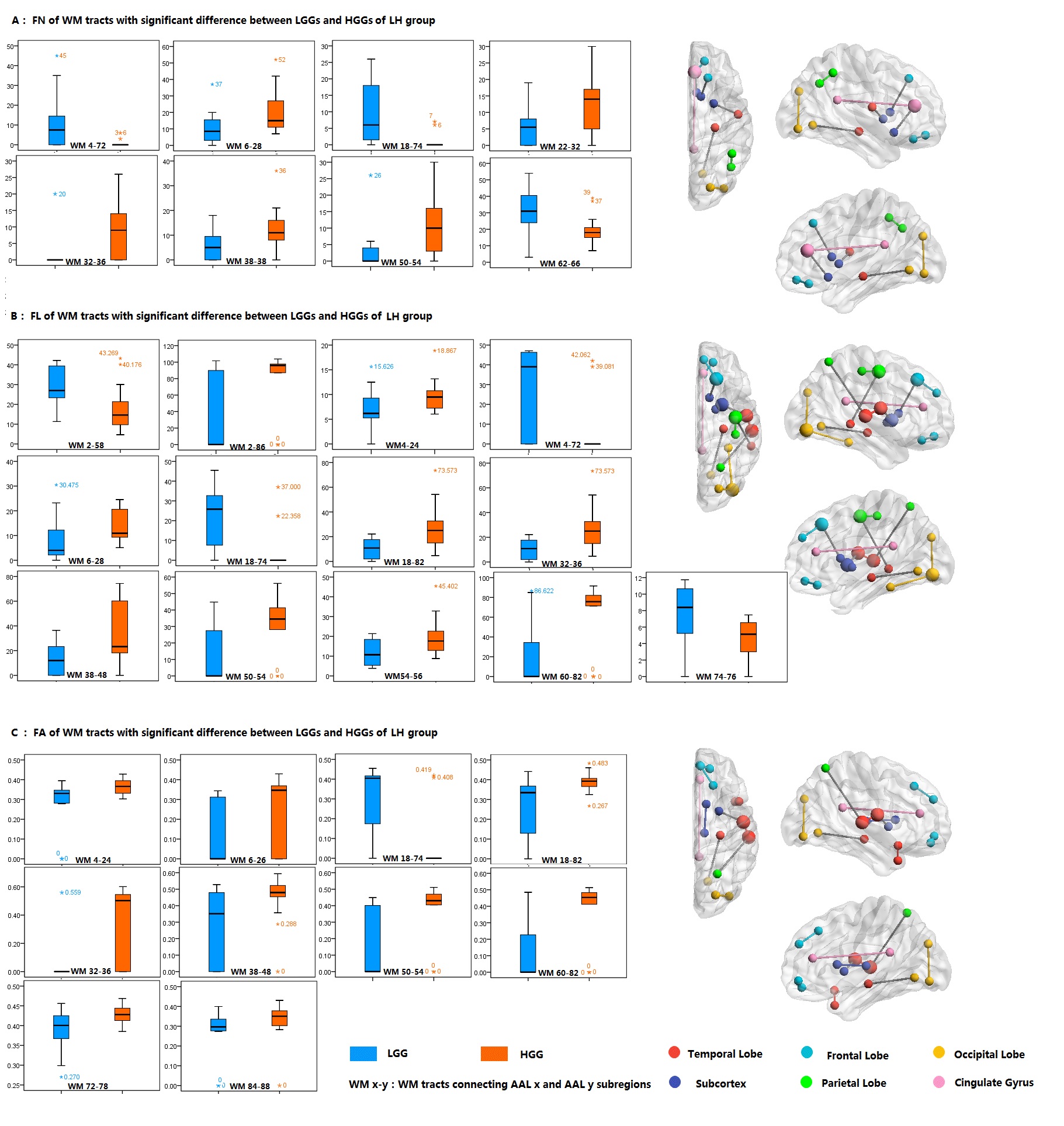
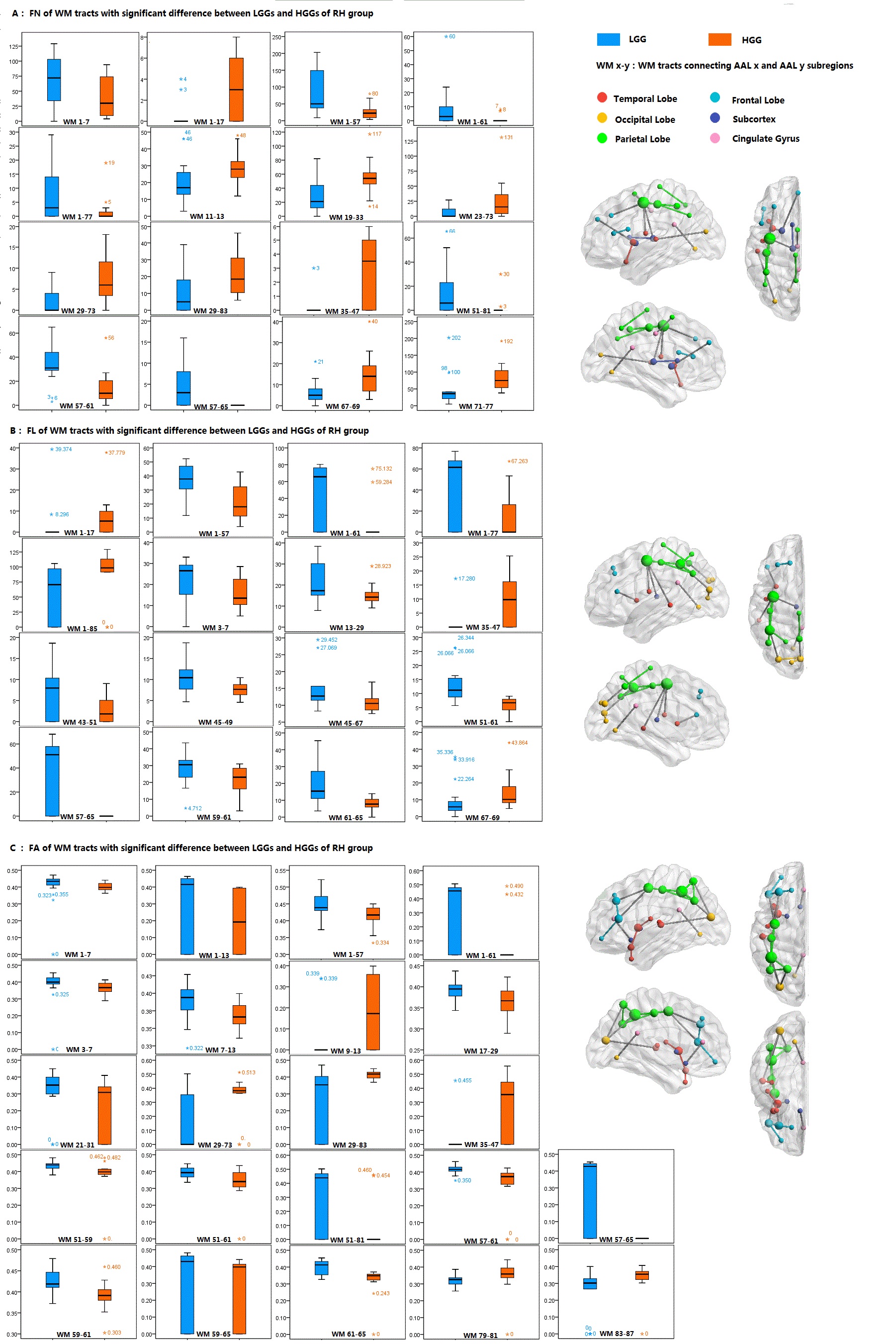
Compared with LGGs, HGGs had discriminate remote effect on the WM tracts of contralesional hemisphere. WM tracts of HGG group showed significantly higher FN, FA and FL than that of LGGs (p<0.05) in the connections between 1) superior and inferior occipital gyri and 2) hippocampus and lingual gyrus) for the LH gliomas. HGG group showed the loss of SC between frontal operculum and putamen, while LGG group lost the SC between superior and posterior cingulate gyri for the subjects with LH glioma. For subjects with RH glioma, WM tracts connecting precentral and postcentral gyri of HGGs showed significantly higher diffusion metrics relative to LGGs (p<0.05). HGG group lost SC connecting 1) precentral gyrus and inferior parietal cortex and 2) postcentral gyrus and angular gyrus, while LGG group lost SC between posterior cingulate gyrus and lingual gyrus for the RH subjects.Discussion
Cerebral tumor triggers neuroplasticity that may underlie the physiologic foundation for functional impairment. The regions with increased diffusion metrics in LH group may suggest that more and stronger SC were formed in the contralesional hemisphere to offset the functional impairment to the memory and motion perception as a result of HGG. For RH group the altered SC mainly involves the parietal lobe and the primary somatosensory center, which may be partially interpretive of the dysfunction in comprehension, verbal memory and somatosensory perception in the HGG group of the RH subjects.5 LH and RH subjects showed difference in the intergroup distribution of the diffusion metrics, possibly oweing to the effect of the functional and structural laterality of human brain.Conclusion
LGGs and HGGs have discriminate remote effect on the contralesional hemisphere with hemispheric specificity in terms of the microstructural network indexed by diffusion metrics.Acknowledgements
The National Basic Research Program of China [2015CB755500] and the National Natural Science Foundation of China [81627901].References
1. Almairac F, Duffau H, Herbet G. Contralesional macrostructural plasticity of the insular cortex in glioma patients: A VBM study. Neurology. 2018;00:1-7.
2. Park J. E, Kim H. S, Kim S. J, et al. Alteration of long-distance functional connectivity and network topology in patients with supratentorial gliomas. Neuroradiology. 2016; 58(3):311-320.
3. Cui Z, Zhong S, Xu P, et al. PANDA: a pipeline toolbox for analyzing brain diffusion images. Front Hum Neurosci.2013; 7(42):1662-5161.
4. Ni S, Liu Y, Li K, et al. Diffusion tensor tractography reveals disrupted topological efficiency in white matter structural networks in multiple sclerosis. Cereb Cortex. 2011;21(11):2565-2577.
5. Mainio A, Hakko H, Niemela A, et al. The effect of brain tumour laterality on anxiety levels among neurosurgical patients. Journal of Neurology, Neurosurgery, and Psychiatry. 2003; 74(9):1278-1282.
Figures