0852
Evaluating white matter microstructure in childhood brain tumour survivors: A combined DTI and MTR approach.1The Hospital for Sick Children, Toronto, ON, Canada, 2University of Toronto, Toronto, ON, Canada
Synopsis
Diffusion tensor imaging has been utilized to study the impact of cancer treatment on white matter microstructure in paediatric brain tumour survivors. We utilized magnetization transfer imaging (MTI), which provides more specific information on myelin, along with DTI, to determine if treatment for paediatric brain tumours has specific or non-specific impacts on white matter structure. When compared to their healthy counterparts, children treated for brain tumours exhibit decreased anisotropy and increased diffusion metrics without any significant differences in MT. This suggests treatment may impact fiber organization rather than myelin structure in this patient population.
Introduction
Cancers of the central nervous system account for approximately 25% of all diagnosed cancers within the paediatric population.1 Treatment includes surgery with or without radiation therapy and/or adjuvant chemotherapy, and exposure to these insults has been shown to negatively impact white matter structure of survivors.1 all studies to date in paediatric brain tumour survivors have employed diffusion tensor imaging (DTI) to evaluate white matter microstructure.2,3 However, measures obtained from DTI are non-specific in regards to tissue properties of white matter.4 Here, we utilized magnetization transfer (MT) imaging, which provides specific information on myelin,5 in addition to DTI, to evaluate whether the impact of paediatric brain tumours and their treatment has a specific impact on myelin, or more non-specific effects on white matter structure.Methods
Forty-five healthy controls (HC) and 50 paediatric brain tumour survivors were recruited for our study from the Hospital of Sick Children (SickKids; Toronto, Ontario, Canada). Of the survivors, 18 were treated with surgery only and 32 were treated with a reduced (2340 cGy) or standard (3060-3940 cGy) dose of radiation to the head and spine along with a radiation boost to either the tumour bed (TB) or entire posterior fossa (PF). Participants were split into three groups: HC, patients treated with surgery only (SUR), and patients treated with surgery and radiation (RAD). Neuroimaging was completed using SickKids Siemens 3.0T MRI scanner. Imaging consisted of a T1 AX 3D MPRAGE Grappa 2 protocol (TI 900 msec; TE/TR 3.91/2300 msec; 160 contiguous axial slices; flip angle 9°; 256 × 224 matrix; FOV 256 × 224 mm; voxel size = 1 mm isotropic), a diffusion weighted single shot spin echo DTI sequence with an EPI readout (30 directions; b = 1000 s/mm2; TE/TR 90/9000 msec; 70 contiguous axial slices; flip angle 90°; 122 × 122 matrix interpolated to 244 × 244; FOV 244 × 244 mm; voxel size = 2 mm isotropic), and a MTRon/off T1 AX FL3D Grappa 2 protocol (TE/TR 5/34 msec; 104 contiguous axial slices; flip angle 10°; 128x128 matrix, interpolated to 256x256; FOV 192x192, voxel size = 1.5 mm isotropic). Fractional anisotropy (FA), axial/radial/mean diffusivity (AD/RD/MD), along with magnetization transfer ratio (MTR) were extracted and compared in a voxelwise manner between groups using tract-based spatial statistics (TBSS) from the Functional MRI of the Brain Software Library (FSL; version 5.0.8). Since anisotropy is known to increase with age,2 age at scan was used as a covariate in all statistical analyses. Further analyses done between patient groups (SUR and RAD) included age at diagnosis as a second covariate.Results
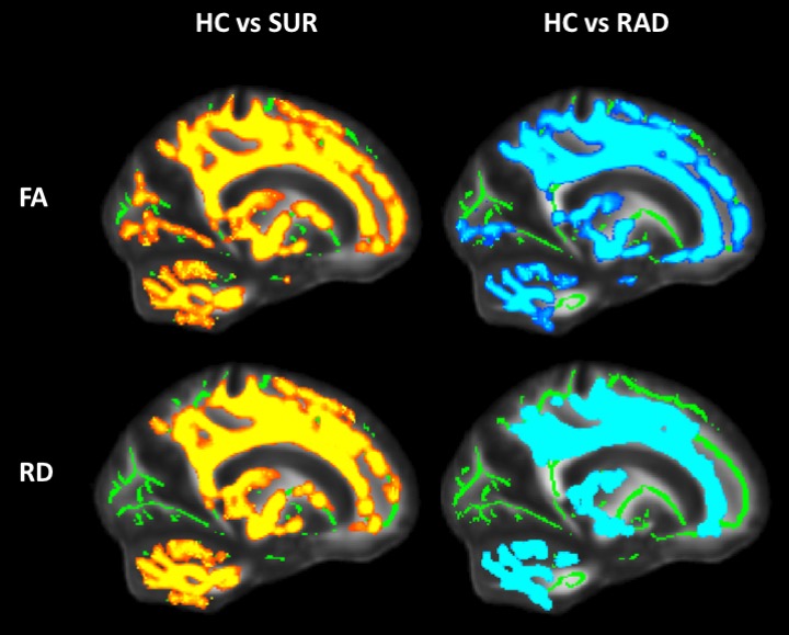
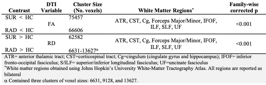
When compared to HC, the patient group (SUR and RAD) exhibited significantly decreased FA (t=2.18; p=0.0012) and increased AD (t≥2.6; p<0.03), RD (t=2.14; p<0.001), and MD (t=1.97; p<0.001) across multiple voxels. There were no significant differences in MTR between groups (p=0.32). To examine the specific impact of treatment with radiation, further analyses between HC and SUR, and HC and RAD were conducted. Both SUR and RAD groups continued to exhibit significantly decreased FA and increased AD, RD, and MD when compared to HC (Figure 1). In both cases, MTR between either patient group and HC did not differ (pHCvsSUR=0.32, pHCvsRAD=0.33). Table 1 displays the white matter clusters that differ between the groups for DTI metrics.Discussion
We have demonstrated that children treated for brain tumours exhibit a less restrictive microenvironment for water diffusion within brain white matter as indicated by increased diffusion and decreased anisotropy. Interestingly, no changes in MTR were found. Based on our findings, we conclude that myelin itself may not be affected by paediatric brain tumours and their treatment. Rather, it appears that treatment could impact other factors that may disrupt fiber organization, including axonal injury, axonal density, fiber packing and fiber orientation.Conclusion
The current study challenges the paradigm that myelin injury is a consequence of paediatric brain tumors and their treatment. MTR should be used as an adjunct to DTI in order to refine interpretation of data within future research in this patient population.Acknowledgements
No acknowledgement found.References
1. Robertson PL. Advances in treatment of pediatric brain tumors. NeuroRx. 2006;3(2):276-291.
2. Asato MR, Terwilliger R, Woo J, Luna B. White matter development in adolescence: A DTI study. Cereb Cortex. 2010;20(9):2122-2131.
3. Khong P-L, Kwong DLW, Chan GCF, et al. Diffusion-tensor imaging for the detection and quantification of treatment-induced white matter injury in children with medulloblastoma: a pilot study. AJNR Am J Neuroradiol. 2003;24(4):734-740.
4. Beaulieu C. The basis of anisotropic water diffusion in the nervous system - A technical review. NMR Biomed. 2002;15(7-8):435-455.
5. Henkelman RM, Stanisz GJ, Graham SJ. Magnetization transfer in MRI: A review. NMR Biomed. 2001;14(2):57-64.
Figures