0841
Improving temporal resolution of 3D Arterial Spin Labeling perfusion imaging by combining CAIPIRINHA encoding and spatio-temporal TGV reconstruction1Institute of Medical Engineering, Graz University of Technology, Graz, Austria, 2Laboratory of FMRI Technology (LOFT), Mark & Mary Stevens Neuroimaging and Informatics Institute, Keck School of Medicine, University of Southern California, Los Angeles, CA, United States, 3Institute of Mathematics and Scientific Computing, University of Graz, Graz, Austria, 4BioTechMed-Graz, Graz, Austria
Synopsis
Pseudo-continuous arterial spin labeling combined with 3D segmented readouts is recommended for acquiring ASL perfusion data. However, the total number of k-space encodings limits the trade-off between motion sensitivity and image blurring. To tackle this problem we implemented an accelerated 3D-GRASE sequence with a time-dependent 2D-CAIPIRINHA sampling pattern to increase the temporal incoherence between averages or PLDs. High quality images can be gained from the under-sampled time series by a variational image reconstruction approach with total-generalized-variation (TGV) regularization in space and time. This allows acquisition of single-shot 3mm isotropic ASL data with whole brain coverage within 1min22sec.
Introduction
The recommended way to acquire arterial spin labeling perfusion data in clinical settings is by using pCASL with 3D segmented readouts.1 For full encoding of the 3D k-space the trade-off between motion sensitivity and image blurring is limited due to the T2 relaxation. To tackle this problem a 2D-CAIPIRINHA2 accelerated 3D-GRASE3 readout was recently employed for single shot acquisition with conventional reconstruction.4,5,6 For retrospective under sampled CAIPIRINHA data with single-PLD a pronounced improvement in image quality was shown by TGV-based reconstruction approach, which incorporated the averaging procedure.7 This study combines and enhances the aforementioned acquisition and reconstruction framework. Firstly, an accelerated 3D-GRASE sequence with a time-dependent 2D-CAIPIRINHA sampling pattern was implemented to increase the temporal incoherence between averages or PLDs. Secondly, the reconstruction algorithm was adapted to incorporate additional temporal regularization on the whole C/L-time series and is therefore capable to handle single- and multi-delay ASL data. This combination allows for single-shot whole brain coverage acquisition with 3mm isotropic resolution in 1min22sec.Methods
Three healthy male volunteers were scanned at a 3T MR system (Skyra, Siemens Healthcare, Germany) using pCASL with 3D-GRASE readout after obtaining informed consent. For the standard fully sampled but segmented acquisition we used the following imaging parameters: matrix=64x64x38, 20% slice oversampling, 3mm isotropic resolution, TE=15ms, EPI-factor=21, TF=23, 6 segments, LD=1800ms, PLD=1800ms resulting in an acquisition time of 5min for five C/L-pairs and one M0-image. Furthermore, a 2D-CAIPIRINHA accelerated 3D-GRASE single-shot ASL acquisition was performed with the same resolution and LD/PLDs as for the segmented acquisition but with a 6-fold acceleration using an adapted CAIPIRINHA 1x6(2) pattern as illustrated in Figure 1. For the accelerated acquisition one M0-image and 20 C/L-pairs were measured in 2min48sec. Additionally, multi-delay acquisition was performed from one subject using 5 PLDs [500,1000,1500,2000,2500] ms and one average for the segmented approach and 10 PLDs [500:250:2750] ms with [2,2,3,3,3,4,4,4,5,5] averages for the single-shot approach. The acquisition time of 5min was matched for both multi-delay acquisitions. All other imaging parameters were the same as for the single-PLD acquisition. Coil sensitivity maps were estimated using ESPIRIT8 from an averaged k-space (acqu.1-6). The C/L-time series were jointly reconstructed by solving the following minimization problem:
$$\min_{c, l} \frac{\lambda_c}{2}\left\|(\mathbf{K}c - d_c)\right\|_2^2 + \frac{\lambda_l}{2}\left\|(\mathbf{K}l - d_l)\right\|_2^2 + \gamma_1(s)TGV_{\alpha1,\alpha0}(l) +\gamma_1(s)TGV_{\alpha1,\alpha0}(c) + \gamma_2(s)TGV_{\alpha1,\alpha0}(c - l)$$
where $$$ c\,$$$and$$$\,l$$$ denote the desired 4D C/L time-series, $$$\lambda_c$$$ and $$$\lambda_l$$$ are the regularization parameters for the control and respective label data. The parameter s controls the weighting between the three TGV functional as described in9, $$$ d_c\,$$$and$$$\,d_l$$$ is the acquired 4D C/L-data, $$$\alpha1\,$$$and$$$\,\alpha0\ $$$ are fixed model parameters.10,11 The operator $$$\mathbf{K}$$$ contains the coil sensitivity maps, the Fourier operator and the undersampling pattern. For the multi-delay dataset CBF was calculated using BASIL.12
Results and Discussion
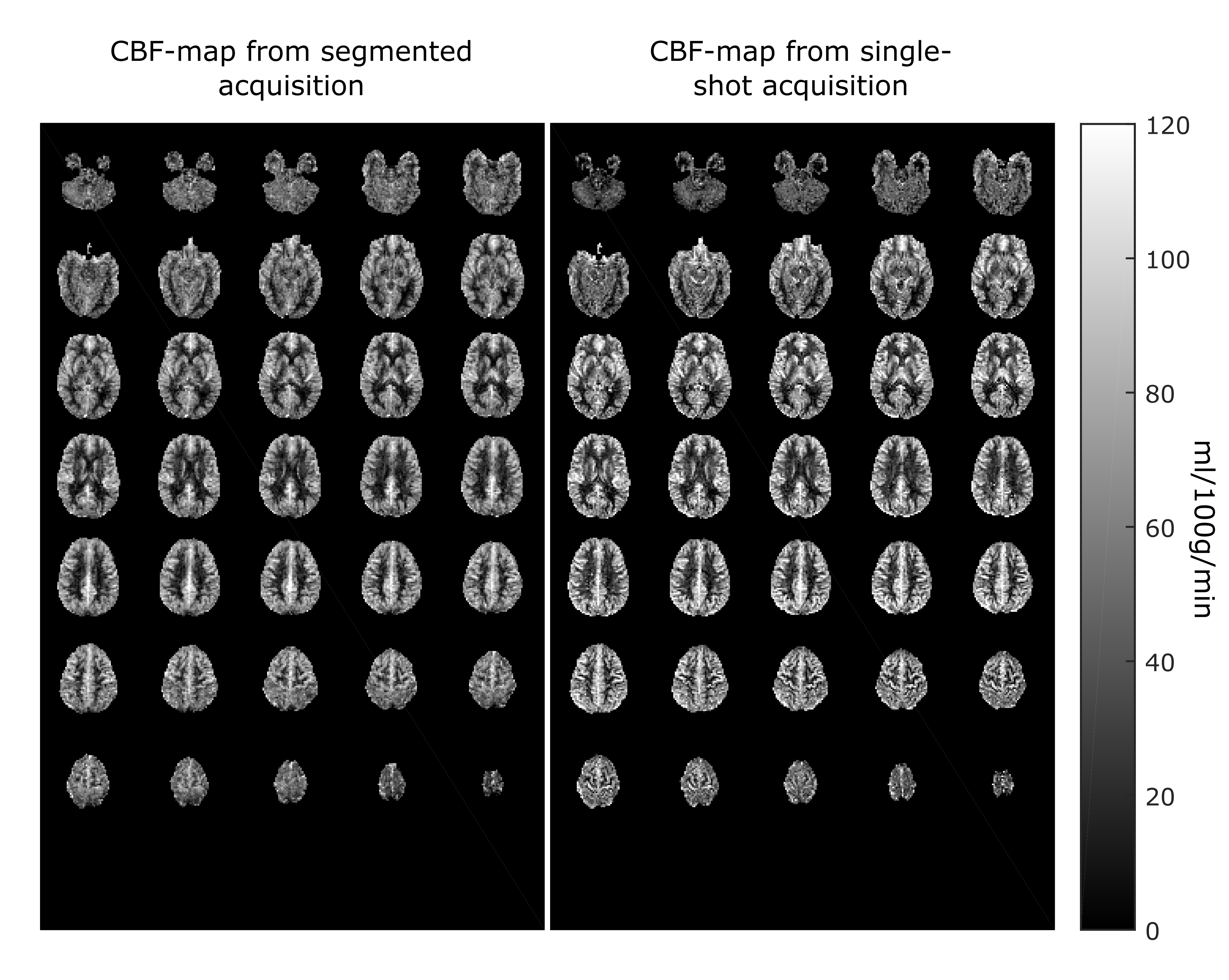
Figure 2 shows the whole brain perfusion map from one representative subject using single-shot acquisition (tacq=2min48sec) and segmented acquisition (6 segments, tacq=5min). The PWIs are in high accordance despite a high acceleration factor and halved acquisition time. Figure 3 shows one slice of each subject from the segmented and single-shot acquisition using different numbers of averages. Also for the lowest number of averages (N=10, tacq=1min 22s) the reconstruction results of the 6-folded single shot-acquisition are in high accordance with the fully sampled data using 6 segments. This is due to the increased incoherence between averages followed by a joint-reconstruction of the C/L-time series. Figure 4 shows the reconstruction results at different PLDs of one representative slice for the segmented and the single-shot acquisition. Figure 5 displays the corresponding whole-brain CBF-map.Conclusion
We combined a single-shot 3D-GRASE pCASL sequence, with a time-dependent 2D-CAIPIRINHA pattern, that changes throughout the acquisition, with a joint spatio-temporal reconstruction approach to improve the temporal resolution and robustness against motion for 3D-ASL data acquisition. The reconstruction framework yields a C/L-time series which makes this approach applicable for single- and multi-delay as well as for functional ASL. For multi-delay ASL the improvement in temporal resolution can be either used to acquire more PLDs, i.e. for a denser sampling of the kinetic information or to increase the number of averages for higher PLDs. Apart from the advantage of increasing the incoherence between averages or PLDs due to the adapted sampling pattern, this approach also allows to estimate the coils sensitivity maps without an additional reference scan by summing up the respectively averages or PLDs. Furthermore, the reconstruction framework can be easily adapted for different readout-schemes. The impact of the method for higher spatial resolutions, different patterns and on patients will be part of future work.Acknowledgements
This work was funded by the Austrian Science Fund "SFB F32-N18". NVIDIA Corporation Hardware grant support.References
1. Alsop DC, Detre JA, Golay X, et al. Recommended implementation of arterial spin-labeled perfusion MRI for clinical applications: A consensus of the ISMRM perfusion study group and the European consortium for ASL in dementia. Magn. Reson. Med. 2015:73:102–116.
2. Breuer FA, Blaimer M, Mueller MF, et al. Controlled aliasing in volumetric parallel imaging (2D CAIPIRINHA). Mag. Reson Med. 2006; 55:549-556.
3. Guenther M, Oshio K, Feinberg DA. Single-shot 3D imaging techniques improve arterial spin labeling perfusion measurements. Magn. Reson. Med.2005:54:491–498.
4. Shao X and Wang D JJ. Single shot high resolution 3D arterial spin labeling using 2D CAIPI and ESPIRiT reconstruction. Proc. Intl. Soc. Mag. Reson. Med. 25 (2017)
5. Ivanov D, Pfeuffer J, Gardumi A, et al. 2D CAIPIRINHA improves accelerated 3D GRASE ASL. Proc. Intl. Soc. Mag. Reson. Med. 25 (2017)
6. Boland M, Stirnberg R, Pracht ED, et al. Accelerated 3D-GRASE imaging improves quantitative multiple post labeling delay arterial spin labeling. Mag. Reson. Med. 26 (2018)
7. Spann SM, Aigner CS, Schloegl M, et al. Acceleration of ASL data acquisition using spatio-temporal TGV Reconstruction, In Proceedings of the 26th Annual Meeting of ISMRM, Paris, 2018
8. Uecker M, Ong F, Tamir JI, et al. Berkeley Advanced Reconstruction Toolbox, Annual Meeting ISMRM, Toronto 2015, In Proc. Intl. Soc. Mag. Reson. Med. 23:2486
9. Spann SM, Kazimierski KS, Aigner CS, et al. Spatio-temporal TGV denoising for ASL perfusion imaging. Neuroimage. 2017;157:81-96
10. Bredies K, Kunisch K, Pock T. Total Generalized Variation. SIAM J Imaging Sci. 2010;3(3):492-526.
11. Knoll F, Bredies K, Pock T, et al. Second order total generalized variation (TGV) for MRI. Magn. Reson. Med. 2011;65:480–491.
12. Chappell MA, MacIntosh BJ, Donahue MJ, et al. Separation of macrovascular signal in multi-inversion time arterial spin labelling MRI. Magn Reson Med. 2010;63(5):1357-1365.
Figures