0836
Automated Analysis of MR Elastography and Quantitative Fat-Water ImagingBogdan Dzyubak1, Jiahui Li1, Sudhakar K. Venkatesh1, Kevin J Glaser1, Alina M Allen2, Meng Yin1, and Richard L. Ehman1
1Radiology, Mayo Clinic, Rochester, MN, United States, 2Gastroenterology, Mayo Clinic, Rochester, MN, United States
Synopsis
Evaluation of liver health with quantitative MRI addresses an issue of major global importance. This work extends ALEC, a previously validated automated method for defining ROIs and reporting liver stiffness from MR elastography images, to reporting fat fraction and R2* from multipoint Dixon images. The tool achieves excellent correlation with an expert reader in 102 clinical exams and allows multiparametric quantitative liver MRI exams to be analyzed in a highly reproducible way within 5 minutes.
Introduction
Nonalcoholic fatty liver disease (NAFLD) is a condition affecting over 20% of the Western population.1 Its effects manifest as liver fibrosis and increased fat deposition, and may lead to cirrhosis and loss of liver function. Fibrosis can be accurately diagnosed with MR elastography (MRE)2 by introducing and imaging acoustic wave propagation in the liver to calculate liver stiffness. Fat content and R2*, a proxy for iron content which is another indicator of NAFLD progression, can be accurately quantified using multipoint Dixon (mDixon) imaging. mDixon acquires multiple echoes with different TEs and decomposes the acquired data into fat, water, in-phase (water+fat), out-of-phase (water-fat), fat fraction (PDFF = fat/(water+fat)), and R2* images. ROIs for MRE and mDixon need to be drawn by experienced readers to avoid artifacts and calculate the parameters accurately and reproducibly. An automated MRE analysis method, called ALEC, was previously presented3 and validated within a clinical practice. This study extends ALEC to analyze mDixon images.Methods
Clinical liver exams of 102 patients containing 4-slice GRE MRE and mDixon (GE IDEAL) acquisitions were retrieved with IRB approval and analyzed with the ALEC+mDixon algorithm summarized in Figure 1. An experienced reader (JL) drew ROIs to calculate liver stiffness, proton density fat fraction (PDFF), and R2*. PDFF measurements were also available from a second reader (SV) in 92 cases and were used to assess inter-reader measurement variability. The algorithm is summarized in Figure 1. For MRE, ROIs were drawn on all 4 acquired slices. For PDFF, the readers located the 8 Couinaud liver segments, drew an ROI in each, and averaged the values. PDFF within the liver is considered to be homogeneous, so another common method is to draw ROIs in a few slices in a large section of the liver, avoiding vessels. The automated method selected 4 slices which had the most homogeneous tissue at the expected liver location for the measurement and drew ROIs which avoided vessels, nonliver tissue, and susceptibility artifacts in those slices. The intraclass correlation coefficient (ICC) was used to compare the stiffness, PDFF, and R2* measurements between ALEC and the primary reader, as well as PDFF between the two readers. Bland-Altman analysis for % differences was also performed for MRE, but not PDFF as fat-fraction values below 5%, which are common, are not considered reliable4 and may be reported qualitatively in the clinic as having “normal fat content.”Results
ALEC’s MRE agreement with the reader was excellent and comparable with an earlier study (difference 0 +/- 8.5%, ICC = 0.976 Figure 2) 3. PDFF measurements also had excellent agreement with the reader (ICC = 0.994 and between readers (ICC>0.999) (Figure 3). R2* agreement was somewhat lower but still very high (ICC = 0.914) (Figure 4). Visual inspection did not detect any automated ROIs that included notable areas outside the liver. An example of the automated MRE+mDixon report is shown in Figure 5.Discussion
The automated ROI tool shows excellent agreement with an experienced reader and analyzes clinical data reliably. mDixon images have much higher contrast and fewer artifacts due to motion, intensity inhomogeneity, and low-resolution blurring than MRE. Thus, the random-walker segmentation refinement step was not necessary for their analysis. The simple local outlier removal method successfully excluded blood vessels (bright lines in the fat images), inferior lung segments (dark crescents in the water image), and susceptibility artifacts (bright haze in R2* images near the heart and lungs). The morphological closing operation ensured that voxels were removed only if they were part of contiguous areas containing outlying intensities. Even cases with the largest algorithm vs. reader PDFF differences did not contain nonliver tissue or artifacts (Figure 5). So, the somewhat larger difference between ALEC and the reader, compared to the difference between readers, is likely attributable to differences in ROI size and location. R2* differences were a bit larger than for PDFF. Unfortunately, no inter-reader reference was available for the R2* data. Using ALEC removes a 10% inter/intra-reader variability when analyzing MRE images and reduces analysis time from 10-15 minute. For PDFF, the inter-reader agreement is already excellent and the analysis takes only about 2 minutes. Nonetheless, the ALEC extension to mDixon offers a substantial workflow benefit by automating the analysis for comprehensive multiparametric liver exams. Further, it provides a natural workflow entry point for tools which predict liver health based on multiparametric data.Conclusions
The new automatic MRE+mDixon ROI analysis algorithm calculates PDFF, and R2* accurately and reliably. The complete ALEC tool allows multiparametric liver health exams to be analyzed easily and reproducibly.Acknowledgements
This work was supported by NIH EB07593, NIH EB001981, K23DK115594References
1. Permutt Z, LE T, Peterson M, Brenner D, Sirlin C, Loomba R. Correlation between liver histology and novel magnetic resonance imaging in adult patients with non-alcoholic fatty liver disease - MRI accurately quantifies hepatic steatosis in NAFLD. Aliment Pharmacol Ther 2012;36:22-29. 2. Muthupillai R, Ehman RL. Magnetic resonance elastography. Nat Med 1996;2(5):601-603. 3. Dzyubak B, Venkatesh S, Manduca A, Glaser J, Ehman R. Automated liver elasticity calculation for MR elastography. J Magn Reson Imag 2016;43(5):1055-1063. 4. Horng D, Hernando D, Reeder S. Quantification of Liver Fat in the Presence of Iron Overload. J Magn Reson Imag 2017;45 (2):428–439.Figures
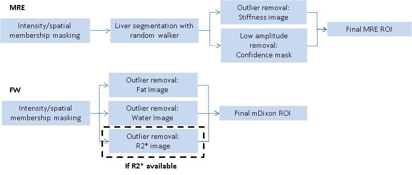
ALEC+mDixon pipeline. Liver segmentation is done
by classifying voxels based on being close to the expected liver position and
similar to its estimated intensity, then morphologically cleaning the mask. For
mDixon, blood vessels are removed by excluding the 20% of the voxels with the
greatest differences from the mean liver fat value. The cleaned mask is then
morphologically closed to bring back isolated pixels and ensure that contiguous
clusters of voxels with big differences are removed. The removal is repeated
for the water image to remove lungs, and to the R2* image to remove
susceptibility artifacts.

Correlation between
ALEC and an experienced reader for the MRE stiffness calculations.
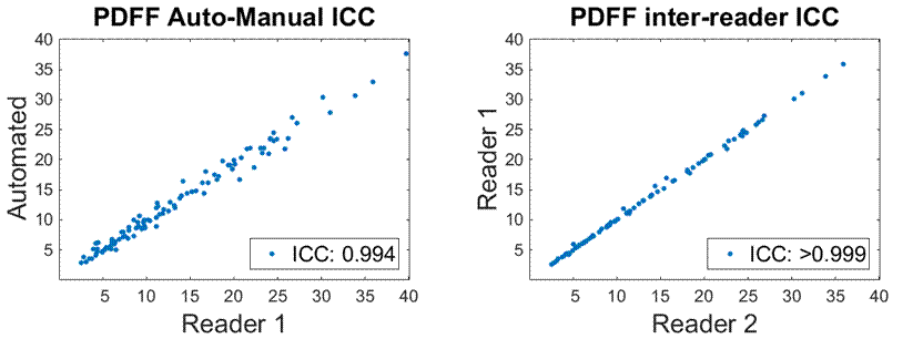
PDFF correlation.
Both the ALEC-reader and reader-reader correlations are excellent. The readers
used a similar analysis method drawing ROIs in the 8 Couinaud anatomical liver
segments. The automated method used a different method, selecting 4 slices with
the largest and most homogeneous liver area. This methodological difference may
explain the lower, but still excellent, correlation of the automated
measurements.
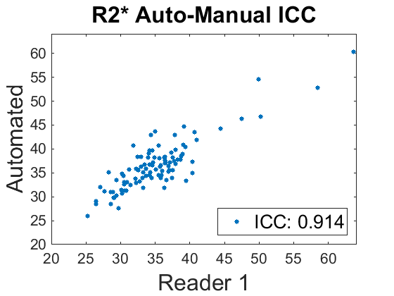
R2* correlation
between ALEC and an experienced reader. A retrospective review of the R2* ROIs
with the biggest differences indicate that they do not appear to contain
artifacts or nonliver tissue. The inter/intra-reader variability of the R2*
measurements has not been assessed and may be higher than PDFF. Further, R2*
may be less homogeneous, causing differences in the ROI locations to have a greater
impact on the reported values.

ALEC report
containing MRE and mDixon ROIs along with stiffness, PDFF, and R2*
measurements. The report is for the case with the largest % difference in PDFF
between ALEC and the primary reader. The automated ROI contains no issues, so
the difference is likely attributable to the use of different slices and regions
within the liver.