0823
Chimeric Mouse Model for MRI Contrast Agent Evaluation1Department of Radiology, Michigan State University, East Lansing, MI, United States, 2Institute of Quantitative Health Science and Engineering, Michigan State University, East Lansing, MI, United States, 3Department of Pharmacology and Toxicology, Michigan State University, East Lansing, MI, United States
Synopsis
While rodents are the primary models for contrast agent evaluation, there is considerable variability in pharmacokinetics of contrast agents in mice vs humans. OATPs play a role in in vivo pharmacokinetics in mice and humans. This study provides evidence that the OATP1B1/1B3 knock-in mouse is a more useful screening tool for novel MRI contrast agents destined for clinical use as compared to the traditionally used wild-type models.
Introduction
Rodents are the primary in vivo models for the evaluation of new pharmaceuticals, including contrast agents, but rodent organ systems do not fully mimic human systems, and may poorly predict how contrast agents will perform in humans. This is demonstrated by the in vivo pharmacokinetics of MRI contrast agents that are transported via hepatic organic anion-transporting polypeptides (OATPs). For example, the FDA-approved contrast agent Gd-EOB-DTPA (Eovist) has ~50% hepatic clearance in both humans and rodents, but Gd-BOPTA (Multihance) has ~50% hepatic clearance in rodents and only ~5% in humans1. Given this drastic difference in pharmacokinetics, using rodents with engineered biological systems that more closely mimic human physiology could streamline evaluation of new MRI contrast agents towards clinical use. Here we characterize a transgenic mouse model that expresses human hepatic OATPs instead of mouse hepatic OATPs, to better mimic human pharmacokinetics of contrast agents in mice.Methods
MSU IACUC approved all animal studies. Wild-type FVB mice, Oatp1A/1B cluster knockout (FVB background), and chimeric OATP1B1/1B3 knock-in mice (Oatp1A/1B cluster knockout FVB background) were from Taconic Biosciences (n=3 per group). Chimeric knock-in mice express OATP1B1/1B3 under the ApoE promoter, restricting expression to the liver. MRI was performed on a 7T Bruker 70/30 BioSpec. T1-weighted RAREst dynamic contrast enhanced (DCE-)MRI was performed (TR=200ms, TE=4.66ms, FOV=38.4x38.4 mm, 8 slices, 1 mm slice thickness, 192x192 matrix). A baseline was established over three acquisitions, and animals were injected via tail vein with a 0.050 mmol/kg contrast agent, followed by saline flush. Images were obtained every minute for 57 minutes (Figure 1). Percent signal enhancement in the liver was calculated for each time point based on an ROI and normalized to baseline. Percent clearance was calculated from area under the curve (AUC) over one hour. In vivo DCE-MRI studies were also carried out using rifampicin to confirm OATP inhibition. To adjust for hepatic blood pool signal enhancement, hepatic signal enhancement in wild-type FVB animals and OATP1B1/1B3 knock-in animals was normalized using Oatp1A/1B knock-out animals as baseline.
Metabolic cage studies were performed to measure hepatic and renal excretion of contrast agent. Feces and urine were collected at 2, 4, 6, 12, 18, and 24 hours. Gadolinium mass per time point and percentage elimination via feces vs kidney urine were calculated using ICP-OES. All statistical analyses were done via two-way ANOVA.
Results and Discussion
Wild-type FVB mice exhibit rapid and sustained hepatic MRI signal enhancement following injection of hepatospecific Gd-EOB-DTPA and Gd-BOPTA, with no hepatic signal enhancement following the injection of non-specific Gd-DTPA (Figure 2a-c). In OATP1A/1b cluster knockout mice, hepatic signal enhancement following injection of Gd-EOB-DTPA and Gd-BOPTA was diminished to 3.5% and 3.9% respectively relative to wild-type FVB mice (Table 1), confirming the importance of OATPs for hepatic uptake. In OATP1B1/1B3 knock-in mice, hepatic signal enhancement following injection of Gd-EOB-DTPA was similar to that of wild-type FVB mice. In contrast, hepatic signal enhancement following injection of Gd-BOPTA was reduced to 18% compared to wild-type mice (Table 1). AUC analysis (Figure 3a) shows non-significant difference between hepatic signal enhancement by Gd-EOB-DTPA in FVB mice vs chimeric OATP1B1/1B3 knock-in mice, and Figure 3b shows significant difference in hepatic signal enhancement by Gd-BOPTA in FVB vs OATP1B1/1B3 knock-in mice. Injection of rifampicin reduced hepatic signal enhancement by Gd-EOB-DTPA in OATP1B1/1B3 knock-in mice to 7.9% relative to FVB mice, confirming pharmacological responsiveness of OATPs to known inhibitors (Table 1).
Figure 4a-d summarizes metabolic cage excretion data. At 24 hours, in wild-type FVB mice, 79% of Gd-EOB-DTPA was recovered in fecal samples and 20% in urine (Table 2). In OATP1B1/1B3 knock-in mice, this elimination profile was nearly identical to that seen in wild-type mice, with 81% elimination via feces and 19% via urine. Gd-BOPTA elimination in wild-type FVB mice was 83% via feces and 16% in urine. This elimination profile was significantly altered in OATP1B1/1B3 knock-in mice, with 32% elimination via feces and 67% via urine. Gd-DTPA elimination in wild-type FVB mice was 10% via feces and 89% via urine, similar to OATP1B1/1B3 knock-in mice (8.6% liver and 91% kidney). The detection of small amounts of gadolinium in feces of mice injected with Gd-DTPA was likely due to contact between urine and fecal samples prior to collection.
Conclusion
Hepatic MRI signal enhancement and elimination of Gd-EOB-BOPTA, Gd-BOPTA, and Gd-DTPA in chimeric OATP1B1/1B3 knock-in mice closely reflects MRI signal enhancement characteristics and clearance in humans, rather than that seen in wild-type mice. The physiological excretion of contrast agents by wild-type FVB and chimeric OATP1B1/1B3 animals is in agreement with the enhancement profiles seen in MRI studies. We conclude that the OATP1B1/1B3 knock-in mouse is a more appropriate model for the evaluation of novel contrast agents than wild-type mice.Acknowledgements
We are grateful to Dr. Bruno Hagenbuch, KUMC, for helpful conversations about OATPs. Grant support from the National Institutes of Health is acknowledged (R01 DK107697 to EMS).References
1. Spinazzi A, Lorusso V, Provano G, Kirchin, M. Safety, tolerance, biodistribution, and MR imaging enhancement of the liver with Gadobenate dimeglumine: Results of clinical pharmacologic and pilot imaging studies in nonpatient and patient volunteers. Academic Radiology 6 (1999) 282-291.Figures
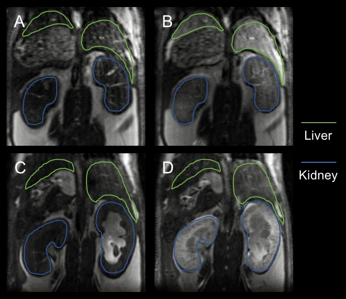
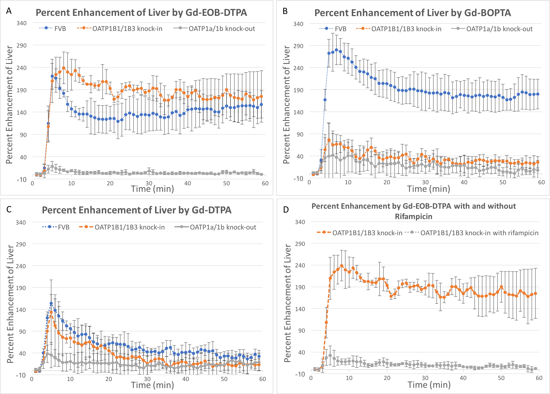
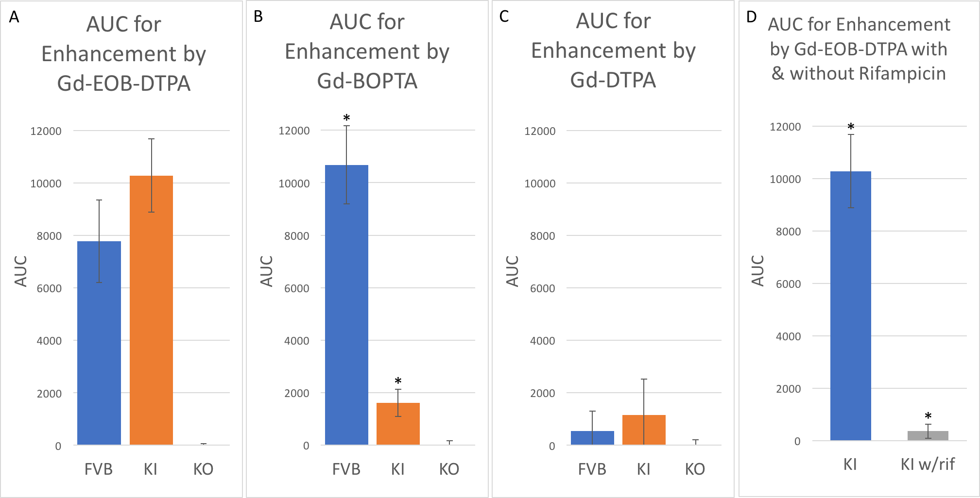
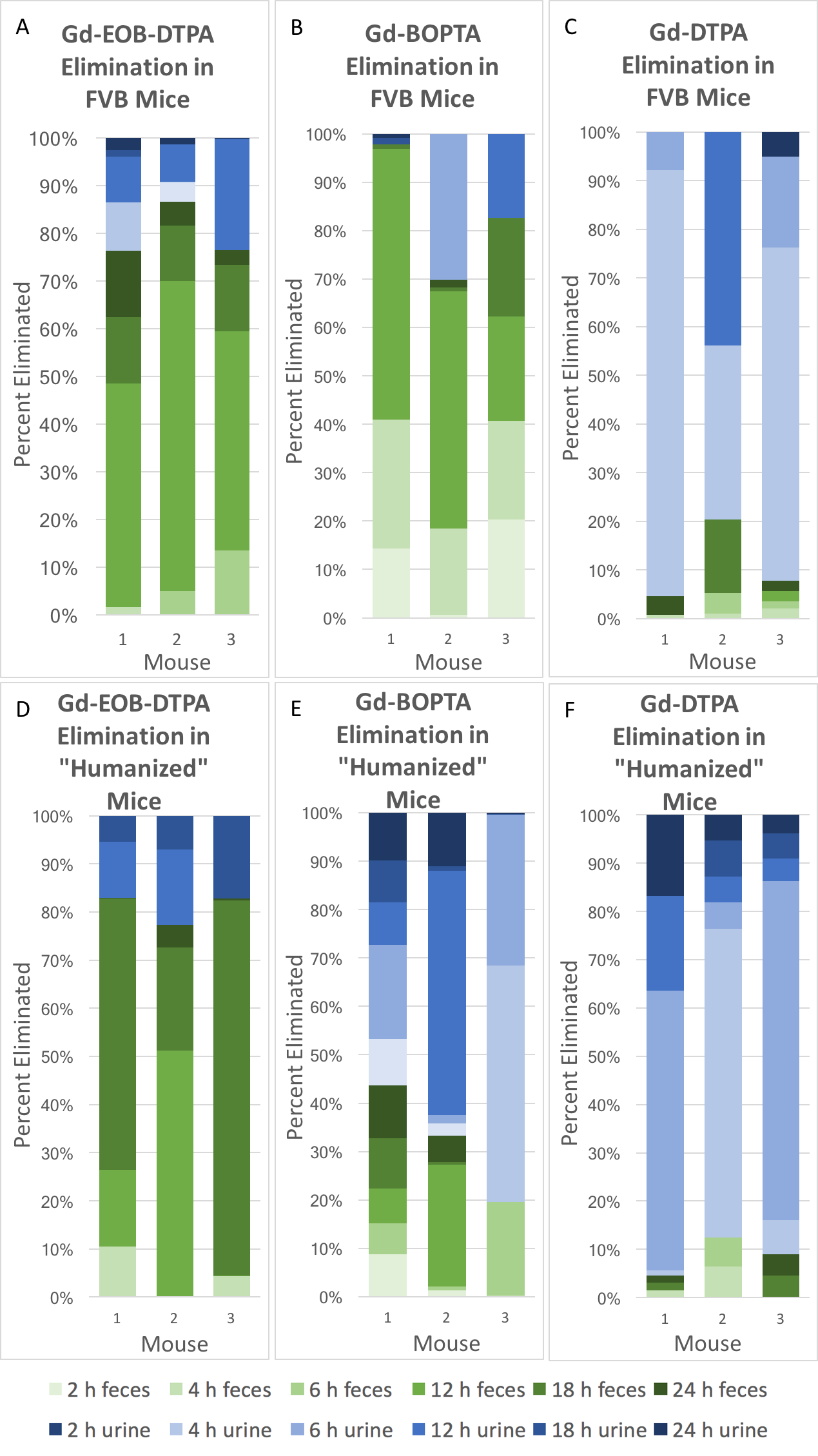
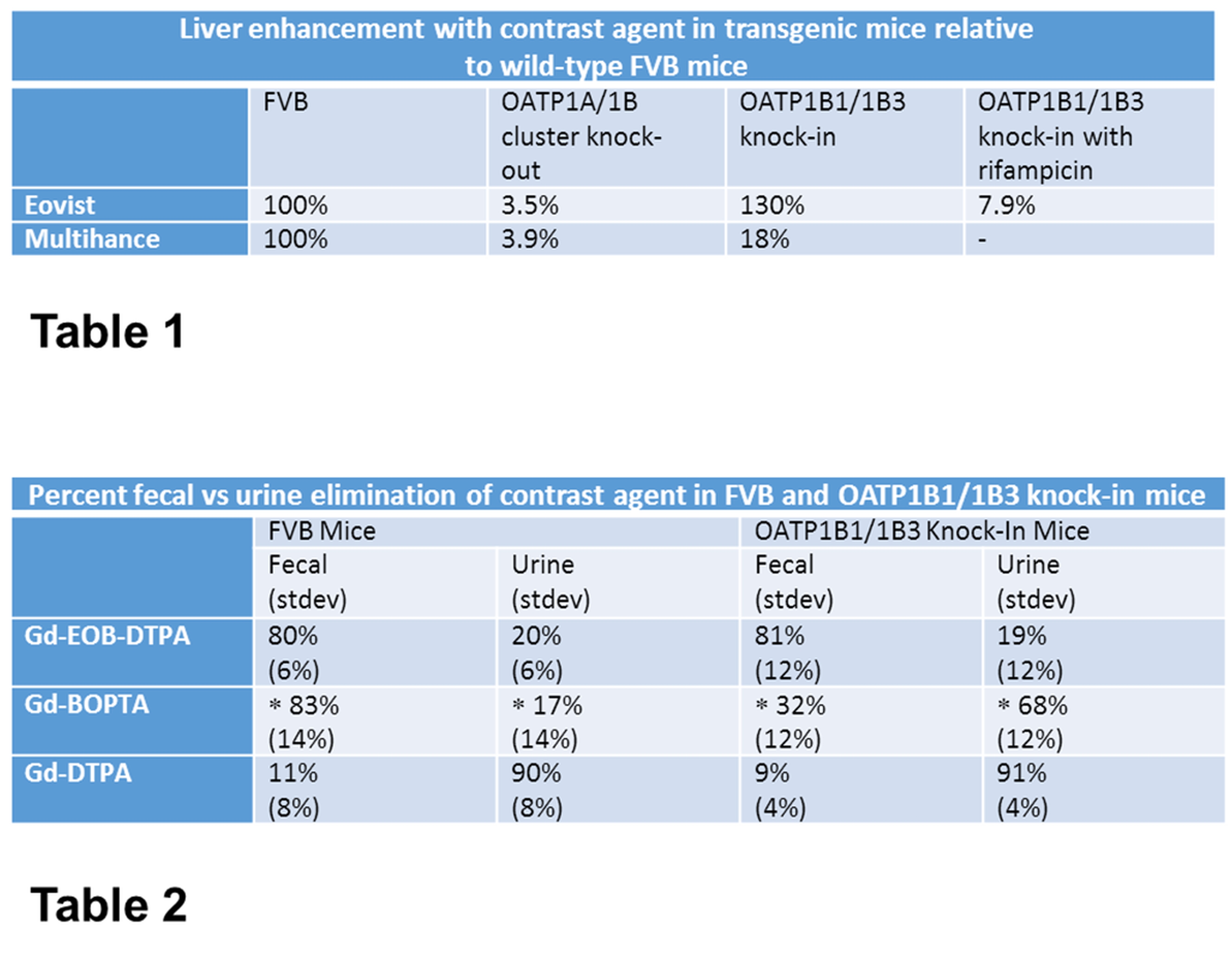
Table 1: Liver enhancement with contrast in OATP1A/1B cluster knock-out and OATP1B1/1B3 knock-in mice as a percentage of liver enhancement in wild-type FVB mice.
Table 2: A comparison of elimination of MRI contrast agent via feces vs urine in wild-types vs “humanized” (OATP1B1/1B3 knock-in) animals. (*P<0.05)