0822
Multimodality OATP1B3-enhanced reporter gene imaging of engineered cells via fluorescence, photoacoustic, and magnetic resonance imaging1Medical Imaging Laboratories, Robarts Research Institute, London, ON, Canada, 2Medical Biophysics, University of Western Ontario, London, ON, Canada, 3Lawson Health Research Institute, London, ON, Canada
Synopsis
Reporter genes can generate information on cell fate in vivo. Multimodality imaging can mitigate limitations of individual imaging modalities; thus, multimodality reporters are highly sought. We present a trimodality reporter gene system based on human Organic Anion-Transporting Polypeptide 1b3 (Oatp1b3) and its ability to take up indocyanine green (ICG) for fluorescence and photoacoustic imaging, as well as gadolinium ethoxy benzyl diethylenetriamine pentaacetic acid (Gd-EOB-DTPA) for MRI. This technology should find utility in tracking cell- and gene-based therapies for preclinical studies, and due to the human-derivation of the reporter gene and already clinically-utilized probes, presents with high translational potential.
Introduction
Reporter genes are invaluable in vivo measurement tools for studying the fate of numerous cell types such as cancer cells, stem cells, and immune cells in preclinical studies and have been recently used in patients. Multimodality reporter systems are particularly useful, as they overcome limitations inherent in a single modality by facilitating non-invasive acquisition of different imaging datasets for a more complete understanding of the biological phenomenon at hand. Human Organic Anion-Transporting Polypeptide 1b3 (Oatp1b3) was recently demonstrated as an MRI reporter gene based on its ability to take up the positive contrast, clinically-used paramagnetic agent gadolinium ethoxy benzyl diethylenetriamine pentaacetic acid (Gd-EOB-DTPA)1. Oatp1b3 was also established as a fluorescent imaging (FLI) reporter gene based on its ability to take up the clinically-used, near-infrared (NIR) fluorescent dye, indocyanine green (ICG)1. Our objective was two-fold: 1) to test the feasibility of this reporter system on a clinical 3-Tesla scanner with clinical Gd-EOB-DTPA doses; and 2) evaluate its use as a photoacoustic imaging (PAI) reporter gene system, as ICG features a high molar extinction coefficient, a relatively low quantum yield and has previously been utilized for PAI2, 3, 4.Methods
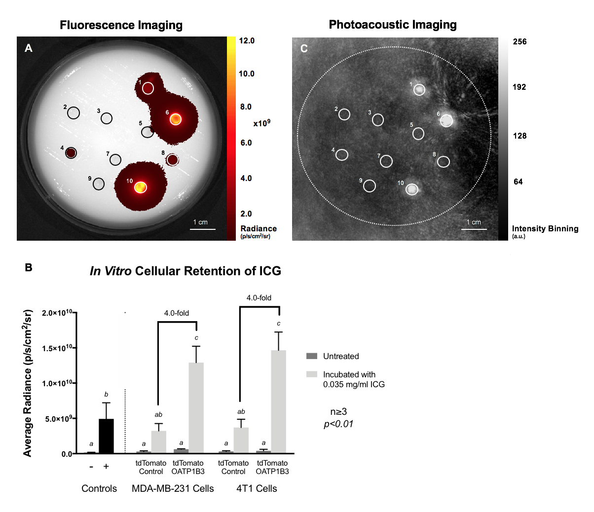
Human (MDA-MB-231) and murine (4T1) cells were engineered with lentivirus encoding tdTomato to generate tdTomato Control cells. An aliquot of these cells was then transduced with a second lentivirus encoding zsGreen and Oatp1b3 to generate tdTomato OATP1B3 cells (Figure 1A). Engineered cells were sorted via flow cytometry with >90% purity (Figure 1B). In vitro FLI, PAI, and MRI were performed on these cells. For FLI, tdTomato Control and tdTomato OATP1B3 cells were incubated with or without 35 μg/ml ICG for 1 hour and imaged with an optical scanner using 780-nm excitation and 845-nm emission filters. The same phantom was imaged with PAI (780-nm excitation) using a custom-built PAI system. R1 relaxation rates of cells incubated with either 6.4 mM Gd-DTPA (control) or 6.4 mM Gd-EOB-DTPA for 90 minutes were measured using a fast spin-echo inversion recovery (FSE-IR) pulse sequence on a clinical 3-Tesla MRI scanner. Mammary fat pads of female mice were implanted with either tdTomato Control (n=6) or tdTomato OATP1B3 (n=5) MDA-MB-231 cells. Tumours were imaged with FLI for tdTomato fluorescence (520-nm excitation, 570-nm emission filters) and ICG fluorescence (780-nm excitation, 845-nm emission filters), as well as NIR-spectrum PAI (680-970 nm, Δλ=5 nm) before and 24-hours after administration of 8 mg/kg ICG. tdTomato FLI was used as a surrogate measure of tumour size to normalize ICG signals.Results
Immunofluorescence staining confirmed the absence of OATP1B3 in tdTomato Control cells and its presence in tdTomato OATP1B3 cells (Figure 1C). Growth rates did not differ between naïve and engineered cell populations with or without ICG incubation (Figure 1D). FLI revealed significantly increased (4.0-fold; p<0.05) FLI signal from tdTomato OATP1B3 cells incubated with ICG relative to tdTomato Control cells (Figure 2A-B). PAI also demonstrated that tdTomato OATP1B3 cells incubated with ICG were readily detectable, but all other treated and untreated controls were not visible (Figure 2C). tdTomato OATP1B3 cells exhibited significantly increased (4.9-fold; p<0.05) R1 rates at 3 Tesla following incubation with Gd-EOB-DTPA, relative to tdTomato Control cells incubated with Gd-EOB-DTPA, and to tdTomato OATP1B3 cells incubated with Gd-DTPA (Figure 3). Normalized FLI signals at ICG wavelengths (780 nm) were significantly higher (27.6-fold; p<0.05) in mice with tdTomato OATP1B3 tumours (n=5) compared to tdTomato Control tumours (n=6) 24-hours after ICG administration (Figure 4). PAI signals were also significantly higher (2.3-fold; p<0.05) in mice with tdTomato OATP1B3 tumours (n=5) compared to tdTomato Control tumours (n=6) 24-hours after ICG administration (Figure 5).Discussion
Molecular imaging aims to detect molecular events in vivo with sensitivity, specificity, and high resolution. To achieve these goals, multi-modality imaging can mitigate inherent limitations present in a single modality. Optical imaging methods such as FLI and PAI offer rapid and cost-effective measures to detect reporter signals from localized engineered cells. MRI offers high resolution, 3D spatial information with detailed anatomical context. Here, we extend previous work on the Oatp1b3 system to describe its use as a multimodality reporter gene system that allows for ICG-enhanced FLI, and now PAI, as well as Gd-EOB-DTPA-enhanced MRI at 3 Tesla to track viable engineered cells in animal models. Importantly, the human derivation of Oatp1b3 relative to non-human reporter genes that raise immunogenicity concerns5, along with longstanding FDA-approval of both ICG and Gd-EOB-DTPA, pave a potential path towards clinical translation for this tri-modality reporter gene system. Future work focuses on in vivo tumour imaging with 3-Tesla MRI and 0.025-mmol/kg clinical Gd-EOB-DTPA dosage.Acknowledgements
No acknowledgement found.References
1. Wu MR, Liu HM, Lu CW, Shen WH, Lin IJ, Liao LW, et al. Organic anion-transporting polypeptide 1B3 as a dual reporter gene for fluorescence and magnetic resonance imaging. FASEB J 2018, 32(3): 1705-1715.
2. Yuan B, Chen N, Zhu Q. Emission and absorption properties of indocyanine green in Intralipid solution. J Biomed Opt 2004, 9(3): 497-503.
3. Philip R, Penzkofer A, Baumler W, Szeimies RM, Abels C. Absorption and fluorescence spectroscopic investigation of indocyanine green. J Photoch Photobio A 1996, 96(1-3): 137-148.
4. Alander JT, Kaartinen I, Laakso A, Patila T, Spillmann T, Tuchin VV, et al. A review of indocyanine green fluorescent imaging in surgery. Int J Biomed Imaging 2012, 2012: 940585.
5. Gschweng EH, McCracken MN, Kaufman ML, Ho M, Hollis RP, Wang X, et al. HSV-sr39TK positron emission tomography and suicide gene elimination of human hematopoietic stem cells and their progeny in humanized mice. Cancer Res 2014, 74(18): 5173-5183.
Figures