0821
In vivo MRI tracking of stem-cell-derived extracellular vesicles1Department of Radiology, Johns Hopkins University, Baltimore, MD, United States, 2FM. Kirby center, Kennedy Krieger Institute, Baltimore, MD, United States, 3Institute for Cell Engineering and Department of Medicine, Johns Hopkins University, Baltimore, MD, United States, 4Department of Biomedical Engineering, Johns Hopkins University, Baltimore, MD, United States
Synopsis
Induced pluripotent stem cell (iPSC) derived extracellular vesicles (EVs) constitute a new class of cell-free regenerative medicine. Non-invasive tracking of the delivery and in vivo distribution of EVs is highly desirable. Traditional labeling methods suffer from either low labeling efficiency or difficulty with purification. Here, we report a new labeling strategy using surface-functionalized magnetic particles, in which the nonencapsulated magnetic particles can be easily separated from the labeled EVs, allowing the preparation of magnetically labeled EVs with high purity. We then demonstrate in vivo tracking of magnetically labeled EVs in the kidney and liver using MRI.
Introduction
Extracellular vesicles (EVs), including exosomes and microvesicles (diameter of 20-1000 nm), are secreted by nearly all cell types for intercellular communication1. Overwhelming evidence shows that EVs secreted by stem cells play an important role in stem cell therapy through paracrine effects and can be used as cell-free substitutes of stem cells. For instance, stem-cell derived EVs exhibit unprecedented therapeutic effects on irreversibly injured kidneys2-5. Non-invasive imaging methods that can track the delivery and in vivo distribution of therapeutic EVs will ultimately be needed for accelerating the clinical translation of EV-based therapies. However, traditional labeling methods for image detection suffer from either low labeling efficiency or difficulty with purification. Our goal was to develop an effective labeling strategy to prepare magnetically labeled induced pluripotent stem cells (iPSCs)-derived EVs and to track their fates using MRI in an acute kidney injury (AKI) mouse model.Methods
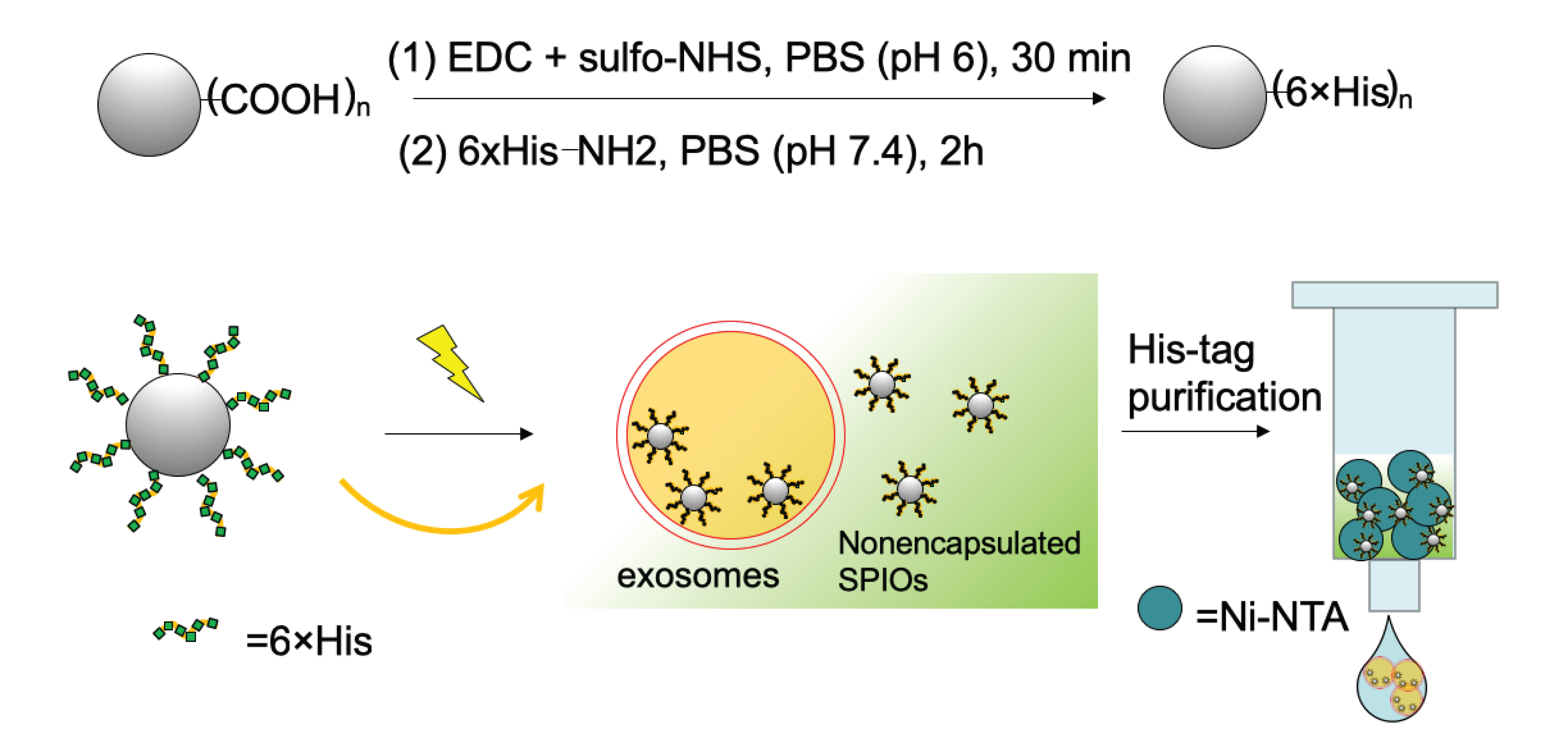
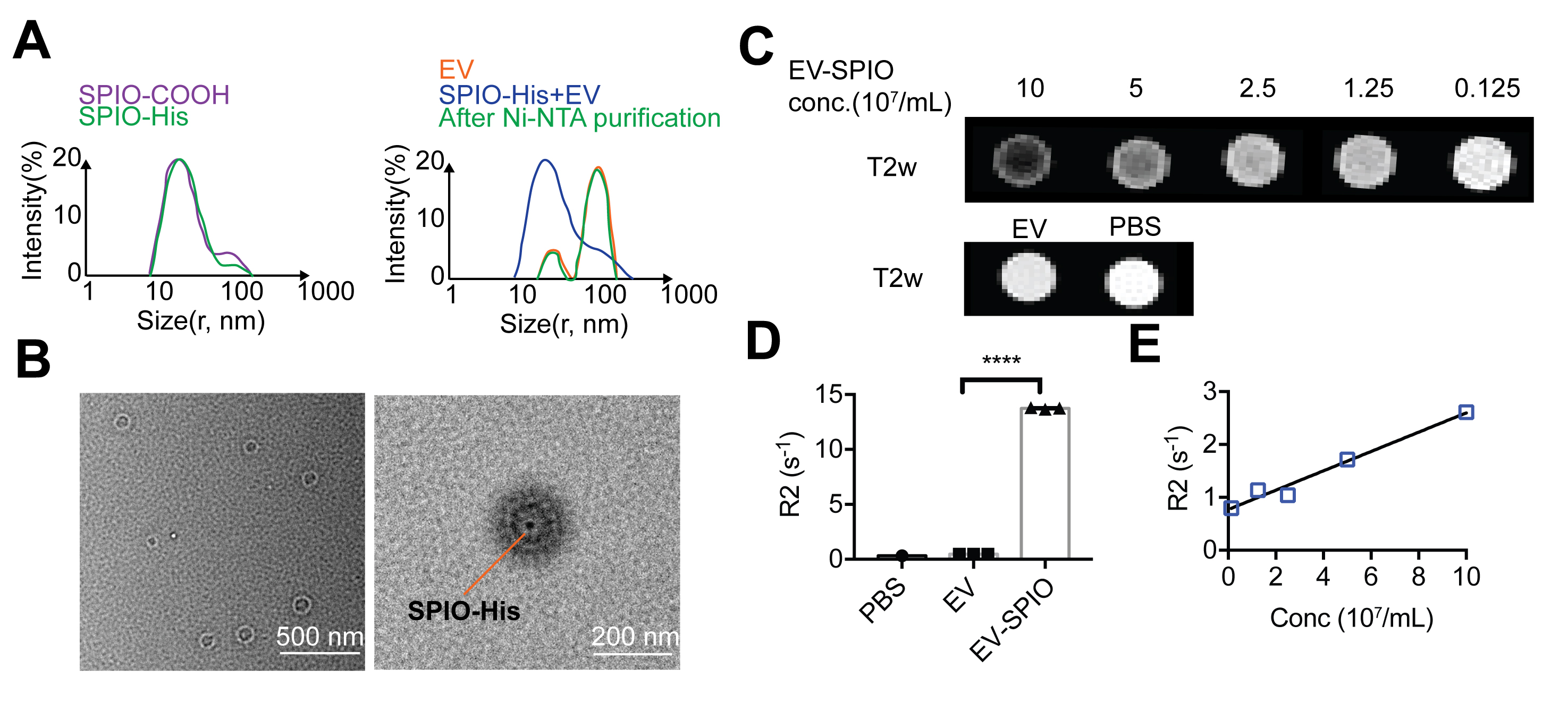
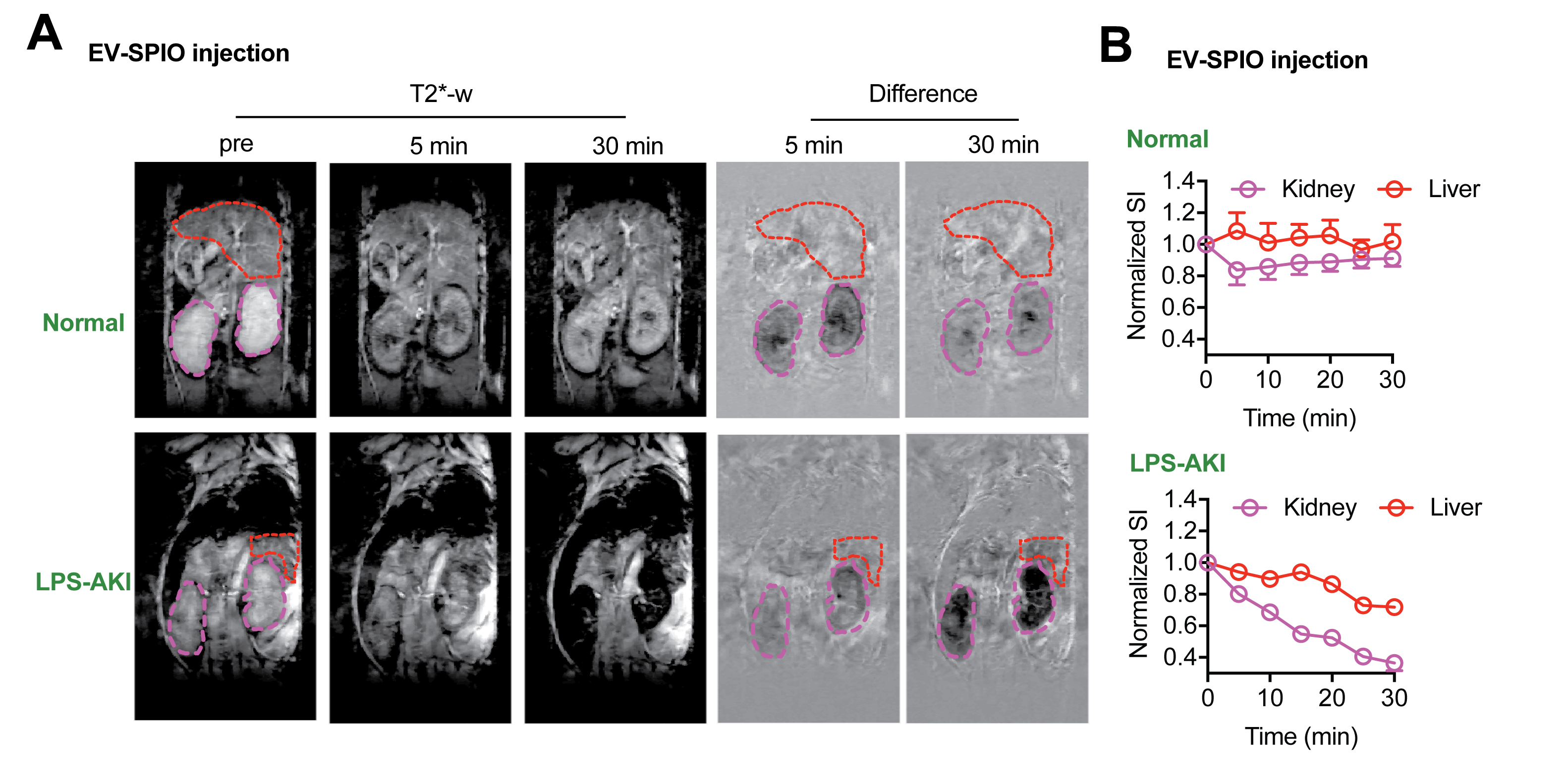
We designed the EVs labeling strategy as illustrated in Fig.1. In brief, we first prepared the Histidine-tag-functionalized superparamagnetic iron oxide nanoparticles (SPIO) using carboxyl-SPIOs (core diameter: 5 nm, Ocean nanotech) and hexa-His peptide (His-tag). iPSC-derived EVs were prepared by concentrating the medium of iPSC culture using Amicon ultra-15 filter column with Ultracel-100 membrane (MilliporeSigma) and further purified by the qEV column (iZON). Fifty uL of the EVs (1011/mL, determined by nanoparticle tracking analysis) were mixed with 15 uL 2mg/mL SPIO-His and electroporation (250V, 100μF, 1000Ω, 1mm cuvette, 2 pulses with 5s interval) was performed using a Gene Pulser Xcell electroporation system (Bio-Rad). The mixture then incubated with 1mL Nickle-charged resin (Ni-NTA) for 30 min with shaking allow adequate binding of his-tagged SPIO to Ni-NTA. The SPIO-labeled EVs then collected by elution. The iron content of labeled EVs were analyzed using ICP-MS, and characterized using dynamic light scattering (DLS) and transmission electron tomography (TEM). The T2-relaxivity of labeled EVs was measured on a Bruker 400 MHz vertical bore scanner using a spin echo method described previously6. The AKI model was prepared by i.p injection of 10 mg/kg lipopolysaccharide (LPS) in C57BL/6J mice (5-8 weeks)7. At 24h after LPS injection, mice were injected (i.v.) with approximately 109 EVs in 200 uL PBS and T2* weighted images were intermittently acquired before and after the injection for 30 minutes using a gradient echo sequence (TR=800 ms, TE=5.8 ms, matrix size=256x128, in plane resolution=0.167x0.280 mm2) performed on 11.7T Biospec scanner (Bruker).Results
The size distribution measured by DLS (Fig. 2A) showed that the His-tag functionalization has negligible effect on the size of SPIO (46.12±14.80 and 43.90±16.51 nm in diameter, for SPIO-COOH and SPIO-His, respectively), readily discriminable from that of EVs (230.04±105.81 nm in diameter). The size of SPIO-labeled EVs (EV-SPIOs) (229.0±35.50 nm) resembled that of unlabeled EVs, indicating the efficient removal of unencapsulated SPIOs in the final product. TEM verified the presence of SPIO-His in EVs (Fig. 2B). The magnetically labeled EVs exhibited strong T2 contrast in vitro (Fig. 2C). The R2 enhancement was 1.828 s-1 for 108/mL EVs (or 16.7 pM), indicating a r2 relaxivity of 1.1 x10¹⁰ s-1mM-1 (per EV). The concentration of iron in EVs was determined to be 155 ng/108 EVs using ICP-MS. In normal mice, the i.v. injected EV-SPIOs demonstrated a rapid accumulation in the kidney, followed by slow clearance over 30 minutes (Fig.3A&B). Interestingly, almost no signal reduction in liver was observed. In contrast, in the AKI mice, T2* images showed that the accumulation of EV-SPIOs was much stronger in the injured kidney. After 30 min, kidneys became almost completely dark, indicative of selective targeting of EVs in the disease site (Fig.3A&B). Meanwhile, liver also demonstrated signal attenuation likely due to the inflammation induced by LPS, which, however, was much less than that in kidneys.Conclusion
We successfully developed an efficient labeling method for preparing SPIO-loaded iPSC-derived EVs. We modified the surface of SPIO with histidine-tags so that the free SPIO can bind to Ni-NTA resin, while those encapsulated in EVs are “protected” inside the vesicles as they lose the contact with the binding resin. Using this labeling strategy we prepared magnetically labeled EVs and used them to track the in vivo distribution of iPSCs-derived EV-SPIOs without interference from free SPIO. Our in vivo MRI results revealed the excellent targeting ability of stem cells-derived EVs to the injured kidneys in the AKI model.Acknowledgements
Supported by NIH grants R01CA211087 and R21CA215860.References
1. Karpman, D., Stahl, A.L. & Arvidsson, I. Extracellular vesicles in renal disease. Nature reviews. Nephrology 13, 545-562 (2017).
2. Collino, F., et al. AKI Recovery Induced by Mesenchymal Stromal Cell-Derived Extracellular Vesicles Carrying MicroRNAs. Journal of the American Society of Nephrology : JASN 26, 2349-2360 (2015).
3. Reis, L.A., et al. Bone marrow-derived mesenchymal stem cells repaired but did not prevent gentamicin-induced acute kidney injury through paracrine effects in rats. Plos One 7, e44092 (2012).
4. Bruno, S., et al. Mesenchymal stem cell-derived microvesicles protect against acute tubular injury. Journal of the American Society of Nephrology : JASN 20, 1053-1067 (2009).
5. Bruno, S., et al. Microvesicles Derived from Mesenchymal Stem Cells Enhance Survival in a Lethal Model of Acute Kidney Injury. Plos One 7(2012).
6. Zhang, J., et al. Triazoles as T2-exchange MRI contrast agents for the detection of nitrilase activity. Chemistry (Weinheim an der Bergstrasse, Germany) (2018).
7. Liu, J., et al. CEST MRI of sepsis-induced acute kidney injury. NMR Biomed. 31, e3942 (2018).
Figures