0816
Chasing True FLAIR: a three-component Magnetic Resonance Fingerprinting approach to synthetic MRI1Department of Physics, University of Pisa, Pisa, Italy, 2IMAGO7 Foundation, Pisa, Italy, 3IRCCS Stella Maris, Pisa, Italy, 4Computer Science, Technische Universität München, Munich, Germany, 5GE Healthcare, Munich, Germany
Synopsis
MR Fingerprinting is a multiparametric imaging technique which allows to obtain several parametric maps of tissue, such as Proton Density and T1/T2 maps, within a single fast acquisition in transient-state. These maps can be used to synthesize a whole set of different contrast-weighted images, potentially substituting an entire conventional protocol. However, these synthetic images suffer from artifacts due to partial volume effects. This is particularly true for FLuid Attenuated Inversion Recovery (FLAIR) images. Here, we modify the signal model to account for CSF and flowing blood, correcting these artifacts, and we compared the resulting synthetic FLAIR to true FLAIR images.
Introduction
Recently, there has been a development of multiparametric quantification techniques, such as QRAPMASTER1 or Magnetic Resonance Fingerprinting (MRF)2, providing maps of Proton Density (PD), T1 and T2 within a single acquisition. These maps can be exploited to obtain any desired contrast-weighted image by using the appropriate signal equation, potentially replacing a whole clinical exam. This approach is called Synthetic MRI. However, both the presence of multiple tissues per voxel and the effect of flowing blood are usually neglected in the signal model, leading to quantification error and to artifacts in the synthetic images. This is particularly evident in synthetic FLuid Attenuated Inversion Recovery (FLAIR)3 images, which are affected from Partial Volume Effect (PVE) due to Cerebrospinal Fluid (CSF) and signal hyperintensities due to flowing blood. It has been shown that CSF PVE can be corrected by using a multi-component signal model4 within an MRF framework. However, previous studies failed to correct for flowing blood: here, we incorporated this effect in the dictionary5, correcting artifacts due to vessels, and we compared the resulting synthetic FLAIRs to true FLAIR6,7 acquisitions.Methods
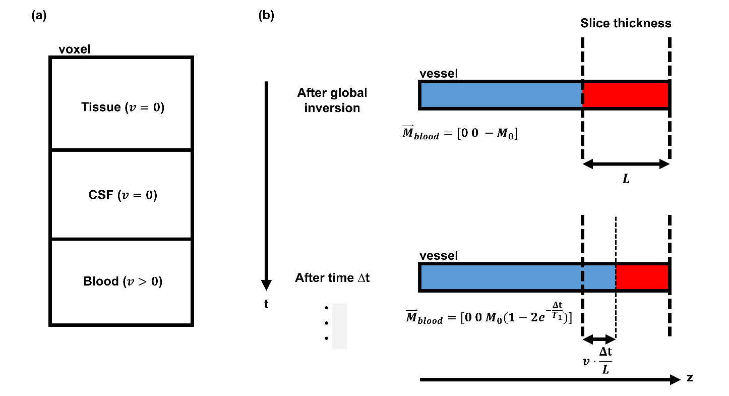
Here, we extended the MRF signal model to account for CSF and flowing blood (see Figure 1a). Therefore, we wrote the dictionary $$$D$$$ as a weighted combination of tissue, CSF and vessel dictionaries $$$D_{T,CSF,v}$$$ :
$$D=w_TD_T+w_{CSF}D_{CSF}+w_vD_v$$
where $$$w_{T,CSF,v}\in\mathbb{R};w_T+w_{CSF}+w_v=1$$$ are the tissue, CSF and vessel fractions. Both tissue (T1 from 0 to 2000ms; T2 from 0 to 300ms) and CSF (T1=3500ms; T2=1500ms) were modelled as static components with velocity $$$v=0$$$, hence their simulation is equivalent to original MRF2. To compute the vessel dictionary, we used a previously introduced simplified model in which a constant scalar velocity $$$v$$$ for the blood is assumed5 (Figure 1b). Velocity values from 0 to 100 cm/s were used for the blood simulation (T1=1500ms; T2=250ms). To perform the reconstruction, a two-step approach was used. In a first matching step, PD, T1, T2, blood velocity, vessel fraction and CSF fraction maps were obtained. Then, the vessel fraction map was cleaned by setting to zero each voxel having velocity $$$v=0$$$. Finally, the matching was repeated fixing blood velocity, vessel and CSF fractions values from the corresponding maps, obtaining corrected PD and tissue T1/T2 maps. Synthetic FLAIR can then be obtained as:
$$S_{FLAIR}=w_T|PD|[1-2exp(-\frac{TI}{T_1})+exp(-\frac{TR}{T_1})]exp(-\frac{TE}{T_2})$$
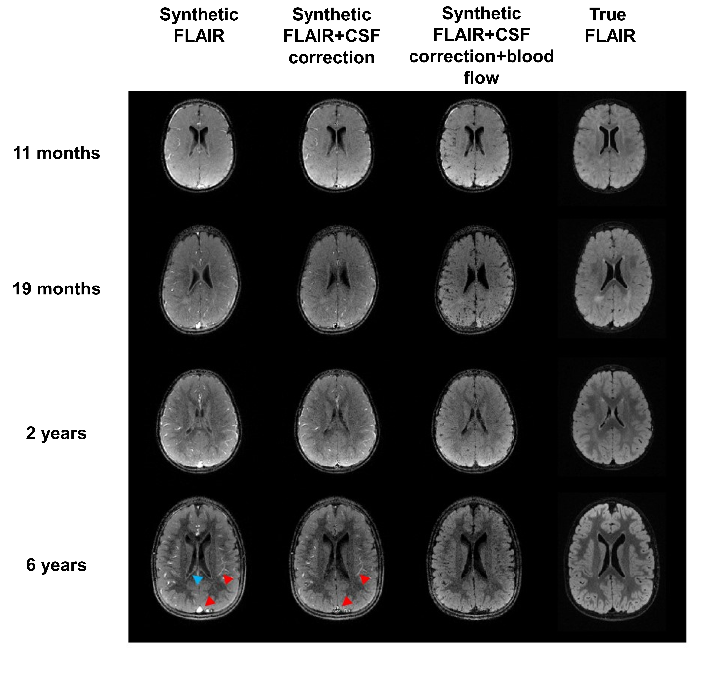
This approach was used to reconstruct previously acquired data from 4 patients (age from 11 months to 6 years) without visible alterations and to compute corresponding synthetic FLAIR images (TI/TR/TE=1883/6000/117ms). Subject ages were chosen to test the technique for different stages of development of the brain. These synthetic images were compared to true FLAIR and to synthetic FLAIR using single-component mode and two-component model accounting only for CSF.
Results
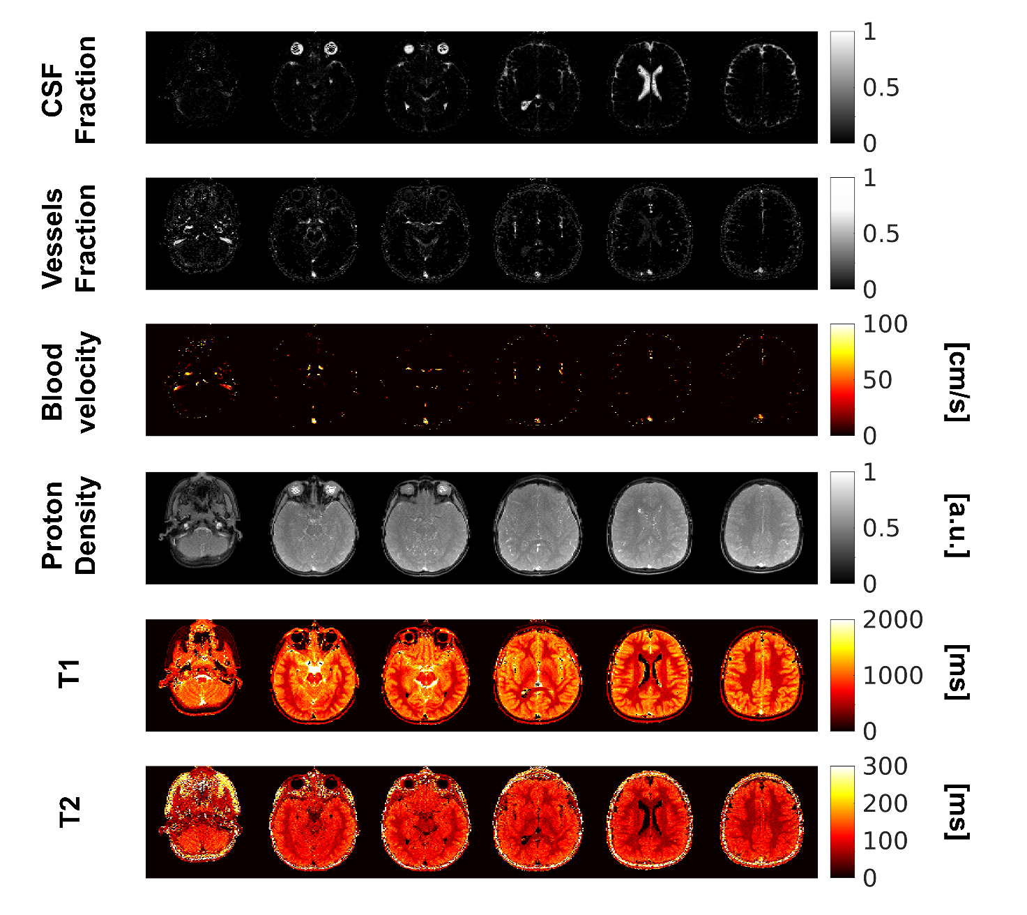
Reconstruction time using the proposed approach was 420s using a CPU with 48 cores. As shown in Figure 2, tissue T1 and T2, blood velocity and PD maps, as well as vessel and CSF fraction maps, were successfully obtained. In particular, CSF fraction map correctly shows structures as the ventricles, while in the vessel fraction map structures as the internal carotid artery, the middle cerebral artery and the superior sagittal sinus are clearly visible. Figure 3 shows the comparison between synthetic and true FLAIRs. It can be seen that our approach is able to suppress the signal from both CSF and vessels achieving a very similar contrast to the true FLAIR. This is true for each subject independently from the subject age (and therefore from the different stages of development of the brain). In contrast, naïve single-component synthetic FLAIR suffers both from PVE artifacts near the ventricles (blue arrow) and inconsistent hyperintensities within the vessels (red arrow), while the two-component model neglecting blood flow is only able to correct PVE near the ventricles.Discussion
Despite of the simplified flow model used in the simulation, which leads to unrealistic velocity values (especially for arteries), the proposed signal model is able to solve the inconsistencies due to the presence of CSF and flowing blood. With respect to previous multi-component approaches4, accounting for blood flow allow to remove signal hyperintensities due to vessels, leading to a contrast which is closer to the true FLAIR. Importantly, no modification of the acquisition pattern is required, thus each previously acquired dataset can be retrospectively corrected. Moreover, the reconstruction time is still short enough to allow clinical use of the technique.Conclusion
We demonstrated that an extended signal model accounting for CSF and blood flow allow to remove artifacts due to these confounding factors and to improve the accuracy of synthetic FLAIR.Acknowledgements
No acknowledgement found.References
1. Warntjes JBM, Leinhard OD, West J, Lundberg P. Rapid magnetic resonance quantification on the brain: Optimization for clinical usage. Magn Reson Med. 2008;60(2):320-329.
2. Ma D, Gulani V, Seiberlich N, Liu K, Sunshine JL, Duerk JL, Griswold MA. Magnetic resonance fingerprinting. Nature. 2013;495(7440):187-92.
3. Tanenbaum LN, Tsiouris AJ, Johnson AN, Naidich TP, DeLano MC, Melhem ER, Quarterman P, Parameswaran SX, Shankaranarayanan A, Goyen M, Field AS. Synthetic MRI for Clinical Neuroimaging: Results of the Magnetic Resonance Image Compilation (MAGiC) Prospective, Multicenter, Multireader Trial. AJNR Am J Neuroradiol. 2017;38(6):1103-1110.
4. Deshmane A, McGIvney D, Badve C, Yu A, Jiang Y, Ma D, Griswold MA. Accurate Synthetic FLAIR Images Using Partial Volume Corrected MR Fingerprinting. In: Proceedings of the 24th Annual Meeting of ISMRM. Singapore; 2016:1909.
5. Gomez PA, Molina-Romero M, Buonincontri G, Menze BH, Menzel MI. Simultaneous Magnetic Resonance Angiography and Multiparametric Mapping in the Transient-state. In: Proceedings of the 26th Annual Meeting of ISMRM. Paris; 2018:63.
6. Hajnal J V, De Coene B, Lewis PD, Baudouin CJ, Cowan FM, Pennock JM, Young IR, Bydder GM. High signal regions in normal white matter shown by heavily T2-weighted CSF nulled IR sequences. J Comput Assist Tomogr. 16(4):506-13.
7. Hajnal J V, Bryant DJ, Kasuboski L, Pattany PM, De Coene B, Lewis PD, Pennock JM, Oatridge A, Young IR, Bydder GM. Use of fluid attenuated inversion recovery (FLAIR) pulse sequences in MRI of the brain. J Comput Assist Tomogr. 16(6):841-4.
Figures