0796
Creating a diffusion tractography-based atlas of human thalamic ventral intermediate nucleus aided by deep learning1Athinoula A. Martinos Center for Biomedical Imaging, Department of Radiology, Massachusetts General Hospital, Harvard Medical School, Charlestown, MA, United States, 2Harvard-MIT Division of Health Sciences and Technology, Massachusetts Institute of Technology, Cambridge, MA, United States, 3Radiological Sciences Laboratory, Department of Radiology, Stanford University, Stanford, CA, United States
Synopsis
Tremor suppression in the hands of patients with essential tremor can be achieved by lesioning the ventral intermediate nucleus (Vim) of the thalamus using transcranial MR-guided focused ultrasound. Recent work has shown that diffusion MR tractography identifies the Vim more precisely and predicts the degree of tremor suppression. Here, we trained a convolutional neural network (CNN) to automatically segment relevant regions-of-interest including the thalamus, red nucleus, dentate nucleus and handknob region for probabilistic tractography to identify the Vim. We applied the CNN to 200 HCP healthy subjects and created a tractography-based atlas of Vim location, which could aid in neurosurgical guidance.
Introduction
Hand tremor compromises essential tremor patients’ activities of daily living and quality of life. Tremor suppression can be achieved by lesioning the ventral intermediate nucleus (Vim) of the thalamus using transcranial MR-guided focused ultrasound (tcMRgFUS).1 The Vim cannot be delineated on standard structural MR images due to its small size and low intrinsic contrast. Diffusion tractography identifies the Vim by mapping tremor circuits (i.e., thalamocortical radiation, dentatothalamic tract). The tractography-identified location has been shown to predict the degree of tremor-suppression following tcMRgFUS.2 An atlas of tractography-identified Vim locations could aid in neurosurgical guidance but would require manual delineation by expert neuroradiologists of relevant regions-of-interest (ROIs) in a large population--a time-consuming and laborious task. Here, we perform probabilistic tractography to identify the Vim in 200 healthy subjects using ROIs automatically segmented by a convolutional neural network (CNN) and create a tractography-based Vim atlas.Methods
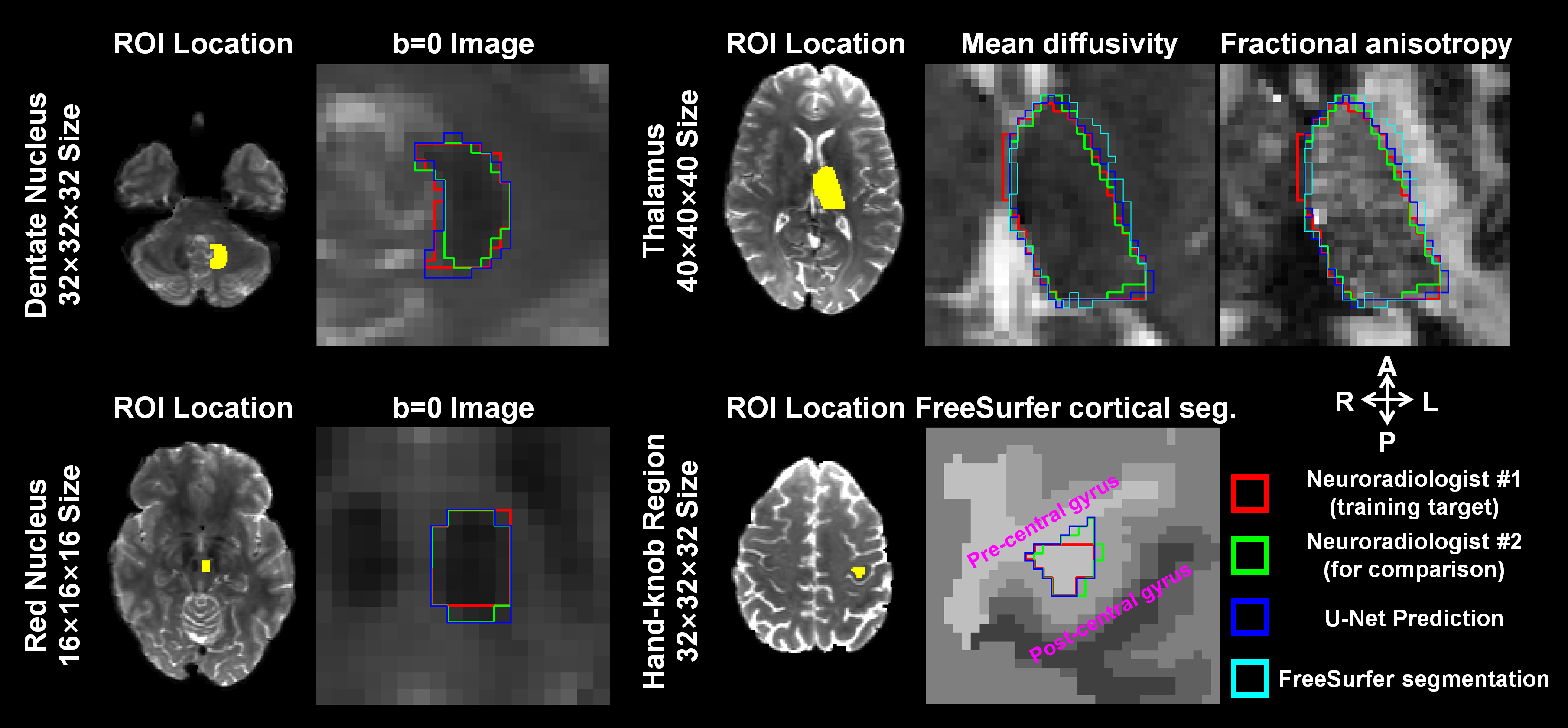
Data. Diffusion MRI data of 236 subjects from the HCP WU-Minn-Oxford Consortium were used (https://www.humanconnectome.org/). Data were acquired at 1.25-mm isotropic resolution using b-values=1000, 2000, and 3000 s/mm2 (90 directions each). DTI fitting on the b=0 and 1000 s/mm2 data produced fractional anisotropy (FA) and mean diffusivity (MD) maps. FreeSurfer segmentation of the T1w data was resampled to diffusion space to obtain binary masks of the pre- and post-central gyrus.
Data Formatting. The following images were used for manual segmentation and served as input to the CNN: (1) FA and MD maps for thalamus; (2) b=0 image for red nucleus (RN) and dentate nucleus (DN); (3) gray/white matter segmentation of the pre- and post-central gyrus for the handknob region (HR). Binary masks of the thalamus, RN, DN and HR from both hemispheres were manually drawn by an expert neuroradiologist for 36 subjects (30 for training, 6 for evaluation) and served as the output of the CNN. ROIs of a single subject from the 6 evaluation subjects were delineated by another neuroradiologist for comparison (Fig.1, green contours).
Manually delineated ROIs of the 30 training subjects were co-registered to MNI152 space and averaged, then co-registered to individual subjects to indicate the location of each ROI. ~60 image blocks per ROI were randomly cropped around the initial ROI locations of the thalamus (size 40×40×40), RN (16×16×16), DN (32×32×32) and HR (32×32×32), flipping training pairs from the right to left hemisphere for joint training.
Network. 3D U-Net3 was used to learn the transform from the input image blocks to the manually segmented ROIs. The U-Net was implemented using the Keras API with a Tensorflow backend, “binary_crossentropy” loss, leaky ReLU activation, 3×3×3 kernels, 4 levels, 64 (thalamus, HR, DN) and 32 kernels (RN) at the highest level and 2× kernels for each lower level. Training was performed on 27 subjects and tested on 3 for 10 epochs (~10 min/ROI).
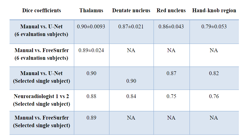
Evaluation. Dice coefficients were computed between manual and U-Net segmentation for the 6 evaluation subjects, between the two neuroradiologists’ delineations for the selected single subject, and between manual and FreeSurfer segmentation for the thalamus.
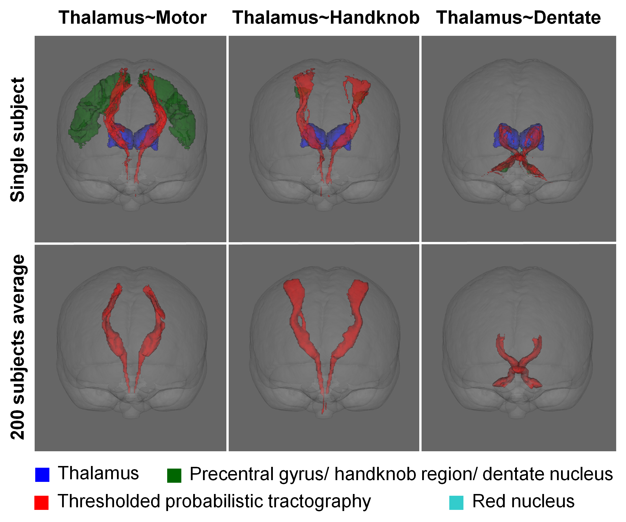
Tractography. Probabilistic tractography was performed on 200 subjects with U-Net segmented ROIs as follows: (1) thalamus as “seed” and ipsilateral sub-cortical white matter of precentral gyrus from FreeSurfer as “target”; (2) thalamus as “seed” and ipsilateral HR as “target”; (3) thalamus as “seed”, the contralateral DN as “target” and ipsilateral RN as “waypoint”.
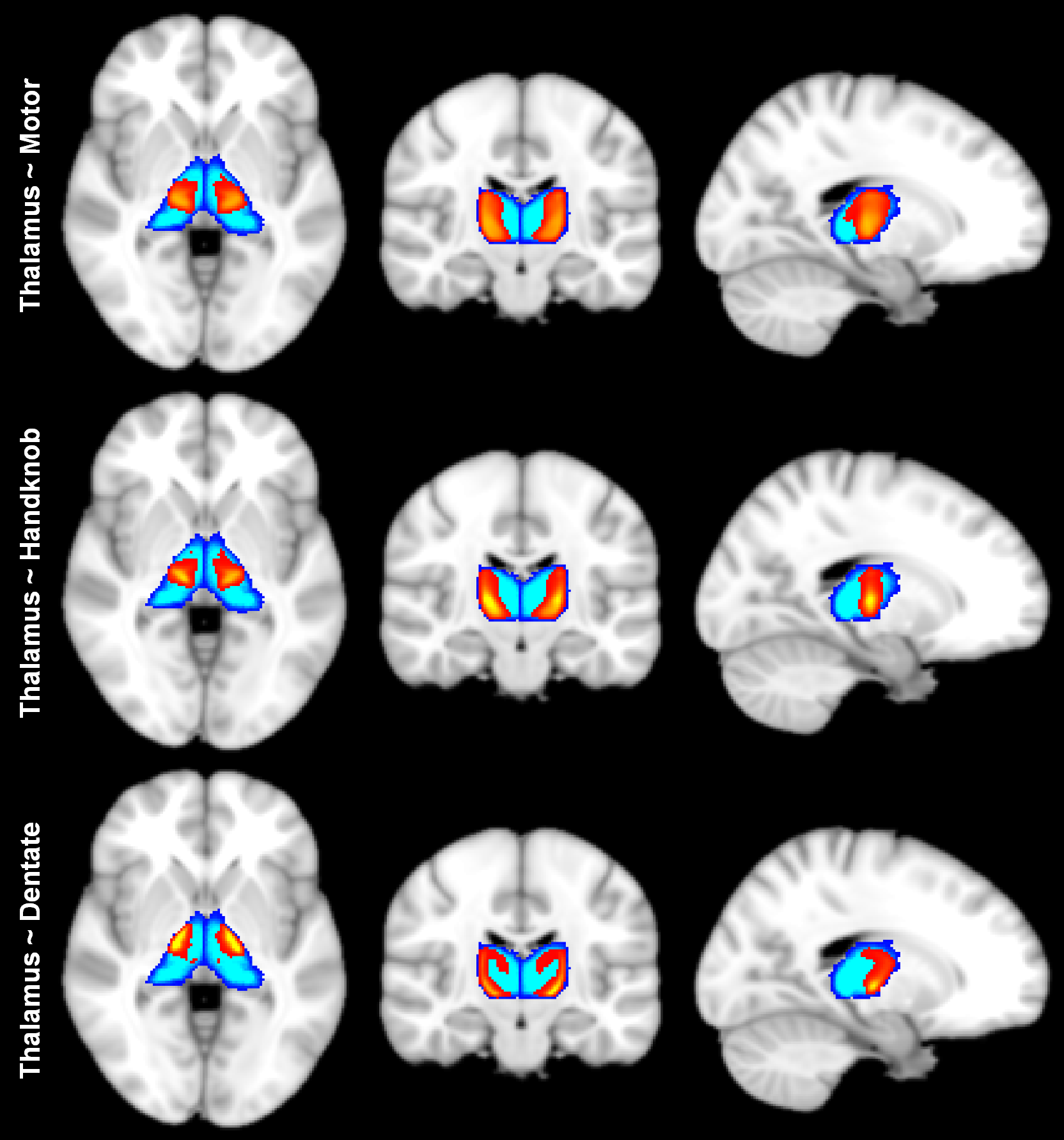
Probability maps of the fiber pathways and normalized streamline count within the thalamus were co-registered to MNI152 space and averaged to provide an atlas of the tractography-identified Vim location and associated tremor circuits.
Results
The U-Net segmentation (Fig.1, green contour) was visually comparable to segmentations from the two neuroradiologists (Fig.1, red+green contours). Dice coefficients between manual and U-Net segmentations were higher than 80% in the 6 evaluation subjects and were higher than the Dice coefficients between the two neuroradiologists in the single selected subject (Table 1).
Figure 2 shows tractography results of a single subject and the average over 200 subjects. The tractography results followed the expected anatomy of the thalamocortical radiation and the dentatothalamic tract.
Figure 3 displays the atlas of the Vim location based on structural connectivity, which could be useful for investigating variations of the Vim in a population and provide a reference Vim location for neurosurgical guidance.
Discussion and Conclusion
We applied a CNN to segment subject-specific brain ROIs for diffusion tractography of tremor circuits. Future work will focus on applying the proposed method to the 1000 HCP subjects to create a more comprehensive atlas of Vim location.Acknowledgements
Funding was provided by the NIH: P41-EB015896, S10-RR019307, K23-NS096056, R01-MH111419, and an MGH Claflin Distinguished Scholar Award.References
[1] Elias WJ, Huss D, Voss T, et al. A pilot study of focused ultrasound thalamotomy for essential tremor. New England Journal of Medicine. 2013;369(7):640-648.
[2] Tian Q, Wintermark M, Elias WJ, et al. Diffusion MRI tractography for improved transcranial MRI-guided focused ultrasound thalamotomy targeting for essential tremor. NeuroImage: Clinical. 2018;19:572-580.
[3] Ronneberger O, Fischer P, Brox T. U-net: Convolutional networks for biomedical image segmentation. Paper presented at: International Conference on Medical image computing and computer-assisted intervention 2015.
Figures