0786
Mitochondrial dysfunction in a rat model of doxorubicin-induced heart failure assessed by hyperpolarized 13C MRS1Physiology Anatomy and Genetics, University of Oxford, Oxford, United Kingdom, 2Oxford Centre for Magnetic Resonance, John Radcliffe Hospital, Oxford, United Kingdom, 3Department of Physics, University of Oxford, Oxford, United Kingdom, 4Department of Biochemistry, University of Cambridge, Cambridge, United Kingdom, 5MRC Mitochondrial Biology Unit, University of Cambridge, Cambridge, United Kingdom
Synopsis
Doxorubicin-chemotherapy can lead to serious cardiac side effects in cancer-patients, culminating in heart failure. Cardiac oxidative stress and impaired energetics are hypothesized to be at the core of this toxicity. We established a clinically relevant rat model of doxorubicin-induced heart failure characterized with CINE MRI by decreased cardiac function. We show here that these functional changes are preceded by a shift from oxidative to anaerobic glucose metabolism measured with hyperpolarized MRS. These changes are likely due to a loss and impairment of mitochondria, which cannot be alleviated with the mitochondrially targeted antioxidant, MitoQ.
Purpose
Doxorubicin (DOX) is a commonly used chemotherapeutic agent for the treatment of a wide range of cancers. However, DOX can also cause serious cardiotoxic side effects leading to heart failure in 5% of patients1 and there are no clinical imaging techniques or biomarkers available to detect this toxicity early enough for therapeutic intervention. The mechanism for this toxicity is not yet fully understood, although mitochondrial oxidative stress and altered cardiac energetics are thought to play a key role in the pathology2. The role of metabolism and metabolic fluxes in the development of energetic impairment and functional decline is, as yet, unknown. Hyperpolarized MRS by dissolution dynamic nuclear polarization of 13C-labelled substrates3 has revolutionized metabolic flux measurements in preclinical models4 and in the human heart5,6. We set out to establish a clinically relevant rat model of DOX-induced heart failure characterized by changes in cardiac function measured with CINE MRI. We used MitoQ7, a mitochondrially targeted antioxidant, to try and prevent oxidative stress, hypothesizing that this would lead to improve cardiac metabolism and function. We assessed cardiac metabolic fluxes in vivo with hyperpolarized MRS and related these to mitochondrial function.Methods
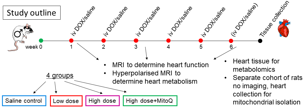
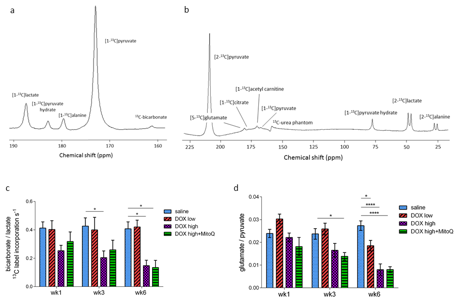
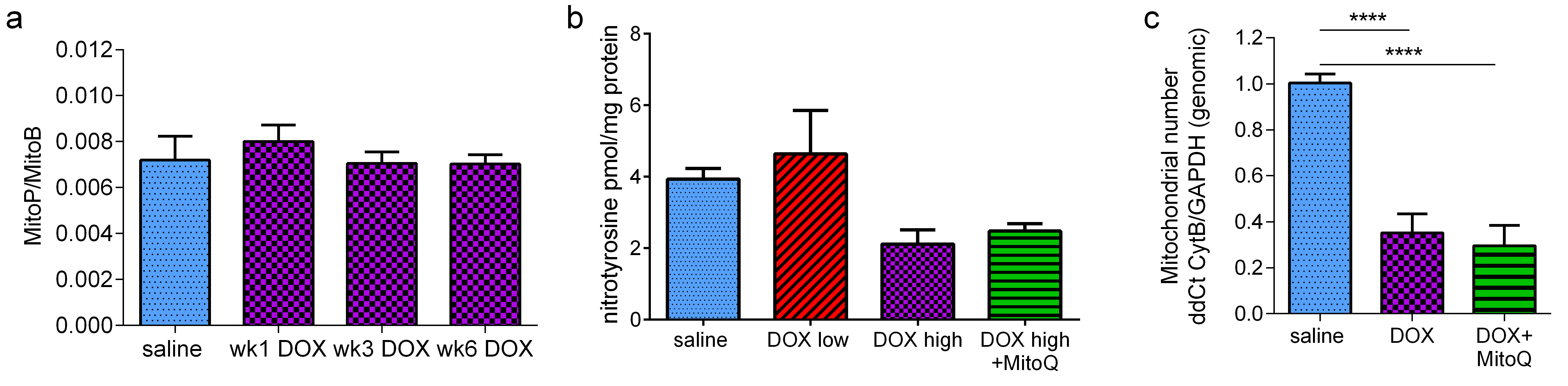
Weight-matched male Wistar rats were spilt into four groups and treated weekly for five or six weeks with i.v. injection of either 4 mL kg-1 sterile saline (n=20), 2 mg kg-1 DOX (Apollo Scientific) dissolved in sterile saline (n=12), 3 mg kg-1 DOX or 3 mg kg-1 DOX+0.5mM MitoQ in drinking water (ad libitum for the duration of the study) (Figure 1). At weeks 1, 3 and 6 following the first dose, functional CINE MR imaging and hyperpolarized [1-13C]- and [2-13C]pyruvate MRS were performed on a 7T spectrometer (Varian) as previously described8. One mL of 80 mM [1-13C]- or [2-13C]pyruvate was injected into the tail vain over 10s. 13C MR spectra were acquired every second for 60s using a 72-mm dual-tuned birdcage volume transmit 1H/13C coil and a 13C two-channel surface receive coil (Rapid Biomedical; 15º hard pulse; 13kHz bandwidth). Multicoil spectra were added in phase, and the first 30s of spectra from appearance of the pyruvate peak were summed and quantified with AMARES/jMRUI9. After the last MRI scan, rats were sacrificed and hearts excised and rapidly snap-frozen. Hearts were extracted with 2:1 Chloroform:Methanol and metabolomic analysis was performed with LC-MS/MS10. A separate cohort of rats was treated as above with either saline, 2 mg kg-1 DOX or 3 mg kg-1 DOX (all n=6). At week 6 rats were sacrificed, hearts excised and subsarcolemmal (SSM) and interfibrillar (IFM) mitochondria isolated. Mitochondrial oxygen consumption was assessed with a Clarke-type oxygen electrode and mitochondrial complex activities were assessed spectrophotometrically as previously described11.Results
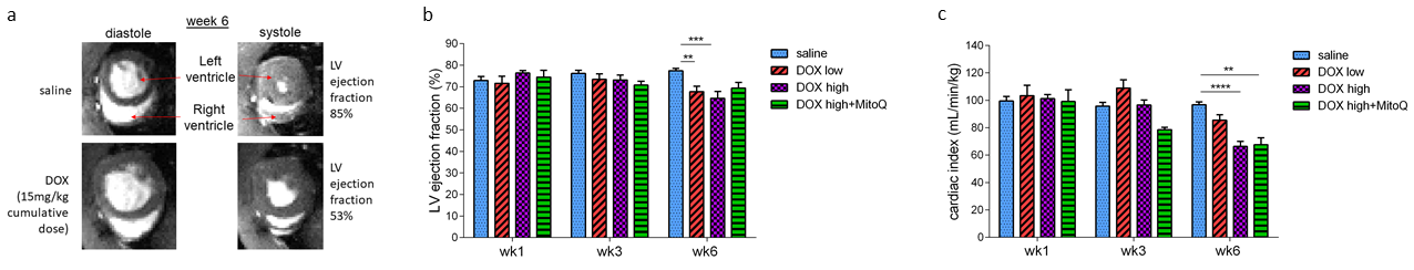
Rats treated with DOX showed a dose-dependent decrease in cardiac function after 6 weeks of treatment that was not prevented by MitoQ treatment (Figure 2). We next assessed cardiac metabolic fluxes with hyperpolarized MRS. In the high dose DOX group, there was a shift away from glucose oxidation towards glycolysis (decreased bicarbonate:lactate ratio) evident from week 3 onward (Figure 3c). In the DOX+MitoQ group this shift occurred at week 6 whilst there was no statistically significant difference in glucose metabolism in the low dose group. In parallel with changes to glucose metabolism, there was a marked decrease of tricarboxylic acid (TCA)-cycle derived glutamate at week 6 in all treatment groups, which was already evident in week 3 in the DOX+MitoQ group (Figure 3d). Metabolomic analysis of heart tissue extracts furthermore showed a decrease in the TCA cycle intermediates citrate, malate and glutamate (data not shown), further supporting the hypothesis that mitochondrial oxidative metabolism is decreased with DOX-treatment. IFM and SSM mitochondria isolated from high dose DOX hearts showed a decreased oxygen consumption with glutamate or palmitoyl-CoA+carnitine as a fuel (Figure 4a-b). No statistically significant difference in oxygen consumption was observed in either IFMs or SSMs from DOX+MitoQ treated hearts compared to saline controls or the high dose DOX group. Electron transport chain (ETC) complex activity assays in IFMs and SSMs revealed a decrease in complex IV activity in both the high dose DOX group and the DOX+MitoQ group (Figure 4c).Discussion and conclusion
We established a clinically relevant rat model of DOX-induced heart failure characterized by decreased left ventricular ejection fraction and decreased cardiac index. Hyperpolarized MRS in these rat hearts revealed a shift from oxidative to anaerobic metabolism mimicked by a decrease in mitochondrial function that could not be alleviated by MitoQ. This research may open up new treatment avenues targeting cardiac metabolism for patients receiving cardiotoxic chemotherapy.Acknowledgements
This work was supported by a British Heart Foundation Immediate Postdoctoral Basic Science Research Fellowship (FS/16/7/31843)References
1. Moslehi JJ. Cardiovascular Toxic Effect of Targeted Cancer Therapies. N Engl J Med. 2016;375(15). doi:10.1056/NEJMra1100265.
2. Tokarska-Schlattner M, Zaugg M, Zuppinger C, Wallimann T, Schlattner U. New insights into doxorubicin-induced cardiotoxicity: The critical role of cellular energetics. J Mol Cell Cardiol. 2006;41(3):389-405. doi:10.1016/j.yjmcc.2006.06.009.
3. Ardenkjaer-Larsen JH, Fridlund B, Gram A, et al. Increase in signal-to-noise ratio of > 10,000 times in liquid-state NMR. Proc Natl Acad Sci U S A. 2003;100(18):10158-10163. doi:10.1073/pnas.1733835100.
4. Timm KN, Miller JJ, Henry JA, Tyler DJ. Cardiac applications of hyperpolarised magnetic resonance. Prog Nucl Magn Reson Spectrosc. 2018;106-107:66-87. doi:10.1016/j.pnmrs.2018.05.002.
5. Cunningham CH, Lau JYC, Chen AP, et al. Hyperpolarized 13C Metabolic MRI of the Human Heart: Initial Experience. Circ Res. 2016;119(11):1177-1182. doi:10.1161/CIRCRESAHA.116.309769.
6. Tyler D, Rider O, Dodd M, et al. Demonstrating the Randle Cycle in Vivo: Assessment of Physiological Alterations in Human Cardiac Metabolism Using Hyperpolarised 13C MR Spectroscopy. In: International Society for Magnetic Resonance in Medicine. Honolulu, Hi, USA; 2017:726.
7. Kelso GF, Porteous CM, Coulter C V., et al. Selective targeting of a redox-active ubiquinone to mitochondria within cells: Antioxidant and antiapoptotic properties. J Biol Chem. 2001;276(7):4588-4596. doi:10.1074/jbc.M009093200.
8. Dodd MS, Ball DR, Schroeder MA, et al. In vivo alterations in cardiac metabolism and function in the spontaneously hypertensive rat heart. Cardiovasc Res. 2012;95(1):69-76. doi:10.1093/cvr/cvs164.
9. Vanhamme L, Van Den Boogaart A, Van Huffel S. Improved Method for Accurate and Efficient Quantification of MRS Data with Use of Prior Knowledge. J Magn Reson. 1997;129(1):35-43. doi:10.1006/jmre.1997.1244.
10. Wang X, West JA, Murray AJ, Griffin JL. Comprehensive Metabolic Profiling of Age-Related Mitochondrial Dysfunction in the High-Fat-Fed ob/ob Mouse Heart. J Proteome Res. 2015;14(7):2849-2862. doi:10.1021/acs.jproteome.5b00128.
11. Heather LC, Cole MA, Tan JJ, et al. Metabolic adaptation to chronic hypoxia in cardiac mitochondria. Basic Res Cardiol. 2012;107(3):268. doi:10.1007/s00395-012-0268-2.
Figures