0775
Validation of DSI compressed sensing reconstruction in ex vivo human brain1Athinoula A. Martinos Center for Biomedical Imaging, Massachusetts General Hospital, Charlestown, MA, United States, 2Harvard-MIT Health Sciences and Technology, Massachusetts Institute of Technology, Cambridge, MA, United States, 3Boston University, Boston, MA, United States, 4Computer Science and Artificial Intelligence Laboratory, Massachusetts Institute of Technology, Cambridge, MA, United States
Synopsis
Compressed sensing algorithms for accelerating DSI acquisitions (DSI-CS) have helped bring DSI into the realm of clinical feasibility. Here, we assess the efficacy of dictionary-based CS methods in reconstructing high resolution ex vivo DSI of human brain blocks, and provide validation of ex vivo DSI-CS with ground truth optical imaging. We find that reconstruction accuracy, computation time and inter-subject dictionary generalizability are comparable to in vivo results, and that SNR appears influential in determining the limit of attainable reconstruction quality. We also show that fiber orientation estimates of reconstructed data are as accurate as fully-sampled estimates at a microscopic level.
Introduction
Diffusion
MRI (dMRI) allows us to study white-matter architecture
non-invasively in
vivo.
Diffusion spectrum imaging (DSI) provides detailed information
helpful for delineating complex intravoxel diffusion patterns1,
but requires lengthy acquisitions that limit its applicability in
vivo.
Recent innovations in parallel imaging2,3,
MRI hardware4,5
and compressed sensing (CS) algorithms have now rendered DSI as a
clinically feasible dMRI protocol6.
CS applied to DSI (DSI-CS) aims to reconstruct the diffusion
probability density functions (PDFs) from sub-Nyquist sampled q-space
using prior knowledge. PDF recovery using adaptive dictionaries and
L2 regularization was shown to yield lower reconstruction error than
CS using prespecified transforms7,8
with computation times orders of magnitude faster than iterative,
dictionary-based CS methods9.
Here, we extend the application of DSI-CS algorithms introduced in 7
to high-resolution ex
vivo
DSI data obtained from human brain blocks. We investigate
reconstruction accuracy with respect to q-space location and SNR, and
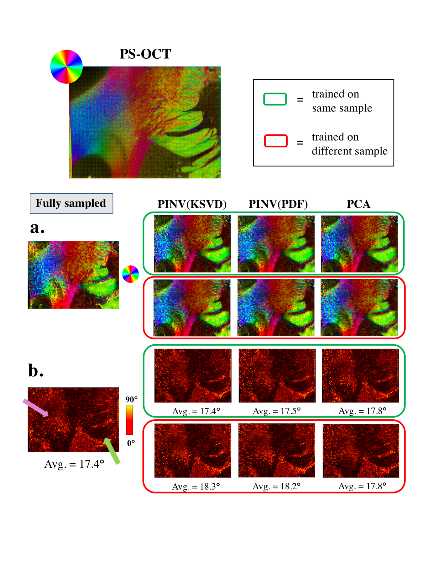
we compare modeled diffusion orientations to fiber orientations
measured directly with Polarization Sensitive Optical Coherence
Tomography (PS-OCT)10,11
providing the first validation of DSI-CS with ground truth optical
imaging.
Methods
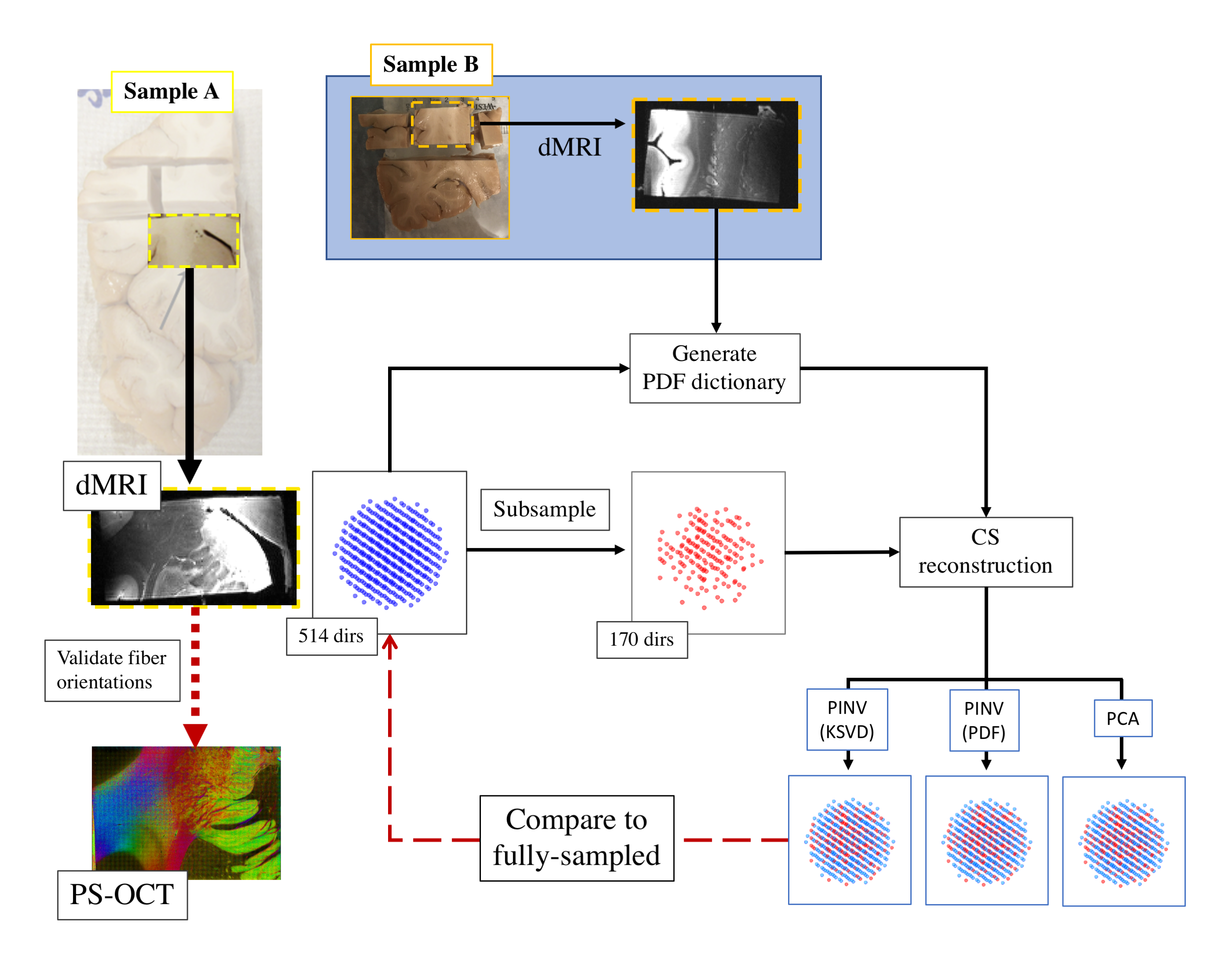
dMRI: We cut two blocks (3x2x2cm) from different anatomical regions of the same ex vivo, fixed human hemisphere (Fig. 1). We imaged each block on a 9.4T Bruker magnet with |G|max=480 mT/m, using a 3D EPI sequence (δ=15ms, Δ=19ms, 514 directions, bmax=40000s/mm2, 0.25mm iso resolution) for full DSI q-space encoding with one b=0 image. A surface coil was used, leading to a dependence of SNR on distance from the coil. This allowed us to investigate the relationship between SNR and reconstruction accuracy.
PS-OCT: Following dMRI, we imaged a 2x1.5x0.5cm section of one brain block with PS-OCT. PS-OCT acquisition and analysis was performed as described previously 10,11, yielding direct measurements of in-plane axonal orientation at 10μm in-plane and 75μm through-plane resolution.
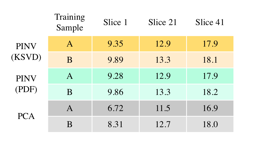
Dictionary Training & Reconstruction: We undersampled the DSI data by an acceleration factor of R=3, and used two L2-based algorithms for CS reconstruction described in 7, with dictionary training sets derived from PDFs from a single slice of fully-sampled data. We compared Principal Component Analysis (PCA) reconstruction, and Tikhonov-regularized pseudoinverse reconstruction using either dictionaries trained with the K-SVD algorithm12 [PINV(KSVD)] or the training PDFs themselves as the dictionary [PINV(PDF)]. We reconstructed slices of sample A using dictionaries trained on either sample A or sample B. We computed the normalized RMSE, in terms of both PDFs and q-space, between the fully-sampled data and those reconstructed with PINV(KSVD), PINV(PDF) and PCA.
Results
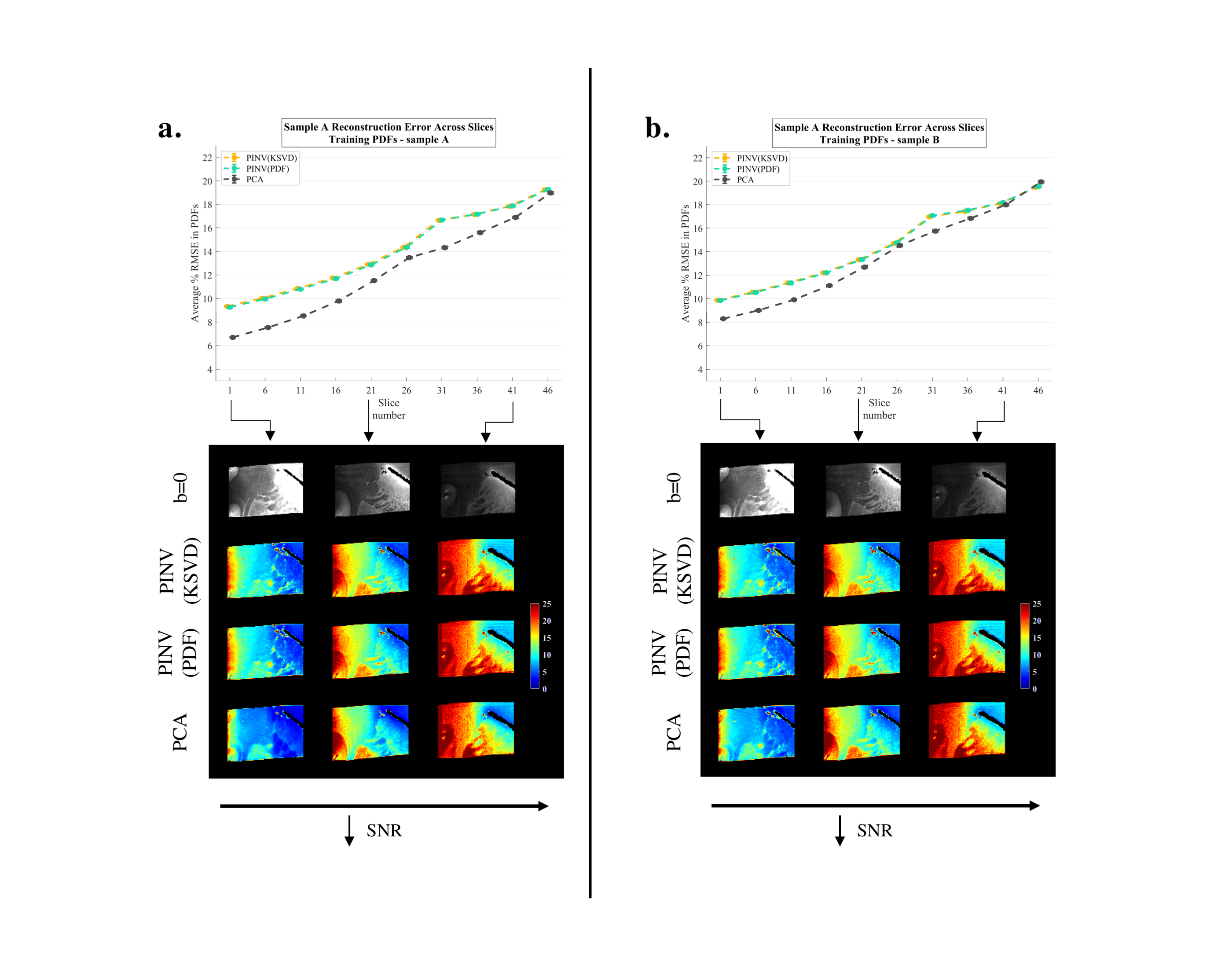
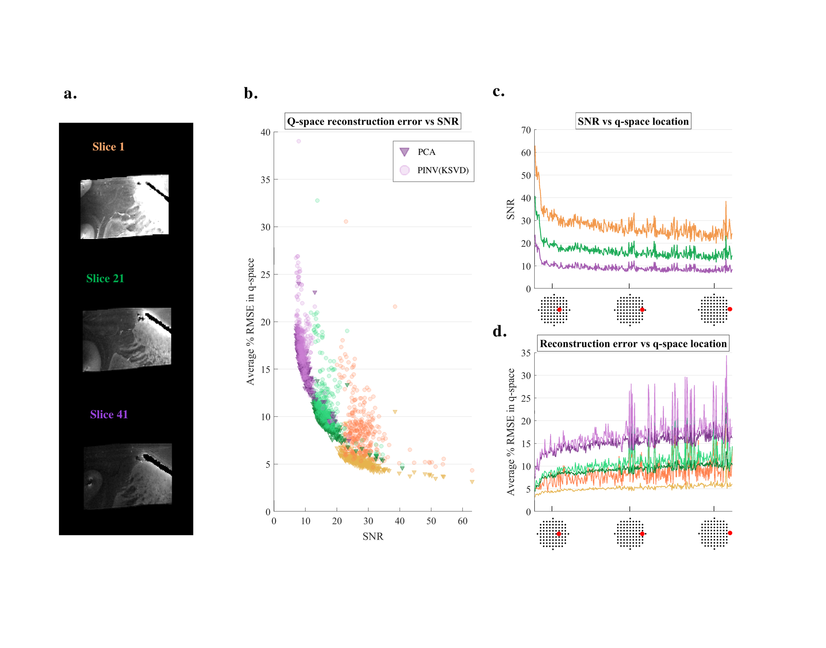
We show the accuracy of reconstructed slices of sample A, using dictionaries trained on sample A (Fig. 2a) or on sample B (Fig. 2b). For three slices, color maps depicting average RMSE in PDFs are shown (Fig. 2a,b). We examined the SNR and RMSE at each undersampled q-space location (Fig. 3c,d), and further evaluated q-space RMSE as a function of SNR (Fig. 3b). We compared in-plane fiber orientation measurements from PS-OCT to diffusion orientation estimates from fully-sampled and reconstructed datasets. We show RGB maps of in-plane orientation angles (Fig. 4a) and heat maps of angle difference between PS-OCT and dMRI orientations (Fig. 4b).Discussion
All reconstruction methods perform similarly across slices, with PCA yielding slightly lower RMSE than the two regularized pseudoinverse methods (Fig. 2a,b). Reconstructions of sample A using dictionaries trained on sample B had errors nearly the same as dictionaries trained on sample A, especially for PINV(KSVD) and PINV(PDF) (Table 1), which supports previous in vivo results7. As expected, we find that RMSE increases with distance in q-space (Fig. 3d). We observe that, as SNR decreases below 20, accuracy deteriorates substantially for all methods (Fig. 3b). Reconstruction errors in PDFs and q-space are similar to those reported previously for lower-resolution in vivo reconstructions7 and with similar computation times (~4 seconds per slice). When compared to PS-OCT, fiber orientations from CS reconstructions show no significant difference in accuracy compared to those from fully-sampled data (Fig. 4).Conclusion
Our ex vivo validation of dictionary-based DSI-CS methods shows that they offer acceleration without compromising the accuracy of the estimated white-matter architecture, with respect to ground truth optical imaging at microscopic resolutions. We quantified the SNR limits of our approach and have begun to investigate its robustness to differences between the training and test data. Our results are promising both for large-scale studies acquiring DSI-CS in vivo6, as well as our aim of building highly accurate models of brain circuitry from ex vivo data. In ongoing work, we are comparing the accuracy of this approach to other (non-Cartesian) q-space sampling schemes.Acknowledgements
This work was funded by NIH grant R01-EB021265.References
1. Wedeen VJ, Hagmann P, Tseng WY, Reese TG, Weisskoff RM. Mapping complex tissue architecture with diffusion spectrum magnetic resonance imaging. Magnetic resonance in medicine. 2005 Dec;54(6):1377-86.
2.Setsompop K, Cohen-Adad J, Gagoski BA, Raij T, Yendiki A, Keil B, Wedeen VJ, Wald LL. Improving diffusion MRI using simultaneous multi-slice echo planar imaging. Neuroimage. 2012 Oct 15;63(1):569-80.
3. Setsompop K, Gagoski BA, Polimeni JR, Witzel T, Wedeen VJ, Wald LL. Blipped‐controlled aliasing in parallel imaging for simultaneous multislice echo planar imaging with reduced g‐factor penalty. Magnetic resonance in medicine. 2012 May 1;67(5):1210-24.
4. Keil B, Blau JN, Biber S, Hoecht P, Tountcheva V, Setsompop K, Triantafyllou C, Wald LL. A 64‐channel 3T array coil for accelerated brain MRI. Magnetic resonance in medicine. 2013 Jul;70(1):248-58.
5. McNab JA, Edlow BL, Witzel T, Huang SY, Bhat H, Heberlein K, Feiweier T, Liu K, Keil B, Cohen-Adad J, Tisdall MD. The Human Connectome Project and beyond: initial applications of 300 mT/m gradients. Neuroimage. 2013 Oct 15;80:234-45.
6. Tobisch A, Stirnberg R, Harms RL, Schultz T, Roebroeck A, Breteler MM, Stöcker T. Compressed Sensing Diffusion Spectrum Imaging for Accelerated Diffusion Microstructure MRI in Long-Term Population Imaging. Frontiers in neuroscience. 2018;12.
7. Bilgic B, Chatnuntawech I, Setsompop K, Cauley SF, Yendiki A, Wald LL, Adalsteinsson E. Fast dictionary-based reconstruction for diffusion spectrum imaging. IEEE transactions on medical imaging. 2013 Nov;32(11):2022-33.
8. Menzel MI, Tan ET, Khare K, Sperl JI, King KF, Tao X, Hardy CJ, Marinelli L. Accelerated diffusion spectrum imaging in the human brain using compressed sensing. Magnetic Resonance in Medicine. 2011 Nov;66(5):1226-33.
9. Bilgic B, Setsompop K, Cohen-Adad J, Yendiki A, Wald LL, Adalsteinsson E. Accelerated diffusion spectrum imaging with compressed sensing using adaptive dictionaries. Magn Reson Med. 2012;68(6):1747-54.
10. Wang H, Black AJ, Zhu J, Stigen TW, Al-Qaisi MK, Netoff TI, Abosch A, Akkin T. Reconstructing micrometer-scale fiber pathways in the brain: multi-contrast optical coherence tomography based tractography. Neuroimage. 2011 Oct 15;58(4):984-92.
11. Grisot G, Jones R, Augustinack J, Boas D, Fischl B, Wang H, Yendiki A. Validation of high angular resolution diffusion MRI models in the human brain with PS-OCT. International Society for Magnetic Resonance in Medicine (ISMRM). 2017.
12. Aharon M, Elad M, Bruckstein A. K-SVD: An algorithm for designing overcomplete dictionaries for sparse representation. IEEE Transactions on signal processing. 2006 Nov 1;54(11):4311.
Figures