0774
High-Resolution Isotropic Diffusion MRI Using Simultaneous Multi-slab (SMSlab) Acquisition1Center for Biomedical Imaging Research, Department of Biomedical Engineering, School of Medicine, Tsinghua University, Beijing, China, 2Radiology, Stanford University, Stanford, CA, United States
Synopsis
3D multi-slab acquisition is an important technique for high-resolution isotropic diffusion MRI. To further accelerate the acquisition, simultaneous multi-slice (SMS) excitation can be combined with multi-slab, termed simultaneous multi-slab (SMSlab). Previously it has been demonstrated that the SMSlab un-folding problem can be solved using a 3D Fourier encoding framework. When applying SMSlab to diffusion MRI, the main challenge is how to simultaneously un-fold the excited multiple slices/slabs and correct the inter-shot phase variations. To achieve this, a delicate navigator acquisition is designed and a POCS-enhanced k-space phase correction method is used in this study.
Introduction
3D multi-slab acquisition is an important technique for high-resolution isotropic diffusion MRI (dMRI) 1-4. To further accelerate the acquisition, simultaneous multi-slice (SMS) excitation can be combined with multi-slab 4-7. As in SMS accelerated 2D multi-shot dMRI, there are two main challenges in simultaneous multi-slab (SMSlab) dMRI: how to un-fold simultaneously excited multiple slices/slabs, and how to correct inter-shot phase variations. First, the un-folding problem can be solved using a 3D Fourier encoding framework 7. Second, the inter-shot phase variations can be recorded by using a delicate navigator acquisition and corrected using a POCS-enhanced k-space method 8.Methods
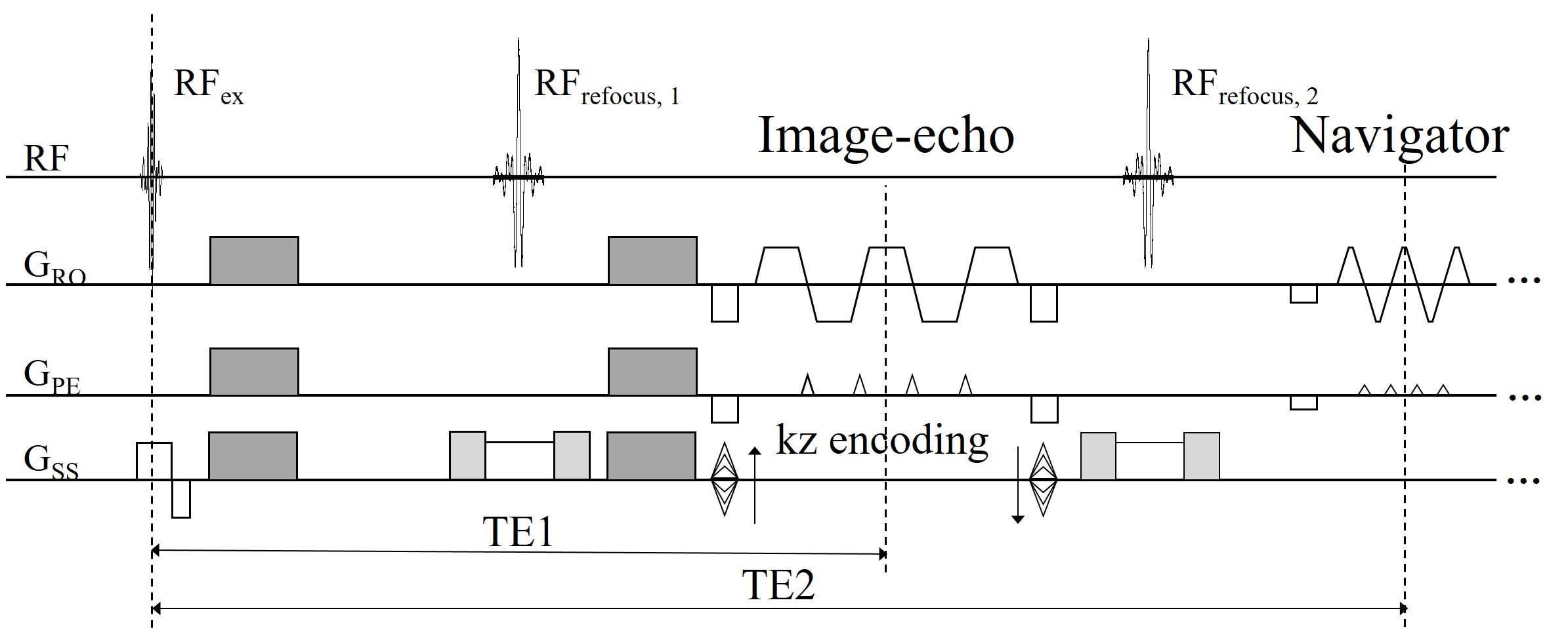
Data acquisition A 3D SMSlab dMRI acquisition sequence is developed (Fig. 1). 4-shot interleaved EPI (iEPI) is used to acquire each kz plane and an extra navigator is acquired subsequently after the image-echo. For the image echo, RF and gradient encoding are used together to form a 3D k-space for SMSlab 7. Assuming that a conventional SMS pulse using RSMS=2 is ,
RFSMS(t)=RF(t)[exp(iω1t)+exp(iω2t)], (1)
then the SMSlab pulse can be written as
RFSMS(t)=RF(t)[exp(iω1t)+exp(i(ω2t-φ))], (2)
where RF(t) is a single-band pulse, ω1 and ω2 are the frequency modulation for each slice/slab, and φ is the inter-slab gap generated phase during the kz encoding. Both the excitation and the first refocusing pulse can be designed using Eq. (2). CAIPI 9,10 is used in the image-echo acquisition for a better use of coil sensitivity.
The 3D SMSlab acquisition strategy for the image-echo cannot be applied for the navigator, as the navigator is a single-shot EPI (ss-EPI) acquisition. Since the 3D SMSlab acquisition requires different RF pulses at different kz planes, thus it is not compatible with the navigator. In this study, the navigator is acquired using the conventional SMS, namely treating each slab as a 2D thick slice. A 2D navigator can record the motion-induced phase of such a slab, as its thickness is about 10 to 20 mm 1-3. However, the CAIPI encoding gradients can generate large phase variation across the slab with thickness from 10 to 20 mm, which may cause image blur 9. Therefore, CAIPI is not used for the navigator acquisition here. Meanwhile, the second refocusing pulse is a little different from Eq. (2), which is
RFSMS(t)=RF(t)[exp(iω1t)+exp(i(ω2t-φ/2))], (3)
All data were acquired on a Philips 3.0T Achieva TX MRI scanner using a 32-channel head coil. 14 slabs using RSMS=2 were acquired in total. For each slab, 12 slices (20% oversampling in the kz direction) were acquired, and adjacent slabs were overlapped by 2 slices. Other main imaging parameters were: FOVxy=220×220 mm2, resolution=1 mm3, partial Fourier factor=0.7 (ky direction) for image-echo, TE1/TE2/TR=91 ms / 187 ms / 1.8 s. Diffusion encoding was applied along 12 directions with b=1000 s/mm2. 2D low-resolution (2 mm3) ss-EPI images were acquired as references. The acquisition time was 18:47 and 2:07 for SMSlab and ss-EPI, respectively. A 2D 4-shot iEPI scan was conducted to estimate the coil sensitivity map.
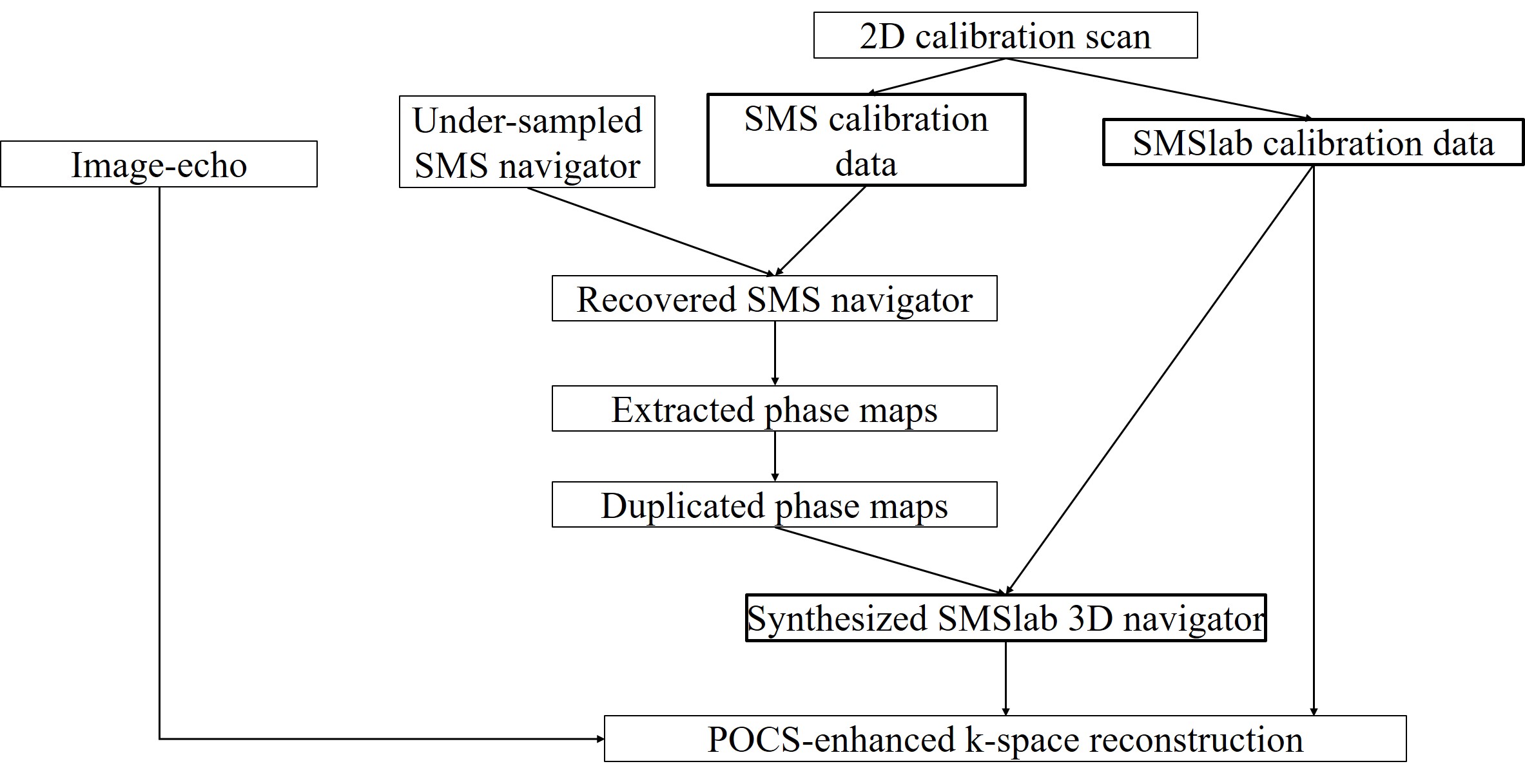
Reconstruction The overall reconstruction pipeline is described in Fig. 2. 1) We used the 2D calibration scan to synthesize two sets of calibration data: one for the navigator recovery (“SMS calibration data”) and one for the image-echo recovery (“SMSlab calibration data”). 2) The under-sampled SMS navigator was recovered using the SMS calibration data. 3) The motion-induced phase maps were extracted from the recovered navigator, duplicated along the slice direction and combined with the SMSlab calibration data to generate the “synthesized 3D navigator” for the SMSlab dMRI. 4) The image echo, the “synthesized 3D navigator” and the SMSlab calibration data were integrated into the POCS-enhanced k-space reconstruction 8 to recover the DW images. After the reconstruction, NPEN 3 was used to correct for the slab boundary artifacts.
Results and Discussion
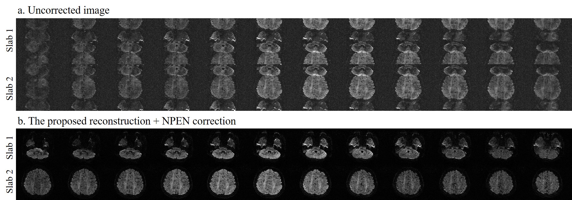
Fig. 3 shows the uncorrected and the reconstructed DW images. Slice aliasing and motion artifacts are observed in the uncorrected images, but are well corrected in the final DW images.
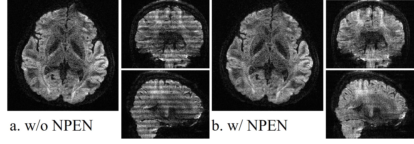
Fig. 4 shows the DW images without and with the NPEN correction, from three different views. It can be seen that NPEN can effectively reduce the slab boundary artifacts in the SMSlab acquisition.
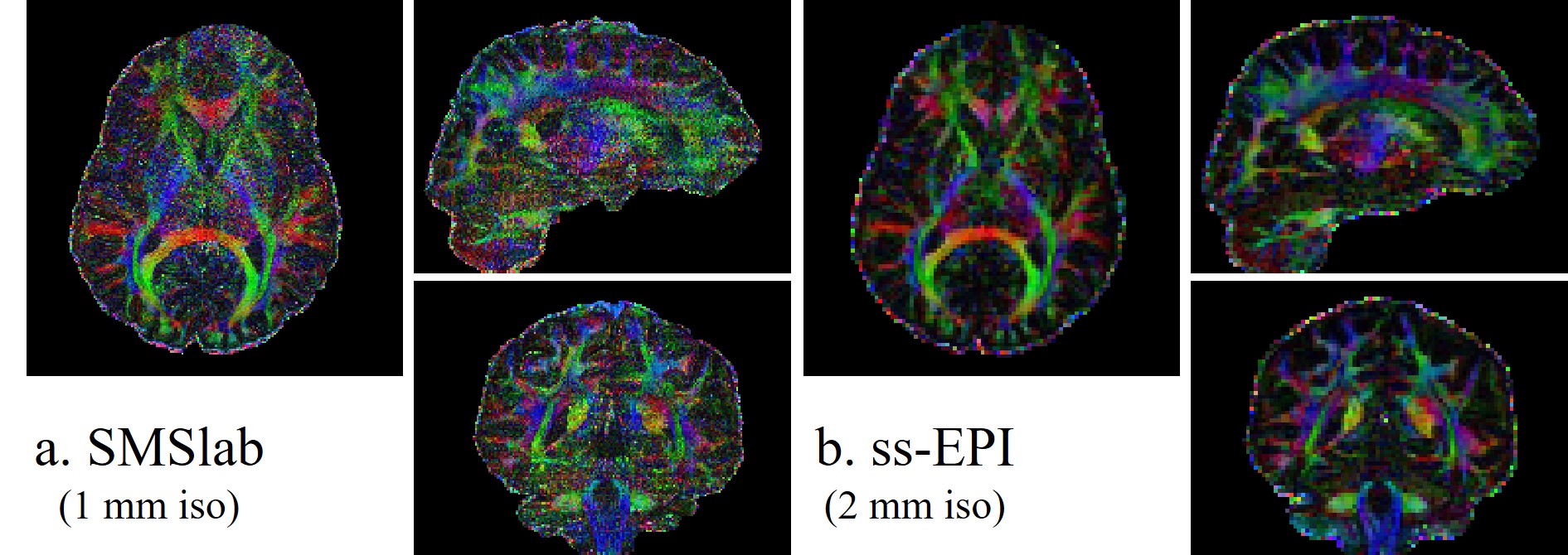
Fig. 5 shows a color-coded FA (cFA) maps comparison between SMSlab and traditional ss-EPI acquisition. The cFA maps from SMSlab is consistent with ss-EPI, but with slightly low SNR.
Conclusion
Based on the recently proposed 3D encoding framework for SMSlab, a delicate navigator acquisition and phase correction method for SMSlab dMRI has been demonstrated. High-resolution isotropic DW images have been reconstructed using the proposed method. An on-going study is to use more advanced RF pulse design, such as the root-flipped method 11, to reduce the pulse duration and thus TE, which can further improve the SNR.Acknowledgements
We would like to thank Drs. Karla L. Miller and Wenchuan Wu from university of Oxford for helpful discussions.References
1. Engstrom M, Skare S. Diffusion-weighted 3D multislab echo planar imaging for high signal-to-noise ratio efficiency and isotropic image resolution. Magn Reson Med 2013;70(6):1507-1514.
2. Van AT, Aksoy M, Holdsworth SJ, Kopeinigg D, Vos SB, Bammer R. Slab profile encoding (PEN) for minimizing slab boundary artifact in three-dimensional diffusion-weighted multislab acquisition. Magn Reson Med 2015;73(2):605-613.
3. Wu W, Koopmans PJ, Frost R, Miller KL. Reducing slab boundary artifacts in three-dimensional multislab diffusion MRI using nonlinear inversion for slab profile encoding (NPEN). Magn Reson Med 2016;76(4):1183-1195.
4. Chang HC, Hui ES, Chiu PW, Liu X, Chen NK. Phase correction for three-dimensional (3D) diffusion-weighted interleaved EPI using 3D multiplexed sensitivity encoding and reconstruction (3D-MUSER). Magn Reson Med 2018;79(5):2702-2712.
5. Frost R, Jezzard P, Porter DA, Tijssen R, Miller K. Simultaneous multi-slab acquisition in 3D multi-slab diffusion-weighted readout-segmented echo-planar imaging. In Proceedings of the 21st Annual Meeting of ISMRM, Salt Lake City, Utah, USA, 2013. Abstract 3176.
6. Bruce IP, Chang HC, Petty C, Chen NK, Song AW. 3D-MB-MUSE: A robust 3D multi-slab, multi-band and multi-shot reconstruction approach for ultrahigh resolution diffusion MRI. Neuroimage 2017;159:46-56.
7. Dai E, Wu Y, Guo H. A 3D k-Space Fourier Encoding and Reconstruction Framework for Simultaneous Multi-Slab Acquisition. In Proceedings of the 26th Annual Meeting of ISMRM, Paris, France, 2018. Abstract 1202.
8. Dai E, Ma X, Zhang Z, et al. A POCS-Enhanced k-Space Reconstruction for 3D Multi-Slab Diffusion Imaging. In Proceedings of the 26th Annual Meeting of ISMRM, Paris, France, 2018. Abstract 5338.
9. Setsompop K, Gagoski BA, Polimeni JR, Witzel T, Wedeen VJ, Wald LL. Blipped-controlled aliasing in parallel imaging for simultaneous multislice echo planar imaging with reduced g-factor penalty. Magn Reson Med 2012;67(5):1210-1224.
10. Breuer FA, Blaimer M, Heidemann RM, Mueller MF, Griswold MA, Jakob PM. Controlled aliasing in parallel imaging results in higher acceleration (CAIPIRINHA) for multi-slice imaging. Magn Reson Med 2005;53(3):684-691.
11. Sharma A, Lustig M, Grissom WA. Root-Flipped Multiband Refocusing Pulses. Magn Reson Med 2016;75(1):227-237.
Figures