0773
An efficient reconstruction by combining tilted-CAIPI with eddy-current calibration for high-resolution distortion-free diffusion imaging using DIADEM1Department of Radiology, Mayo Clinic, Rochester, MN, United States, 2A. A. Martinos Center for Biomedical Imaging, Department of Radiology, Massachusetts General Hospital, Charlestown, MA, United States, 3Department of Electrical Engineering and Computer Science, MIT, Cambridge, MA, United States, 4Harvard-MIT Health Sciences and Technology, MIT, Cambridge, MA, United States
Synopsis
As a variant of multi-shot echo-planar imaging, DIADEM (Distortion-free Imaging: A Double Encoding Method) enables high-resolution distortion-free imaging, but the prolonged scan time can be a major challenge. Recently, a novel parallel imaging approach, termed tilted-CAIPI, was suggested to unfold highly accelerated DIADEM data and to substantially reduce the acquisition time. However, the reconstructed image still suffers from eddy-current-induced geometric distortions in diffusion-weighted data. In this work, it is demonstrated that the DIADEM reconstruction becomes more efficient and practical after combining the tilted-CAIPI with the eddy-current calibration approaches.
Introduction
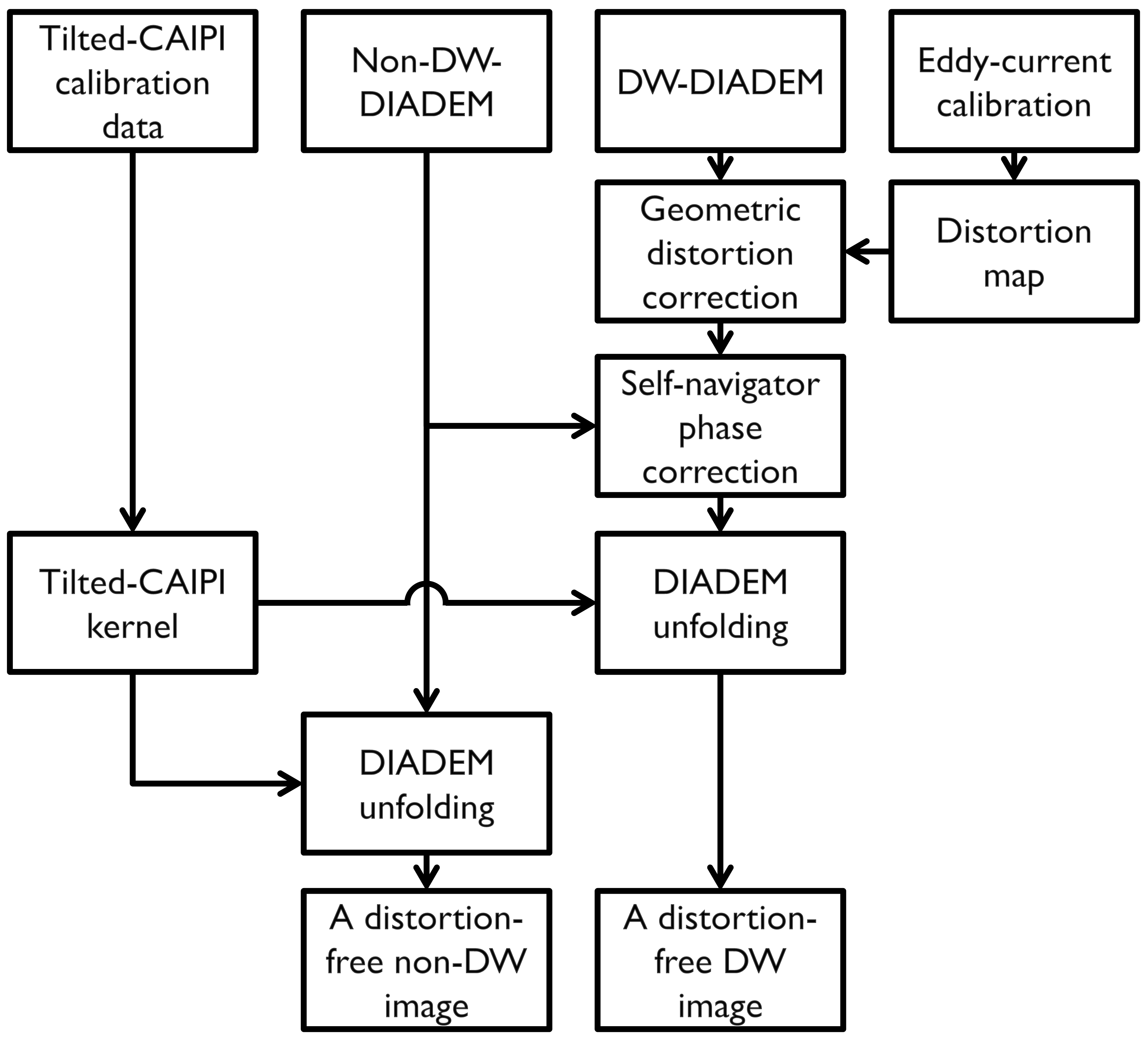
As a variant of multi-shot echo-planar-imaging (EPI), DIADEM (Distortion-free Imaging: A Double Encoding Method) enables high-resolution distortion-free imaging, but the prolonged scan time is a major challenge for this approach to be adapted for practical use1,2. Recently, a novel parallel imaging approach, termed tilted-CAIPI3, has been suggested to substantially reduce the acquisition time. The approach is able to unfold highly accelerated DIADEM data, and a distortion-free image can be calculated from DIADEM data, even when acquired with a small number of shots. However, the tilted-CAIPI k-space reconstruction kernels have to be recalculated for each diffusion-weighted (DW)-DIADEM reconstruction to account for the shot-to-shot phase variations, which is very time-consuming. Moreover, if eddy-current-induced geometric distortion is not negligible in DW-DIADEM data, the reconstructed image still suffers. The eddy-current correction can be incorporated into tilted-CAIPI reconstruction, but at a cost of further computational complexity and reliance on eddy current estimates from post-processing. In this work, it is demonstrated that a more efficient tilted-CAIPI reconstruction can be achieved by combining it with an eddy-current calibration approach4.Theory and Methods
For a DIADEM dataset, neglecting finite sampling, discretization, and T2* blurring effects, the multi-shot, spin-warp (SW) phase-encoded EPI images without Inon-DW(y,ks) and with DW gradient IDW(y1,ks) can be approximately described, at any readout position, respectively as:
Inon-DW(y,ks)=I(y)×exp(-iγ∆B(y)tTE)×exp(-iksy), where y=s+∆(s)suscep (1)
and
IDW(y1,ks)=I(y1)×exp(-i(γ∆B(y1)tTE+∆Ømotion(y1,ks)))×exp(-iksy1), where y1=s+∆(s)suscep+∆(s)eddy. (2)
Diffusion gradient eddy-current effects ∆(s)eddy are calibrated in advance using a phantom and obtained by subtraction between the two distortion maps calculated from the DIADEM data with and without DW gradient (i.e. ∆(s)DW and ∆(s)suscep)4:
∆(s)eddy=∆(s)DW-∆(s)suscep (3)
The geometry between the non-DW and DW images is matched after the eddy-current correction (ECC) using the calibrated distortion map ∆(s)eddy, which results in y1=y in Eq. 2. Because of the matched geometry, the phase variations between shots are simply estimated by division of the DW images by the non-DW images.
Ưmotion(y,ks)=arg(IDW(y,ks)/Inon-DW(y,ks)) (4)
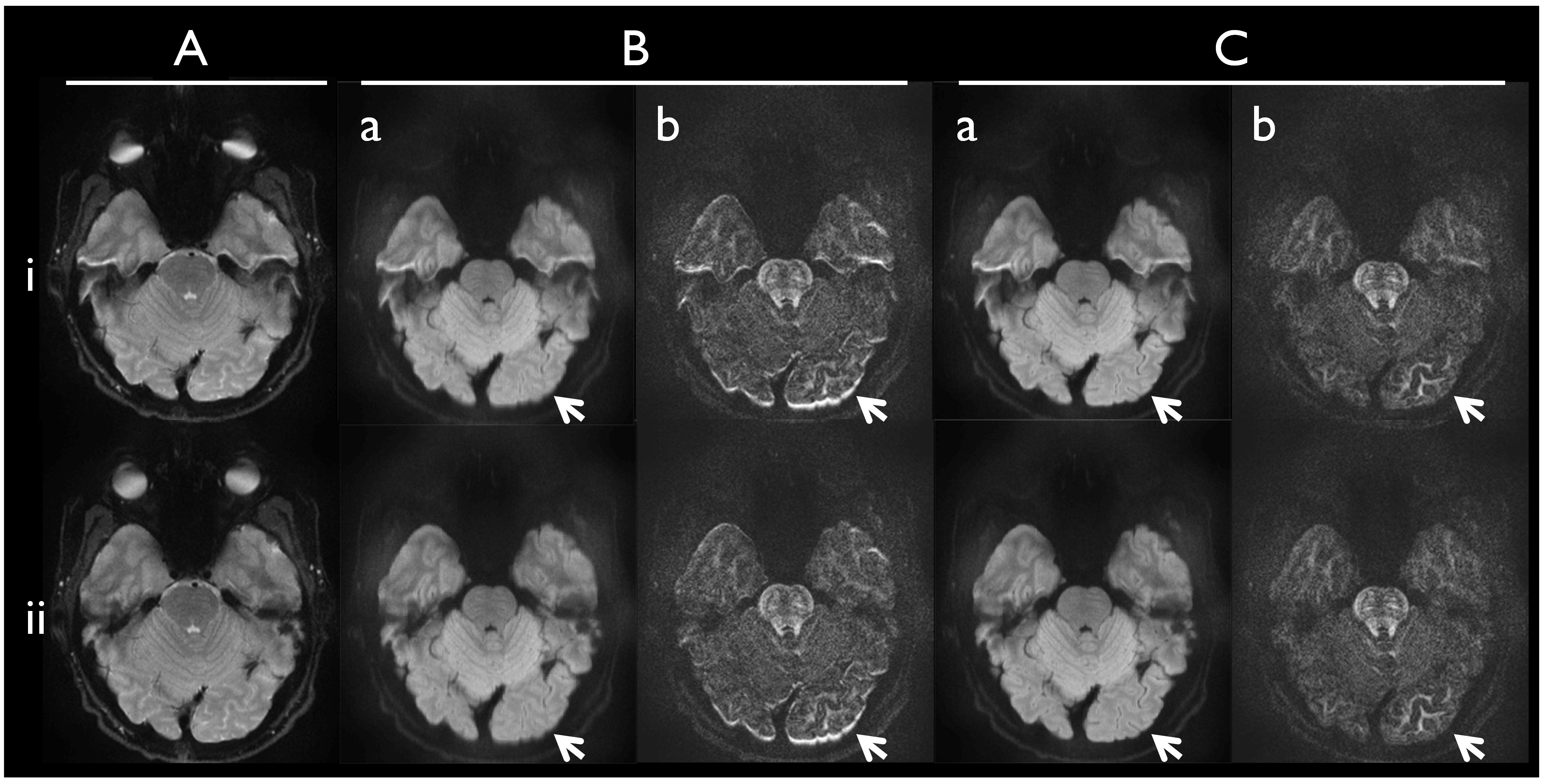
After compensating the phase variations directly to the DW-DIADEM data (fully-sampled along y), both the geometry and the phase are matched and a tilted-CAIPI kernel is applied for both the non-DW and the DW-DIADEM data reconstruction along ks. Finally, a distortion-free image I(s) is calculated from each DIADEM data in the SW-PE dimensions (s). A phantom calibration and two healthy in-vivo scans were performed using an 8-channel coil (Invivo, Gainesville, FL) on a compact 3T5-7 with concomitant field compensation8,9 and higher-order gradient non-linearity correction10. Imaging parameters (Fig. 3) of the in-vivo and eddy-current calibration scans were identical, except for the acceleration factor (32 in in-vivo and 16 in the calibration to unfold the DIADEM data without tilted-CAIPI reconstruction). After the tilted-CAIPI reconstruction of the DIADEM data without and with ECC, the images in the EPI phase-encoding (PE) I(y) and the SW-PE I(s) dimensions were obtained from the unfolded DIADEM data and the corresponding DTI scalars were calculated using FSL11 for comparisons.
Results and Discussion
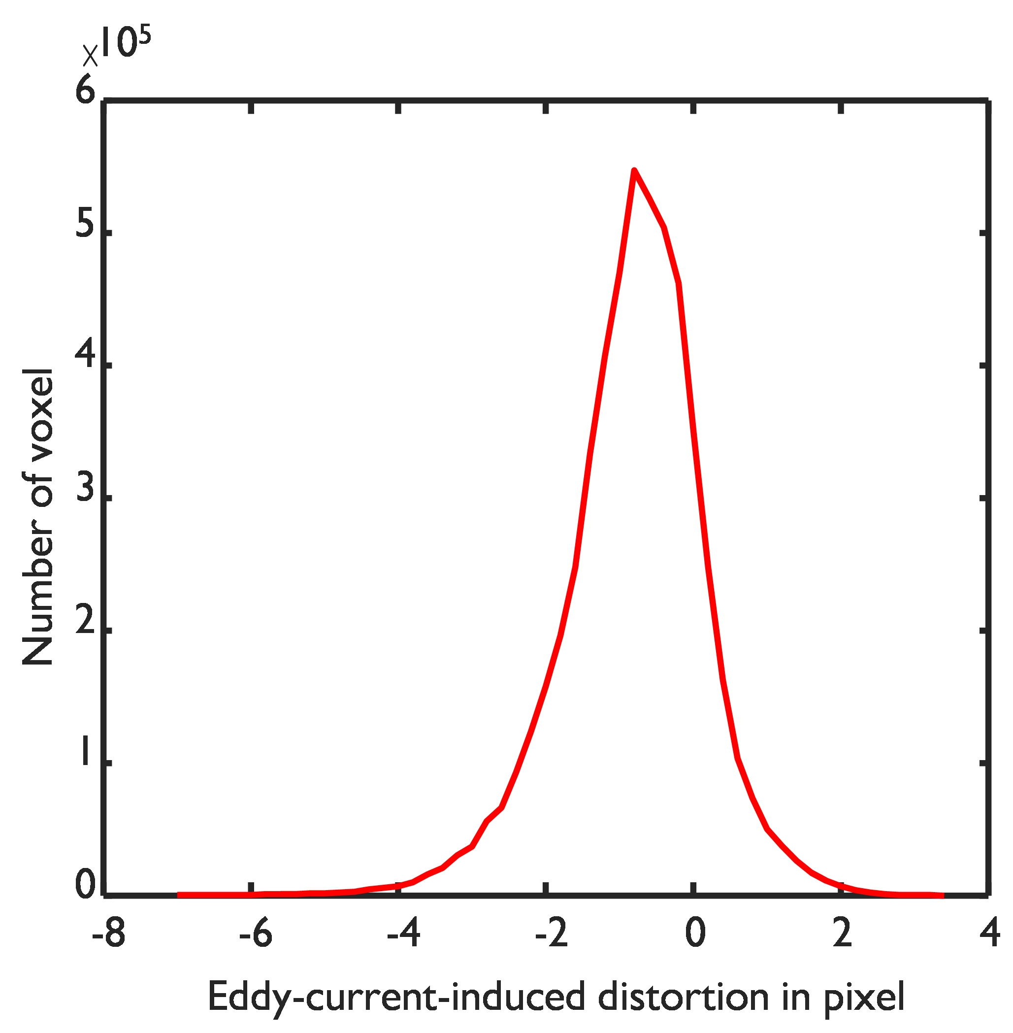
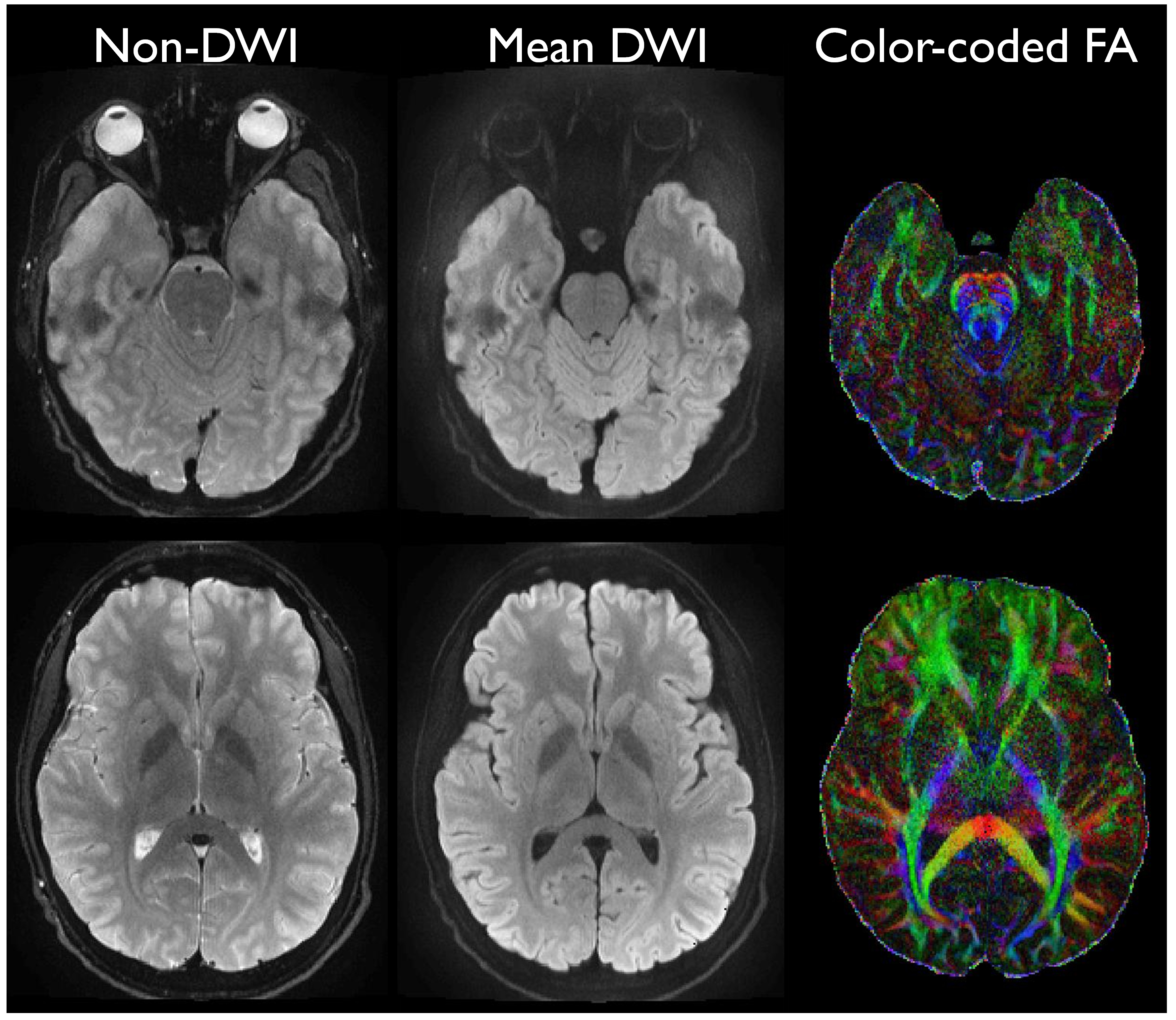
Non-negligible eddy-current-induced distortions ranging from -7.3 to 3.6 pixels were measured by the phantom calibration on the compact 3T (Fig. 2). After the tilted-CAIPI reconstruction, varying eddy-current-induced distortions still appeared in DW-images calculated from the unfolded DW-DIADEM data in both the EPI-PE (Fig. 3B-i) and the SW-PE dimensions (Fig. 3B-ii). In the proposed scheme, reliable distortion correction was performed by the phantom calibration approach before the tilted-CAIPI reconstruction and significant distortion effects were not observed in the final reconstructed images (Figs. 3C-i and 3C-ii). Therefore, the proposed reconstruction scheme could strongly mitigate both susceptibility-induced and eddy-current-induced distortions, even for the highly accelerated DIADEM data (Fig. 4). In addition, a single set of tilted-CAIPI kernels was used for both the non-DW and DW-DIADEM reconstruction without updating the kernel. Compared to other post-processing methods of eddy-current-induced distortion correction such as in FSL11, the calibration data in this work can be incorporated into the in-line reconstruction on the scanner for realtime correction. In previous study3, negligible level of distortions was obtained with a high in-plane acceleration factor of 4. However, this may not be same on different gradient systems and limits the flexibility of choosing the acceleration factor when combined with multi-band imaging12. Therefore, the proposed combination will allow increased flexibility and be more practical for clinical use.Conclusion
This study demonstrates the feasibility and advantage of combining the tilted-CAIPI reconstruction with eddy-current calibration to unfold the highly accelerated DIADEM data for high-resolution distortion-free diffusion imaging. Since the eddy-current calibration approach not only makes the tilted-CAIPI reconstruction more efficient, but also supports the in-line reconstruction to unfold the highly accelerated DIADEM data, the combination will be beneficial for practical use of high-resolution diffusion imaging using DIADEM.Acknowledgements
This work was supported by NIH U01 EB024450-01 and R01 MH116173. The authors would like to thank Erin Gray for her help in collecting the data.References
1. In MH, Posnansky O, Speck O, High-resolution distortion-free diffusion imaging using hybrid spin-warp and echo-planar PSF-encoding approach. Neuroimage. 2017 Mar 1;148:20-30. doi: 10.1016/j.neuroimage.2017.01.008.2. In MH, Tan ET, Trzasko JD, Shu Y, Tao S, Gray EM, Houston J, Bernstein MA. Distortion-free high-resolution diffusion imaging on a compact 3T MRI with high-performance gradients within a clinically feasible scan time. ISMRM 2018 p. 1203
3. Dong Z, Wang F, Reese TG, Manhard MK, Bilgic B, Wald LL, Guo H, Setsompop K. Tilted-CAIPI for highly accelerated distortion-free EPI with point spread function (PSF) encoding. MRM 2018 doi: 10.1002/mrm.27413.
4. In MH, Posnansky O, Speck O. PSF mapping‐based correction of eddy‐current‐induced distortions in diffusion‐weighted echo‐planar imaging. MRM 2016;75(5):2055-2063.
5. Lee S-K, Mathieu J-B, Graziani D, et al. Peripheral nerve stimulation characteristics of an asymmetric head-only gradient coil compatible with a high-channel-count receiver array. MRM 2016;76:1939–1950. doi: 10.1002/mrm.26044.
6. Foo TK, Laskaris E, Vermilyea M, Xu M, Thompson P, Conte G, Van Epps C, Immer C, Lee SK, Tan ET. Lightweight, compact, and high-performance 3 T MR system for imaging the brain and extremities. MRM 2018. doi: 10.1002/mrm.27175.
7. Weavers PT, Shu Y, Tao S, Huston J 3rd, Lee SK, Graziani D, Mathieu JB, Trzasko JD, Foo TK, Bernstein MA. Compact three-tesla magnetic resonance imager with high-performance gradients passes ACR image quality and acoustic noise tests. Med Phys 2016; 43:1259-64.
8. Weavers PT, Tao S, Trzasko JD, Frigo LM, Shu Y, Frick MA, Lee SK, Foo TK, Bernstein MA. B0 concomitant field compensation for MRI systems employing asymmetric transverse gradient coils. Magn Reson Med 2018; 79: 1538-1544.
9. Tao S, Weavers PT Trzasko JD, Shu Y, Huston J , Lee SK, Frigo LM, Bernstein MA. Gradient pre-emphasis to counteract first-order concomitant fields on asymmetric MRI gradient systems. Magn Reson Med. 2017; 77:2250-2262.
10. Tao S, Trzasko, J, Gunter J, Weavers P, Shu Y, Huston J, Lee SK Tan E, Bernstein MA. Gradient nonlinearity calibration and correction for a compact, asymmetric magnetic resonance imaging gradient system. Phys Med Biol. 2017 21;62:N18-N31.
11. FSL package, https://fsl.fmrib.ox.ac.uk/fsl/fslwiki/FSL
12. Setsompop K, Gagoski BA, Polimeni JR, Witzel T, Wedeen VJ, Wald LL. Blipped‐controlled aliasing in parallel imaging for simultaneous multislice echo planar imaging with reduced g‐factor penalty. MRM 2012;67(5):1210-1224.
Figures