0770
Achieving sub-mm clinical diffusion MRI resolution by removing noise during reconstruction using random matrix theory1Radiology, NYU School of Medicine, New York, NY, United States
Synopsis
We show how to achieve the benefits of inline-scan averaging for reducing thermal noise and lowering the Rician noise floor prior to image reconstruction, albeit in inequivalent diffusion MRI (dMRI) acquisitions. For that, we identify and remove the pure-noise principal components in joint coils x q-space x voxels dMRI data, as they follow the universal Marchenko-Pastur distribution. The method is demonstrated on 0.8mm isotropic voxels for b=1000 and 2000 protocol (3T), showing an increase of SNR and decrease of the Rician noise floor by 5-fold. We discuss applications in dMRI parameter estimation, tractography and functional neurosurgery.
Introduction
In diffusion MRI (dMRI), we develop various creative ways to suppress the signal by the diffusion weighting, in an attempt to reveal the rich sub-voxel tissue structure. The more we push the diffusion weighting, the more we can potentially see, – if not for low SNR and the proximity to the Rician floor that “squashes” all interesting signal features. Here we reveal a large untapped reserve of SNR and the possibility of lowering the Rician noise floor, available in routine clinical dMRI. For that, we design a rigorous method to remove most of thermal noise before image reconstruction, giving us the benefits of inline-scan averaging1 albeit in inequivalent q-space acquisitions.Methods
Principle
We$$$\,$$$rely$$$\,$$$on$$$\,$$$the$$$\,$$$ability$$$\,$$$to$$$\,$$$separate pure-noise principal components when our high-dimensional measurement is redundant, i.e., when it has a small number $$$P$$$ of significant principal components relative to the size of the $$$N\times M$$$ data-matrix $$$X$$$. While thermal noise makes the data-matrix full-rank, in the limit $$$N,M\to\infty$$$ we can use the asymptotic random matrix theory result2 to identify3 and remove4 the pure-noise components in the eigenvalue histogram of the covariance matrix $$$XX’$$$.
Implementation
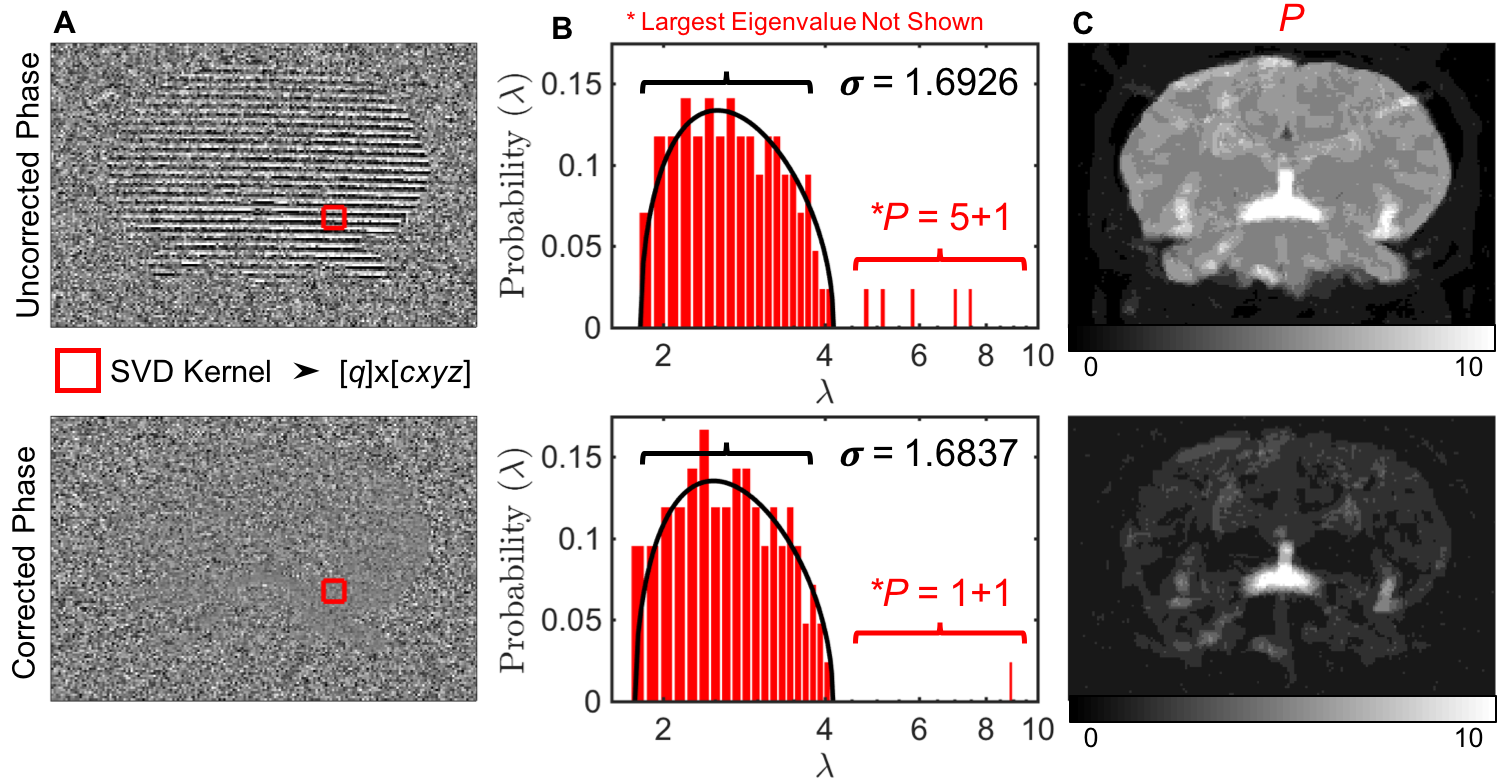
Recently, we developed the MP-PCA method3,4 at the level of diffusion (or functional) weighted images. Here, we utilize the extra redundancy in the joint coils x q-space x voxels data, which allows us to precisely identify and remove noise-only components [Figs.1-2], and then recombine the denoised coil data, thereby achieving extra SNR increase and Rician floor decrease, Fig.3. We first decorrelate signals from multiple receive coils [Fig1] using the noise covariance matrix measured using a separate noise scan, such that noise statistics in the “rotated” coil basis becomes independent identically-distributed Gaussian, and MP theory2 applies. We estimate $$$P$$$ and noise-level $$$\sigma$$$ self-consistently, as compared to LLR5,6 approaches. Estimating and unwinding the coil phase helps reduce the number $$$P$$$ of significant components and increase redundancy, Fig.2. We then use standard parallel-imaging reconstruction7,8 and adaptive combine (AC)9 to generate images with ~5-fold reduced Rician noise floor and signal variation.
Applications
The$$$\,$$$pulsed-gradient$$$\,$$$spin-echo$$$\,$$$(PGSE)$$$\,$$$diffusion$$$\,$$$product sequence$$$\,$$$was$$$\,$$$used$$$\,$$$to$$$\,$$$acquire$$$\,$$$phantom$$$\,$$$(1,below)$$$\,$$$and$$$\,$$$2$$$\,$$$brain$$$\,$$$datasets$$$\,$$$(2-3,below)$$$\,$$$on$$$\,$$$a$$$\,$$$3T$$$\,$$$Siemens$$$\,$$$Prisma$$$\,$$$system$$$\,$$$with$$$\,$$$a$$$\,$$$20$$$\,$$$channel$$$\,$$$head/neck$$$\,$$$coil,$$$\,$$$of$$$\,$$$which$$$\,$$$16$$$\,$$$coil$$$\,$$$elements$$$\,$$$were$$$\,$$$enabled$$$\,$$$during$$$\,$$$scan$$$\,$$$and$$$\,$$$used$$$\,$$$for$$$\,$$$reconstruction.
(1)$$$\,$$$A$$$\,$$$room-temperature$$$\,$$$water$$$\,$$$phantom$$$\,$$$(SNR=35$$$\,$$$at$$$\,$$$b=0),$$$\,$$$Fig.3(B,D,E,F),$$$\,$$$was$$$\,$$$acquired$$$\,$$$with$$$\,$$$(2mm$$$\,$$$isotropic$$$\,$$$resolution),$$$\,$$$no$$$\,$$$parallel$$$\,$$$imaging,$$$\,$$$no$$$\,$$$partial$$$\,$$$Fourier,$$$\,$$$TR=4200ms,$$$\,$$$TE=152ms$$$\,$$$with$$$\,$$$12$$$\,$$$b-shells$$$\,$$$(6$$$\,$$$directions$$$\,$$$each)$$$\,$$$up$$$\,$$$to$$$\,$$$b=4000.
(2)$$$\,$$$The brain of a 25 y/o female volunteer (SNR 6-10 at b=0) was acquired with 1mm isotropic resolution [Figs.1-4], no parallel imaging, partial$$$\,$$$Fourier = 5/8,$$$\,$$$TR=4000ms,$$$\,$$$TE=93ms,$$$\,$$$with 10$$$\,$$$b=0,$$$\,$$$20$$$\,$$$directions of$$$\,$$$b=[500,1000,1500,2000]$$$\,$$$s/mm2.
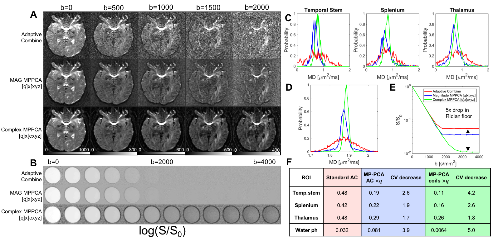
(3)$$$\,$$$A full brain of a 34 y/o male volunteer (SNR=13-15 at b=0) was imaged with 0.8mm isotropic resolution [Fig.5], TR=3100 ms, TE=75 ms, 10 b=0, 20 b=1000, 40 b=2000, partial Fourier 5/8, Grappa=2, no multiband. This was acquired with 5 segments of 26 slices that were fused in post-processing. We compared these reconstructions between standard 2-dimensional AC, magnitude MP-PCA on AC3,4, and the proposed complex (i.e, multicoil-level) MP-PCA reconstruction. Diffusion images were processed via the DESIGNER10 pipeline, and ODFs and probabilistic tractography was generated and visualized with MRTRIX311.
Results & Discussion
Application of MP-PCA before non-linear transformations, such as trapezoidal re-gridding, partial Fourier, or Grappa which all distort12 the original perfectly Gaussian noise statistics, allows for a far more precise separation between signal and noise [Figs.1-2] than previously reported.3,4$$$\,$$$Fig.2$$$\,$$$shows that we achieve perfectly Gaussian statistics of residuals down to 5 standard deviations, i.e., the histogram$$$\,p(r)\,$$$of normalized residuals $$$r$$$ for the coil images is Gaussian down to the ~1ppm tail relative to its peak. If any anatomy were to be removed, it would show up in the deviation of$$$\,\ln\,p(r)\,$$$vs$$$\,r^2\,$$$from a perfectly straight line, Fig.1D.
Fig.3 shows that we increase the precision, and create a larger dynamic range for dMRI parameter estimation, up to a 5-fold drop in the Rician noise floor in water phantom. The b-range for which the functional form$$$\,\ln\,S/S_0=-bD\,$$$for perfectly Gaussian diffusion in water is observable increased from ~0-700 to ~0-2000 s/mm2 [Fig.3E]. Comparing mean diffusivity (MD) derived from the Gaussian regime, we observed an increase in precision over the AC recon [Fig3.C,D,F]. At this SNR level, some degree of Rician bias was always present for AC reconstructions at all b values, resulting in an artifactual lowering of MD. Attempting to correct for the Rician bias after AC (magnitude MPPCA, Figs.3-4),$$$\,$$$was not enough to completely undo the effect of bias. With complex MP-PCA reconstruction, the MD variablity in the brain ROIs decreased by up to 4.2-fold, and bias in MD for all ROIs disappeared by shifting towards higher values that were consistent with literature10[Fig.3C,D;Fig.4].
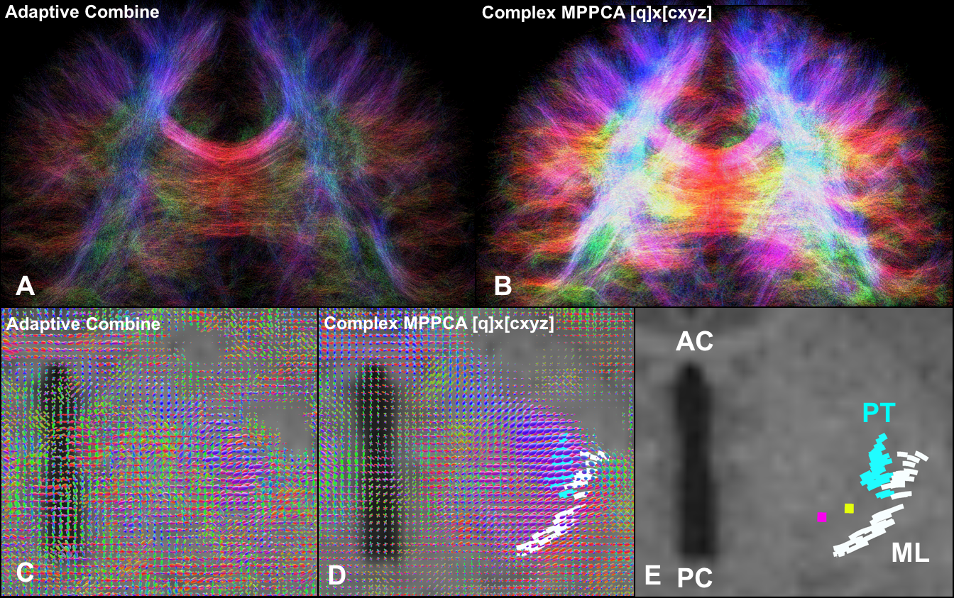
High-resolution$$$\,$$$dMRI$$$\,$$$was$$$\,$$$identified$$$\,$$$as$$$\,$$$a$$$\,$$$critical$$$\,$$$biomarker$$$\,$$$for$$$\,$$$delineation13$$$\,$$$of$$$\,$$$the$$$\,$$$ventral-intermediate$$$\,$$$nuclei$$$\,$$$(VIM)$$$\,$$$in$$$\,$$$Parkinson’s$$$\,$$$disease$$$\,$$$and$$$\,$$$essential$$$\,$$$tremor.$$$\,$$$We$$$\,$$$were$$$\,$$$able$$$\,$$$to$$$\,$$$acquire$$$\,$$$a$$$\,$$$full$$$\,$$$brain$$$\,$$$high-b$$$\,$$$acquisition$$$\,$$$at$$$\,$$$0.8mm$$$\,$$$isotropic resolution in 22 minutes [Fig.5], with sufficient SNR to identify the medial leminiscus and pyramidal tract pathways to deduce the location of the VIM; the AC reconstruction was unable to resolve these pathways and yielded unusable fiber orientations “squashed” by the Rician floor with such small voxels.
Conclusion
We$$$\,$$$show$$$\,$$$how$$$\,$$$to$$$\,$$$enjoy$$$\,$$$the$$$\,$$$benefits$$$\,$$$of$$$\,$$$inline-scan$$$\,$$$averaging$$$\,$$$for$$$\,$$$reducing$$$\,$$$thermal$$$\,$$$noise$$$\,$$$and$$$\,$$$lowering$$$\,$$$Rician$$$\,$$$noise$$$\,$$$floor$$$\,$$$prior$$$\,$$$to$$$\,$$$image$$$\,$$$reconstruction,$$$\,$$$but$$$\,$$$instead$$$\,$$$of$$$\,$$$averaging$$$\,$$$by$$$\,$$$exploiting the redundancy in coils and inequivalent q-space acquisitions. The MP-PCA denoising+reconstruction method is straightforwardly generalizable to other multi-dimensional acquisitions, such as fMRI, perfusion, and transverse relaxation.Acknowledgements
No acknowledgement found.References
1. Treit S et al. High resolution in-vivo diffusion imaging of the human hippocampus. Neuroimage 2018;182:479-487.
2. Marchenko VA et al. Distribution of eigenvalues for some sets of random matrices. Mathematics of the USSR-Sbornik 1967;1(4):457.
3. Veraart J et al. Diffusion MRI noise mapping using random matrix theory. Magn Reson Med 2016;76(5):1582-1593.
4. Veraart J et al. Denoising of diffusion MRI using random matrix theory. Neuroimage 2016;142:394-406.
5. Zhang T et al. Accelerating parameter mapping with a locally low rank constraint. Magnetic Resonance in Medicine 2014;73(2):655-661.
6. Trzasko J et al. Local versus global low-rank promotion indynamic MRI series reconstruction. In Proceedings of the 19thAnnual Meeting of ISMRM, Montreal, Canada, 2011. p. 4371.
7. Pruessmann KP et al. SENSE: sensitivity encoding for fast MRI. Magn Reson Med 1999;42(5):952-962.
8. Griswold MA et al. Generalized autocalibrating partially parallel acquisitions (GRAPPA). Magn Reson Med 2002;47(6):1202-1210.
9. Walsh DO et al. Adaptive reconstruction of phased array MR imagery. Magn Reson Med 2000;43(5):682-690.
10. Ades-Aron B et al. Evaluation of the accuracy and precision of the diffusion parameter EStImation with Gibbs and NoisE removal pipeline. Neuroimage 2018;183:532-543.
11. Tournier JD et al. MRtrix: Diffusion tractography in crossing fiber regions. International Journal of Imaging Systems and Technology 2012;22(1):53-66.
12. Sprenger T et al. Real valued diffusion-weighted imaging using decorrelated phase filtering. Magn Reson Med 2017;77(2):559-570.
13. Sammartino F et al. Tractography-Based Ventral Intermediate Nucleus Targeting: Novel Methodology and Intraoperative Validation. Mov Disord 2016;31(8):1217-1225.
14. Benabid AL et al. Long-term suppression of tremor by chronic stimulation of the ventral intermediate thalamic nucleus. Lancet 1991;337(8738):403-406.
Figures
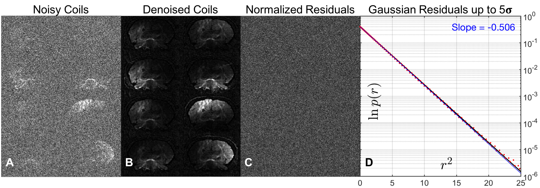
Joint coils$$$\times\,q$$$-space MP-PCA. A typical coil combination shown for$$$\,8/16\,$$$coils for$$$\,b=2000\,s/mm^2$$$, before (A) and after MP-PCA (B), when combining$$$\,q$$$-space points with coils in an$$$\,N=3\times3\times3\,$$$patch, and (C) maps of residuals$$$\,r=\delta y/\sigma\,$$$normalized by the estimated noise$$$\,\sigma$$$. Note the absence of any anatomy in the residual map. (D) Histogram (red), of normalized residuals$$$\,r\,$$$(Re{$$$r$$$}$$$\,$$$and$$$\,$$$Im{$$$r$$$}$$$\,$$$pooled from all coils) is perfectly Gaussian over $$$5$$$ standard deviations, (blue line ln probability vs $$$r^2$$$). Unit normal pdf$$$\,e^{-r^2/2}/\sqrt{2\pi}\,$$$is shown as reference (black line).