0763
T-Hex: Spiral sampling on a tilted hexagonal grid1Institute for Biomedical Engineering, ETH Zurich and University of Zurich, Zürich, Switzerland, 2ETH, Zürich, Switzerland, 3Skope Magnetic Resonance Technologies, Zurich, Switzerland
Synopsis
In this work, we show a stack of spirals on a tilted hexagonal grid. This trajectory is used for fast 3D acquisitions with long readouts and combines the optimal hexagonal 3D sampling with the high acquisition efficiency of spirals. Isotropic 2.8 mm whole-brain coverage is achieved in 200 ms relying on cg-SENSE reconstruction, without the need for non-linear regularization.
Introduction
Rapid MR scanning of 3D volumes is aimed at in applications like fMRI with high temporal resolution (1). 3D Fourier encoding lends itself to this task: it provides high SNR due to data acquisition from the entire volume, while being amenable to undersampling by optimal parallel imaging acceleration in all three dimensions.
However, existing 3D encoding strategies require trade-offs between the following desirable characteristics:
- average k-space speed close to the maximum given by the gradient amplitude limit
- uniform-density, near-isotropic sampling in all indirect dimensions
- smooth T2* weighting across k-space for a benign PSF
Stacks of EPIs (2) do not cover k-space as time-efficiently as spirals. Cones, concentric shells, and the yarnball trajectory (3, 4) are inefficient in that they oversample the k-space center or poles.
Recently, these obstacles have been overcome by tilted hexagonal (t-Hex) stacks of spirals, which acquire an arbitrary number of adjacent k-space planes within one shot (5). Thus, 3D k-space is covered extremely efficiently and exhibits a smooth T2* weighting. In this work, t-Hex is extended to higher spatial resolution as well as ultra-high-speed cases.
Methods
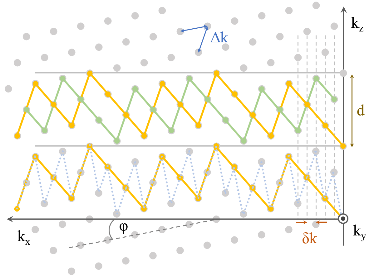
T-Hex: The hexagonal grid (6,7) formed by a conventional stack of spirals is tilted by
$$\varphi = tan^{-1}(\dfrac{P-2}{P*\sqrt{3}})$$
where P is the number of k-space planes visited by each shot (Fig.1) and defines the band thickness d. By introducing interleaved readout trains, rational numbers of planes per shot (i.e. 3/2 = 3 planes per 2 shots) can be achieved. This imparts more flexibility to the method and can take it to higher spatial resolutions.
Combining t-Hex on the other hand with a spiral-in readout allows for higher temporal resolutions (Figure 3 and 4).
Hardware:
- Philips 7T Achieva system
- 32 channel head array (Nova Medical)
- 16 19F NMR field probes mounted on the head array and a dedicated MR acquisition system (8)
Sequence: A GRE sequence with T2*-contrast was implemented. K-space trajectories were designed for an isotropic resolution and whole-brain coverage (FOV= 24x24x12 cm3). Resolution is defined via the extent of an equivalent spherical k-space volume.
- 1 mm resolution, 36 shots, TAQ = 41 ms, TE = 15 ms, TR = 75 ms, spiral-out, 1.5 planes per shot
- 2.8 mm resolution, 5 shots, TAQ = 24 ms, TE = 25.5 ms, TR = 40 ms, spiral-in, 4 planes per shot
Image reconstruction was performed by an iterative cg-SENSE reconstruction (9) extended to 3D, including multi-frequency-interpolation (10,11) for static off-resonance correction and based on the concurrently monitored trajectories (12). Off-resonance and coil sensitivity maps were computed from a multi-echo, spin-warp pre-scan (6 echoes, TE 2-7 ms, 2x2x2 mm3 resolution).
Results
See Figures 2 and 5.Discussion and Conclusion
The spiral t-Hex k-space acquisition scheme favorably combines core requirements for fast 3D imaging: The hexagonal grid guarantees a uniform (under-)sampling pattern, its apt tilt provides a smooth T2* weighting and the spiral pathway ensures a high k-space speed.
Unlike common spiral imaging, time cannot be redistributed among different shots by mere rotation of the same undersampling spiral arm due to the distinguished site of blipping. However, in this work, a way of interleaving differently shaped t-Hex spirals is found to arrive at an arbitrary ratio of covered k-space planes per shot. 1mm resolved whole-brain coverage is achieved within 2.7 s at an image quality attractive for high-spatial-resolution fMRI. The approach is nevertheless limited by the blipping itself: The more planes each shot traverses, the larger the blips have to be. And the more interleaving shots there are, the more overhead for other sequence modules such as RF-excitation and spoiling there is.
In the high-resolution case, t-Hex was applied to a spiral-out trajectory. Likewise, the scheme can be taken to other readout strategies such as the more conventional EPI, catching however the same time loss compared to spirals as in 2D. For an optimal use of acquisition time in long-TE sequences for BOLD imaging, we also applied t-Hex to a spiral-in trajectory. Thereby, 2.8 mm resolved whole-brain images could be acquired within 200 ms. The resulting images exhibit largely decent quality, especially in areas vulnerable to dephasing artefacts such as the frontal lobes. Only slight ringing occurred, likely due to residual fat signal, that would not confound BOLD analysis.
To achieve appealing image quality the reconstruction was based on the expanded signal model described above.
The present work follows up on the new tilted hexagonal sampling scheme and pushes it forward in the two important application cases of high spatial and high temporal resolution.
Acknowledgements
No acknowledgement found.References
1. Feinberg DA, Moeller S, Smith SM, Auerbach E, Ramanna S, Glasser MF, Miller KL, Ugurbil K, Yacoub E. Multiplexed Echo Planar Imaging for Sub-Second Whole Brain FMRI and Fast Diffusion Imaging Valdes-Sosa PA, editor. PLoS ONE 2010;5:e15710. doi: 10.1371/journal.pone.0015710.
2. Poser BA, Koopmans PJ, Witzel T, Wald LL, Barth M. Three dimensional echo-planar imaging at 7 Tesla. NeuroImage 2010;51:261–266. doi: 10.1016/j.neuroimage.2010.01.108.
3. Irarrazabal P, Nishimura DG. Fast Three Dimensional Magnetic Resonance Imaging. Magn. Reson. Med. 1995;33:656–662. doi: 10.1002/mrm.1910330510.
4. Stobbe RW, Beaulieu C. A Looping Trajectory for Single-Shot 3D Imaging. In: Proc. Intl. Soc. Mag. Reson. Med. Vol. 19. ; 2011.
5. Engel M, Kasper L, Barmet C, Schmidt T, Pruessmann KP. Proceedings of the ISMRM 2017. Proc. ISMRM 2018 [Internet]
6. Mersereau RM. The Processing of Hexagonally Sampled Two-Dimensional Signals. Proc. IEEE 1979;67:930–949.
7. Engel M, Kasper L, Pruessmann KP. Rapid 3D imaging with multiplanar spirals. Proc. ISMRM 2017 [Internet].
8. Dietrich BE, Brunner DO, Wilm BJ, Barmet C, Gross S, Kasper L, Haeberlin M, Schmid T, Vannesjo SJ, Pruessmann KP. A field camera for MR sequence monitoring and system analysis. Magn. Reson. Med. 2016;75:1831–1840. doi: 10.1002/mrm.25770.
9. Pruessmann KP, Weiger M, Börnert P, Boesiger P. Advances in sensitivity encoding with arbitrary k-space trajectories. Magn. Reson. Med. 2001;46:638–651. doi: 10.1002/mrm.1241.
10. Man L-C, Pauly JM, Macovski A. Multifrequency interpolation for fast off-resonance correction. Magn. Reson. Med. 1997;37:785–792. doi: 10.1002/mrm.1910370523.
11. Barmet C, Tsao J, Pruessmann KP. Sensitivity encoding and B0 inhomogeneity – A simultaneous reconstruction approach. In: Proceedings of the ISMRM. ; 2005. p. 682.
12. Barmet C, Wilm BJ, Pavan M, Katsikatsos G, Keupp J, Mens G, Pruessmann KP. Concurrent higher-order field monitoring for routine head MRI: an integrated heteronuclear setup. In: Proceedings of the 18th Annual Meeting of ISMRM, Stockholm, Sweden. ; 2010. p. 216.
Figures