0758
Spread-spectrum MRI: acceleration of image acquisition using locally modulated magnetic fieldsKlaus Scheffler1,2, Jonas Bause1, Ali Aghaeifar1, Theodor Steffen1, Bernhard Schölkopf1, and Alexander Loktyushin1
1Max Planck, Tuebingen, Germany, 2University of Tuebingen, Tuebingen, Germany
Synopsis
We introduce the principles of a novel approach for the acceleration of signal acquisition that is based on a rapid and unique modulation of localized magnetic fields superimposed to the conventional linear gradient-based spatial encoding.
Introduction
Spread-spectrum MRI is based on rapid dynamic (up to MHz) and local modulation of magnetic fields produced in local current loops. Current localized gradient encoding techniques such as PatLoc, O-space imaging and FRONSAC (1-3) rely on static or quasi-static (<kHz) local magnetic field profiles that are essentially constant (compared to the Larmor frequency) during each MR echo acquisition. In spread-spectrum MRI, these fields are modulated dynamically during signal acquisition to imprint local and distinct signal characteristics into the spin arrangement, which can be interpreted as a unique fingerprint onto confined regions within the object. Spread-spectrum MRI distributes or spreads the basic bandwidth of gradient-encoded resonance frequencies along the encoding axis using distinct carrier frequencies (or other time courses such as orthogonal noise patterns) originating from a certain spatial portion of the object. This spatially unique information can then be used to resolve aliasing in undersampled acquisition, and thus to drastically boost imaging speed.Methods
Local magnetic fields, for example, can be generated by a set of current loops that are placed close to the object. An example is shown in Fig. 1 that consists of 8 independent magnetic field coils that are placed on a cylinder (5cm x 5 cm, 25 windings). A temporal current variation induces a locally varying magnetic field that modifies the local Larmor frequency of the magnetization. In Fig. 1, a 1 kHz and 1 A current produces a 1kHz varying local magnetic field that generates a locally confined frequency modulation of the free induction decay, visible as side bands. Different frequencies and phases can be applied to each coil separately during the acquisition of the MR signal to improve localization. Using this additional local information, the conventional imaging process can be accelerated. In Fig. 2 we illustrate the effect of injecting currents in different local B0 coils on the image reconstructed with inverse Fourier transform without taking into account additional encoding components due to an oscillating field. However, in the presence of local oscillating magnetic fields, a linear model for image reconstruction can be used. S(t) is the measured signal and m(r) the image to be reconstructed. Gx, Gy and Gz are the linear gradients, and Bc(r) are the local sensitivities or B0-field maps of the coils driven with a sine wave of amplitude A. In order to reconstruct the image m, a linear system has to be solved. E is the linear operator that aggregates the exponential encoding terms and performs the summation (integration) over the spatial domain. To reconstruct the image, we solve the following regularized optimization problem: The regularization coefficient sets the weight of the total variation term TV that penalizes high-frequency artifacts in the reconstruction. An extension of the model to the case of accelerated acquisition is straightforward and involves decreasing the number of rows in the matrix E subject to the spectral undersampling pattern.Results
Fig. 3 shows an example where the local field modulations have been used to accelerate image acquisition. Fig. 3 (reference) is the corresponding reference. In Fig 3 (2-fold undersampling) k-space of the 2D gradient echo sequence was 2-fold undersampled resulting in a N/2 ghosting artifact. Application of alternating currents of 3A and 5kHz all channels with a phase shift of 45° between channels during each readout period of the gradient echo sequence, and using the reconstruction algorithm shown above gives the 2-fold accelerated image shown in Fig. 3 (spread spectrum MRI). Additionally, we performed a simulation experiment where we evaluated reconstruction performance at different acceleration factor, modulation frequency and current combinations. In Fig. 3 bottom right, we show normalized root mean square errors between the reconstructed images and the reference.Discussion
In preliminary experiments, we could demonstrate an acceleration factor of 2 with only minor imaging artifacts. The proposed method is essentially based on an increased acquisition bandwidth, as local frequency modulations will increase the spectral bandwidth of MR signals. Therefore, image acquisition acceleration based on spread spectrum MRI will go with a decrease in SNR proportional to the square root of acquisition time. However, optimal arrangements of local B0 coils and combination of spread spectrum MRI with Parallel Imaging based on local B1 coils might open novel and currently unforeseen possibilities and applications.Acknowledgements
Reinhart Koselleck-Projekt DFG SCHE 658References
1. Hennig J, Welz AM, Schultz G, Korvink J, Liu Z, Speck O, Zaitsev M. Parallel imaging in non-bijective, curvilinear magnetic field gradients: a concept study. Magma;21(1-2):5-14. (2008) 2. Wang H, Tam LK, Constable RT, Galiana G. Fast Rotary Nonlinear Spatial Acquisition (FRONSAC) Imaging. Magn Reson Med 2016 March ; 75(3): 1154–1165 Stockmann JP, Ciris PA, Galiana G, Tam LK, Constable RT. O-Space images: highly efficient parallel imaging using second-order nonlinear fields as encoding gradients with no phase encoding. Magn Reson Med 2010; 64(2):447–56Figures
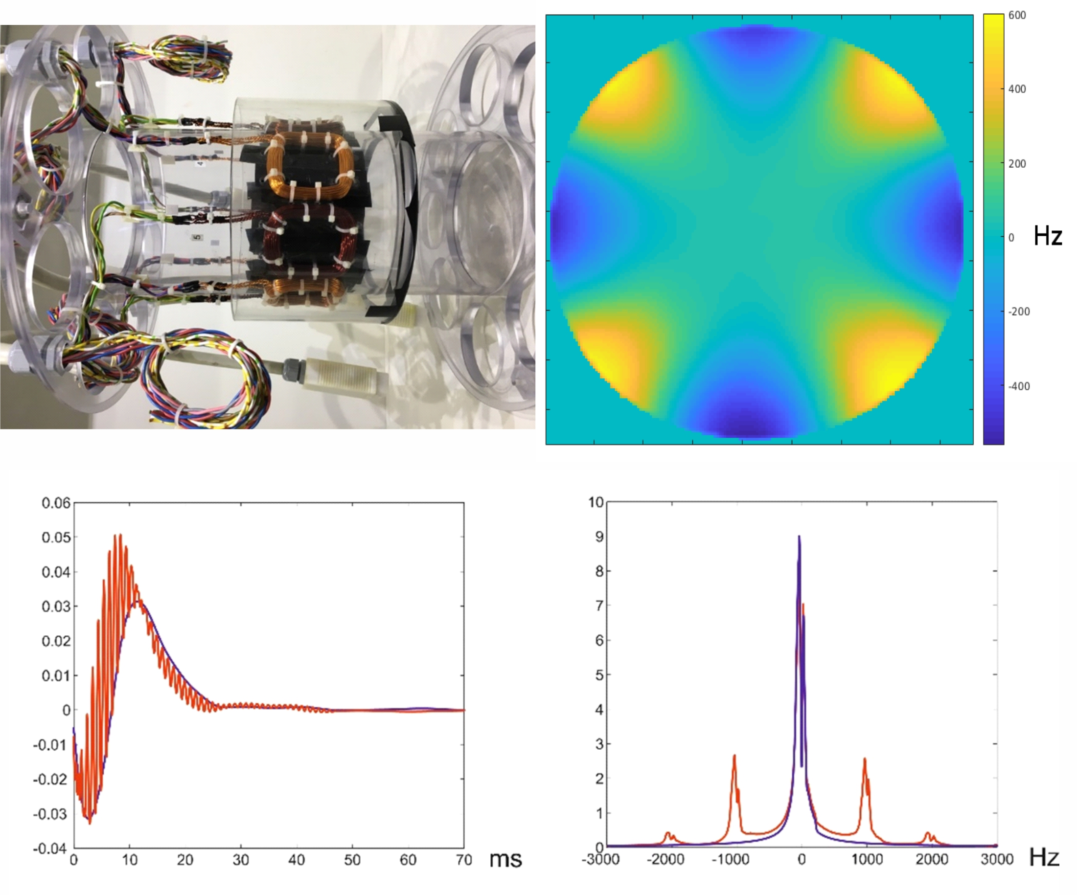
Fig. 1: Top: Arrangement of
8 local B0 coils and corresponding B0 field maps. Bottom left: The blue curve shows a free induction decay
acquired at a constant magnetic field without applying any currents to the
local coils. Bottom right is the corresponding spectrum showing a peak around 0
Hz. Application of a varying magnetic field of 1 KHz via the local coil
superimposes an oscillation onto the free induction decay. This produces 1 kHz
separated side lobes in the corresponding spectra shown right.
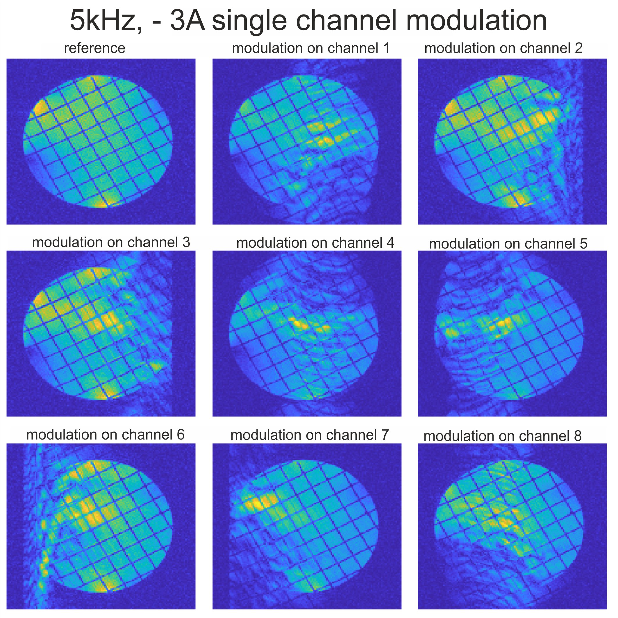
Fig. 2: Application
of alternating currents (5kHz at 3 A) to each single channel of the coil arrangement
shown in Fig. 1. If reconstructed with a conventional Fourier transform, local
spreading artifacts are generated along the readout direction.
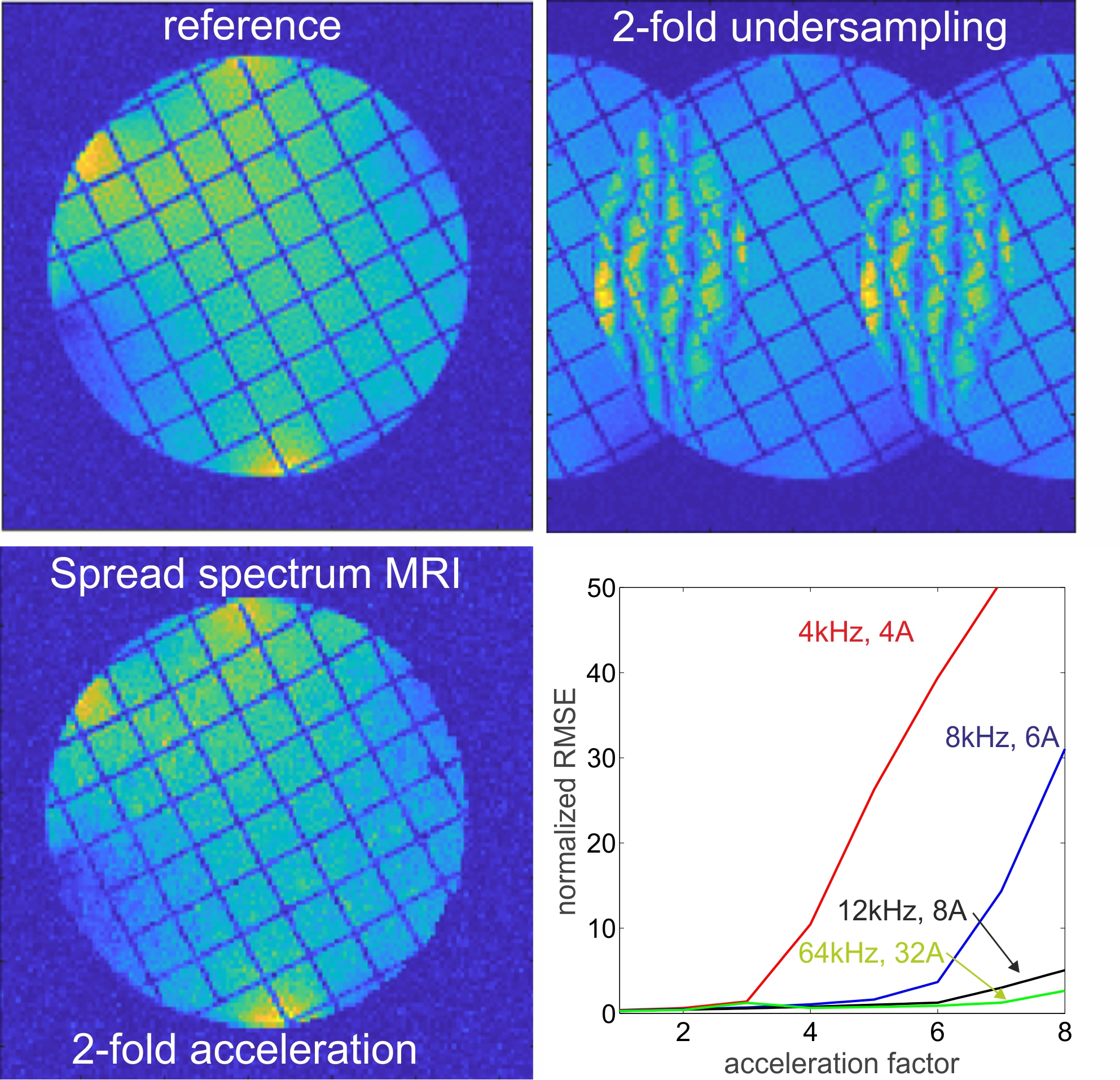
Fig. 3: Top
left is the fully sampled reference image without application of alternating
magnetic fields. Top right shows the result of 2-fold undersampling (along
phase encoding direction) without application of alternating local fields. Left
bottom is again 2-fold undersampled combined with 5kHz and 3A modulation
currents to each local coil and a phase shift of 45° between the individual
channels. Bottom right shows normalized root mean square errors (percentage) between
reconstructions of simulated acquisitions and the reference image as a function
of acceleration for different frequencies and current amplitudes.