0746
High resolution T2-prepared MRI enables non-invasive assessment of CSF flow in perivascular spaces of the human brain1Radiology, Leiden University Medical Center, Leiden, Netherlands, 2Mathematical Sciences, University of Southampton, Southampton, United Kingdom, 3LKEB, Leiden University Medical Center, Leiden, Netherlands
Synopsis
Recently, flow of cerebrospinal fluid (CSF) has been shown to play an important role in the transport of brain metabolites, ushering in the concepts of glymphatics and intramural periarterial drainage. Failure of waste-drainage has been linked to incurable neurodegenerative diseases such as Alzheimer’s disease and cerebral amyloid angiopathy. The lack of a non-invasive imaging technique for the investigation of perivascular drainage mechanisms in the human brain strongly hinders the assessment of brain clearance in human subjects. In this study, we present the first non-invasive technique to visualize the CSF-flow along the middle cerebral artery (MCA), as well as along perivascular spaces of smaller arteries in the human brain.
Introduction
Despite its high metabolic rate, the brain does not have a classic lymphatic system for the clearance of interstitial fluid and waste products from its intracerebral regions. In recent years, the flow of cerebrospinal fluid (CSF) has been shown to play an important role in the transport of brain metabolites, ushering in the concepts of glymphatics1 and intramural periarterial drainage (IPAD)2. Failure of waste-drainage has been linked to incurable neurodegenerative diseases such as Alzheimer’s and cerebral amyloid angiopathy3. The lack of non-invasive imaging techniques for the investigation of perivascular drainage mechanisms in the human brain greatly hinders the assessment of brain clearance in human subjects.
Here, we propose the use of ultra-high field MRI with a high spatial resolution, long TE readout in combination with a T2-prepared (T2prep) module with crusher gradients to visualize the CSF-flow along the middle cerebral artery (MCA), as well as along perivascular spaces of smaller arteries in the human brain. This mirrors the more traditional DTI approach with which Harrison et al.4 assessed CSF-flow in the spaces surrounding the MCA in the rat brain.
Methods
Four healthy subjects (age=23±3 years) were scanned at 7T (Achieva, Philips, The Netherlands) using a 32-channel head-coil. All provided written informed consent for this IRB-approved study. High-resolution (0.45mm3 isotropic, FOV=210×181×43mm), 3D images were acquired with a turbo-spin echo sequence. A very long echo time was applied to measure only the CSF signal (TE=227ms, TR=3047ms, TSE-factor 104). First, we validated the use of a long-TE readout to visualize the CSF compartments around arteries as proposed by Harrison et al4. Subsequently, a T2prep-module with velocity-encoded gradients of 1cm/s was included. Seven sets of images were acquired to create a DTI-like tensor: one without crushers and six with crushers applied in different directions.
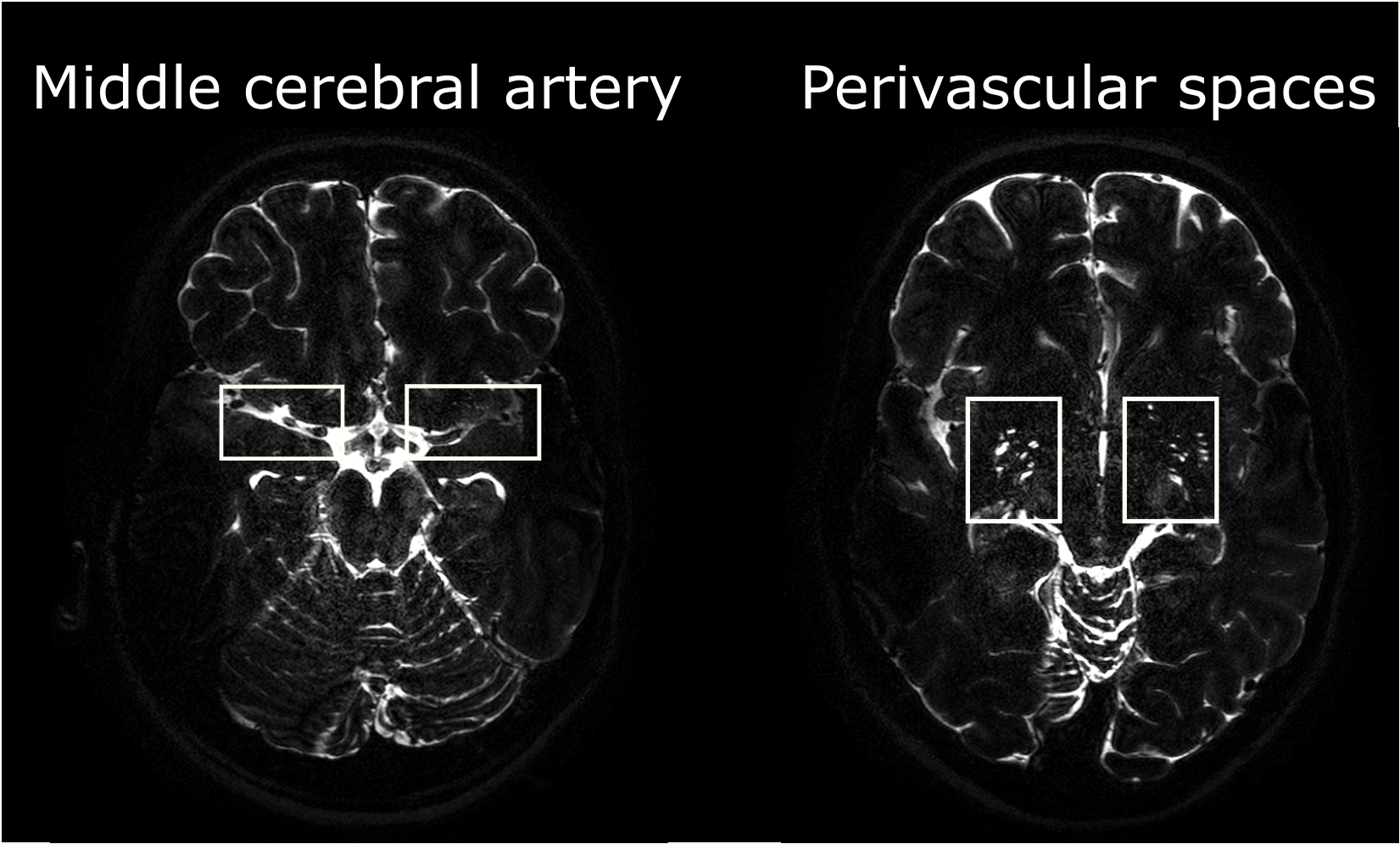
Intra-subject images were registered using Elastix5 and DTI postprocessing6 was performed using Matlab to compute diffusion-like parameters such as the principal CSF-flow direction and the fractional anisotropy (FA); the results were visualized with Paraview7. Analysis was first focused on the larger fluid compartments around the MCA and subsequently on the perivascular spaces of smaller arteries (see Figure 1).
Results
The long-TE approach combined with a high-resolution readout enabled to visualize CSF-flow around large vessels, such as the MCA, but also within perivascular spaces (Figure 1). Other signals coming from the blood and from the brain tissue are attenuated, i.e. the CSF-signal has successfully been isolated.
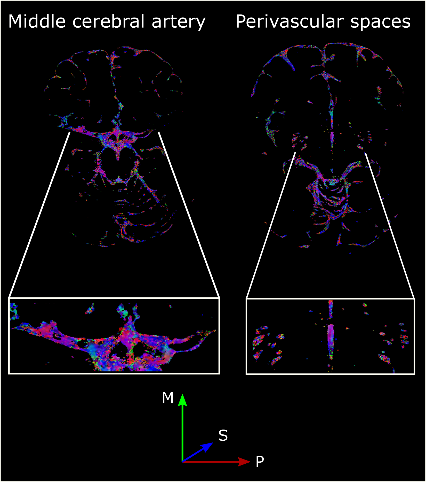
The introduction of velocity-encoded gradients crushed 12±4% (average±SD across volunteers) of the CSF-signal in the MCA. The color-coded FA map (Figure 2) around the MCA shows a main direction predominantly oriented along the phase-encode direction (red), corresponding to the vessel direction, although some directionality along the slice-direction (blue) can be observed as well.
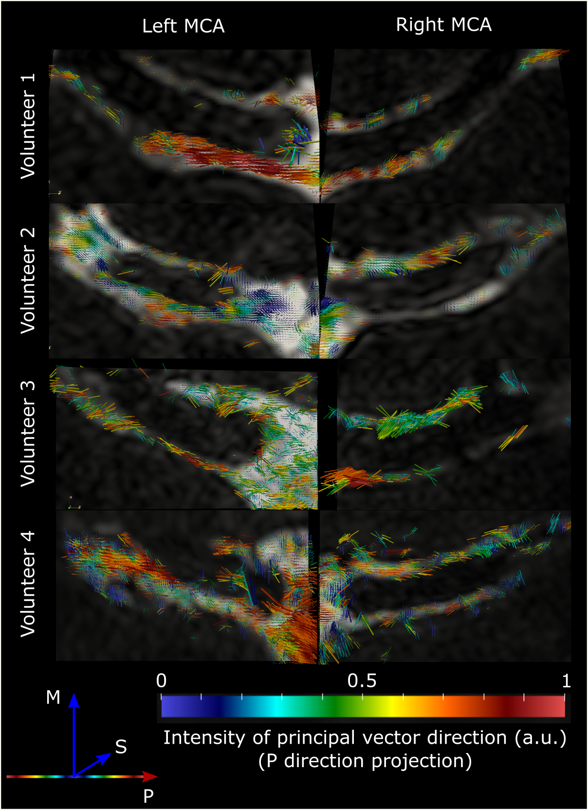
To better understand the orientation of the CSF-movement, the principal direction vector was represented (Figure 3), showing that CSF is moving along the vessel wall of the MCA. Even though the main direction is along the vessel, a swirling pattern of the CSF around the MCA can also be observed, particularly in volunteers 3 and 4.
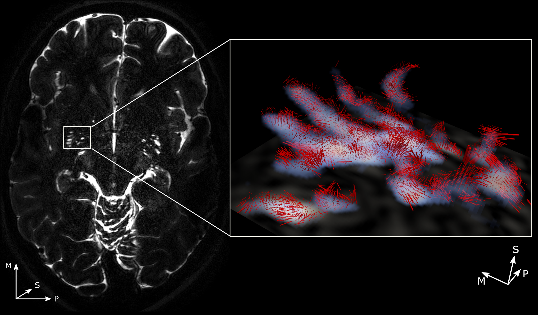
In the perivascular spaces of smaller vessels, it is more difficult to observe clear patterns (Figure 4): while in some perivascular spaces a global CSF-flow along the vessel can be observed, in others the flow direction is almost perpendicular to the vessel.
Discussion
The use of a T2-prep module instead of traditional diffusion approaches let to several advantages: it enabled the use of a TSE readout for which the image quality improved compared to an EPI readout, especially at 7T. No consistent image quality could be obtained when acquiring data with the more traditional spin echo EPI readout with diffusion gradients around the refocusing pulse (data not shown). The fact that the flow-prepared magnetization was stored in the longitudinal plane (slower relaxation rate) further facilitated the use of 3D readout sequences as opposed to multi-slice readouts of traditional DTI.
For the first time, CSF-movement was measured non-invasively in perivascular spaces of human subjects. The 3D-dataset consists of a huge amount of data that calls for more extensive exploration, with more advanced tools in order to confirm fluid flow patterns, especially in the perivascular spaces of smaller arteries. Moreover, in future studies, we will evaluate whether lower velocity encoding values help enhance the sensitivity of the technique in perivascular spaces.
Ultimately, it would be highly interesting to compare these measurements done in healthy, young volunteers with data acquired in patients suffering from cerebral amyloid angiopathy, Alzheimer’s disease or idiopathic normal pressure hydrocephalus.
Acknowledgements
This work is part of the research programme Innovational Research Incentives Scheme Vici with project number 016.160.351, which is financed by the Netherlands Organisation for Scientific Research (NWO).References
1. Iliff JJ, Wang M, Liao Y, et al. A paravascular pathway facilitates CSF flow through the brain parenchyma and the clearance of interstitial solutes, including amyloid β. Sci Transl Med. 2012;4(147):147ra111.
2. Albargothy NJ, Johnston DA, MacGregor-Sharp M, et al. Convective influx/glymphatic system: tracers injected into the CSF enter and leave the brain along separate periarterial basement membrane pathways. Acta Neuropathol. 2018;136(1):139-152.
3. Tarasoff-Conway JM, Carare RO, Osorio RS, et al. Clearance systems in the brain-implications for Alzheimer disease. Nat Rev Neurol. 2015;11(8):457-70.
4. Harrison IF, Siow B, Akilo AB, et al. Non-invasive imaging of CSF-mediated brain clearance pathways via assessment of perivascular fluid movement with diffusion tensor MRI. Elife. 2018;7:e34028. Published 2018 Jul 31. doi:10.7554/eLife.34028
5. Klein S, Staring M, Murphy K, et al. Elastix: a toolbox for intensity based medical image registration. IEEE Trans. Med. Imaging 2010;29:196–205.
6. Kroon, D.-J., (2008). DTI and Fiber Tracking. [online] Available at: https://www.mathworks.com/matlabcentral/fileexchange/21130-dti-and-fiber-tracking
7. Ayachit, Utkarsh, The ParaView Guide: A Parallel Visualization Application, Kitware, 2015, ISBN 978-1930934306
Figures