0735
Accelerated SAR Computations by Exploiting Sparsity in the Anatomical Domain1Leiden University Medical Center, Leiden, Netherlands, 2Delft University of Technology, Delft, Netherlands
Synopsis
Accelerated SAR computations can improve awareness on actual RF exposure levels, which are known to depend heavily on subject anatomy. In this work, we accelerate SAR computations by reformulating the underlying electromagnetic field equations in terms of anatomical differences rather than the entire anatomy itself. This yields a “sparse” representation of the target anatomy, reducing problem complexity without compromising accuracy.
Purpose
RF exposure is well-known to vary significantly among subjects, depending on anatomical parameters such as tissue distribution, body habitus and positioning within the RF coil.1 The main bottleneck in the assessment of specific absorption rate (SAR), or RF heating in general, is in the numerical computation of RF fields as a function of anatomy. Many attempts have been made on accelerating this type of problem, for example by using boundary integral methods or surface integral equations, which lend itself more for modeling homogeneous regions or RF coils.2,3
In this work, we present a numerical approach to simplify SAR calculations by reformulating the underlying electromagnetic field equations in terms of anatomical differences with respect to a database model, rather than solving for the entire anatomy itself. This yields a “sparse” representation of the anatomy, reducing the number of unknowns without compromising accuracy. Furthermore, by exploiting a threshold operation we can further reduce the number of unknowns, effectively compressing the problem.
Methods
Theory
Building on electromagnetic scattering theory, we express the total electric field $$$E$$$ for a given body model as a field perturbation with respect to a “background” body model, viz.$$ E=(\mathbb{I}-\mathbb{G}\chi)^{-1}E^{\tt b}.$$
Here, $$$E^{\tt b}$$$ denotes the background electric field and $$$\chi=\varepsilon-\varepsilon^{\tt b}$$$ denotes the electric susceptibility of the target body model with respect to the background model. $$$\mathbb{G}$$$ denotes the Green's tensor which accounts for the response of the heterogeneous background and is characterized in advance during an offline stage of the algorithm.
Configuration and Implementation
As a proof-of-concept, a 7T neuroimaging scenario was modeled on a very coarse 10 mm isotropic grid in order to test the approach within available computational resources. A 30-cm diameter 16-rung high-pass birdcage was loaded with the head models “Duke” and “Ella”.4 Incident fields in the empty coil were calculated using XFdtd (v7.4, Remcom, State College, PA) and fed into a custom iterative solver implemented using GMRES in Matlab (r2016a, Mathworks inc., MA, USA) to compute the total fields in both models. 10-g averaging of the SAR distributions was obtained using an iterative kernel growing method.5
"Ella" was taken to be the target model for which SAR is to be computed, while assigning “Duke” as the background model. Its response was evaluated by computing field-responses for each of the edges within the background model. This resulted in a total of ~14,000 edges, which took approximately 12 hours to evaluate using a -30 dB convergence criterion on a single workstation.
Exploiting sparsity
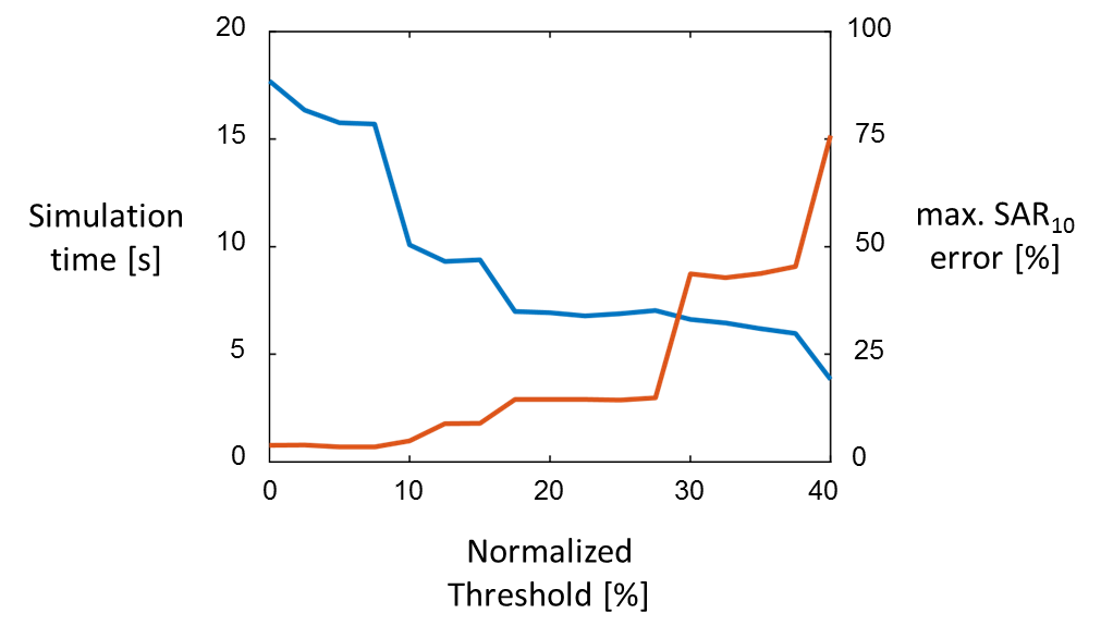
By thresholding $$$\mid\chi\mid$$$, we can reduce the number of unknowns further, while preserving the dominant perturbations in the RF field. By removing the corresponding entries in $$$\mathbb{G}$$$, we can substantially reduce the size of the system matrix, leading to faster computations.
Results
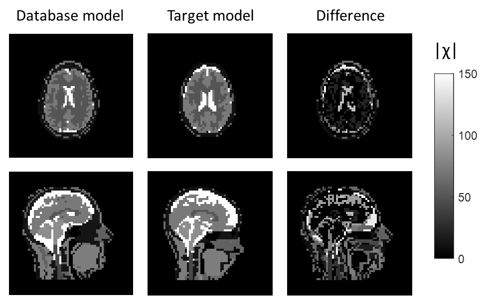
Figure 1 shows the database body model “Duke”, target model “Ella” and their anatomical difference in terms of their electric susceptibility.
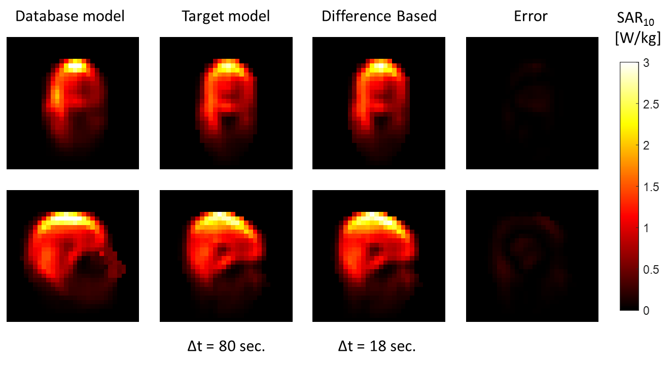
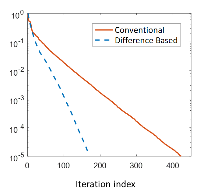
Figure 2 shows the SAR10 maps obtained in these models, where results in the proposed approach were obtained approximately four times faster, without compromising accuracy. A very small error of ~3% can be observed, which is due to error accumulation from the offline calculations. This can be improved by increasing their convergence threshold. The improved convergence rate of the iterative solver is shown in Figure 3.
Finally, the thresholding procedure enables further acceleration of the method of up to an order of magnitude, at the cost of a ~15% error in the maximum SAR10 values.
Discussion/Conclusion
A novel approach on SAR computations was presented which casts the computation of RF field in terms of anatomical differences with respect to a pre-characterized database model. This allows for reducing the number of unknowns, improving the convergence rate of numerical solvers. This approach can potentially become useful in online SAR assessments, where a database body model can serve as a background model.
The current benchmark of the method was performed on a male and female body model, which are quite different from each other. Having multiple body models in a pre-characterized database would increase the chances of obtaining a better starting point, further accelerating this approach. An important challenge is the scalability of the method. Where the current 10 mm assessment resulted in a matrix G of 1.5 GB in single precision, in a corresponding 5 mm model the number of edges would increase by a factor of ~8, and memory requirements by a factor of ~64. Future work should therefore include model compression to improve scalability of this approach within realistic computational resources.
Acknowledgements
This project was funded by the Dutch Technology Foundation (TTW) project 16820.References
1. Murbach M, Neufeld E, Kainz W, Pruessmann KP, Kuster N. Whole-body and local RF absorption in human models as a function of anatomy and position within 1.5T MR body coil. Magn Reson Med 2014;71:839–45. doi: 10.1002/mrm.24690.
2. van den Bergen B, Stolk CC, van den Berg JB, Lagendijk JJW, van den Berg CAT. Ultra fast electromagnetic field computations for RF multi-transmit techniques in high field MRI. Phys Med Biol 2009;54:1253–1264. doi: 10.1088/0031-9155/54/5/010.
3. Villena JF, Polimeridis AG, Eryaman Y, et al. Fast Electromagnetic Analysis of MRI Transmit RF Coils Based on Accelerated Integral Equation Methods. IEEE Trans Biomed Eng [Internet] 2016;63:2250–2261. doi: 10.1109/TBME.2016.2521166.
4. Christ A, Kainz W, Hahn EG, et al. The Virtual Family—development of surface-based anatomical models of two adults and two children for dosimetric simulations. Phys Med Biol 2010;55:N23-38. doi: 10.1088/0031-9155/55/2/N01.
5. Kuehne A, Seifert F, Ittermann B. GPU-Accelerated SAR Computation with Arbitrary Averaging Shapes. In: Proceedings of the 20th Annual Meeting of ISMRM, Melbourne, Australia. ; 2012. p. 2735.
Figures