0734
MRI RF Heating: The Effect of Vasculature Detailedness on Numerical Temperature Increase Estimations1IT'IS Foundation, Zurich, Switzerland, 2Swiss Federal Institute of Technology (ETH), Zurich, Switzerland
Synopsis
The numerical assessment of the radiofrequency (RF)-induced local temperature increase inside a patient undergoing magnetic resonance imaging (MRI) diagnostics is state-of-the-art in MRI safety studies. In light of the continuous improvement in the resolution of anatomical models, we investigated the impact of the level of detail in the vasculature models on estimates of temperature increase. Results show that the difference of the peak temperature increase for the investigated high-exposure scenario is in the order of 20%. Future investigations should broaden the studied exposure scenarios and consider vascular convection.
Introduction
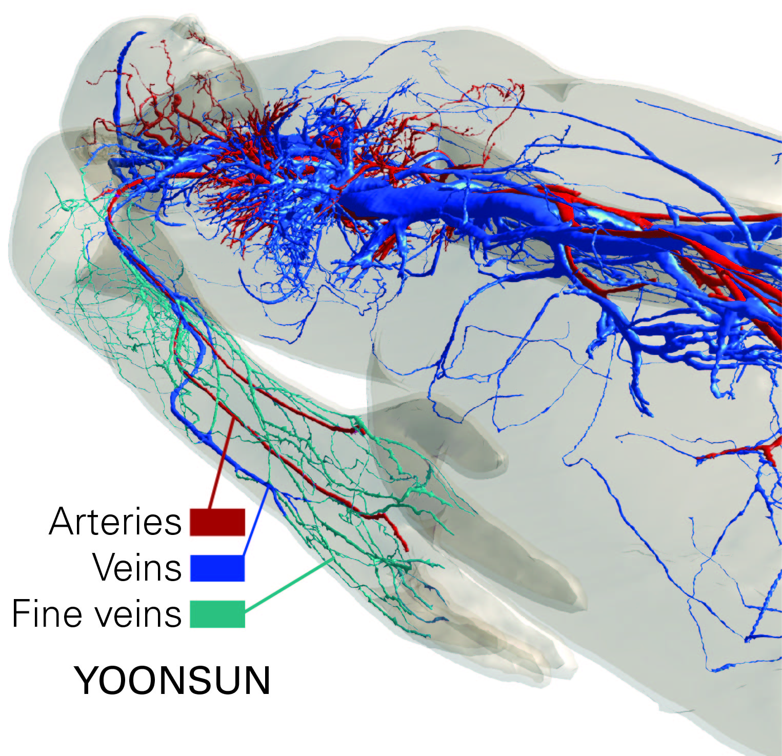
Magnetic resonance imaging (MRI) radiofrequency (RF) safety with regard to associated induced tissue heating can be studied by means of simulations involving computational anatomical models. These thermal simulations are becoming more and more important in academia and regulatory safety assessment sectors, yet they require a large data set of thermal and physiological parameters, including the tissues' thermal conductivity and blood perfusion. A further requirement is that the anatomical human model studied is sufficiently detailed. The latest generation of the Virtual Population (ViP)1 includes YOONSUN V3.12, a female model with unprecedented detail in nerve and vasculature anatomy. While the impact of microvasculature is typically handled through a homogeneous and distributed heat-sink term, the thermal impact of large vessels is typically simulated by imposing a Dirichlet boundary condition3 (37°C) – corresponding to the assumption that such vessels effectively cool their surfaces. We investigated the influence of the level of detail in the vasculature model with regard to its effect on RF-induced temperature increase estimations, by comparing simulations with the newly available extremely detailed YOONSUN vasculature to simulations where the YOONSUN vessels with diameters <2 mm are not treated as boundary conditions. This provides insight into the influence of the vessel tree detail on thermal modelling as well as likely allows estimation of an upper bound on the impact of medium-sized vasculature.Methods
ViP model YOONSUN was investigated in the abdominal MRI imaging position at 3T, which constitutes a high exposure scenario4 with a thermal hotspot in the right forearm. The Pennes bioheat equation5 has been applied with thermo-physical and -physiological parameters from the literature6. The medium-sized vessels with diameters <2 mm in the comparison-simulation were cut off and assigned the properties of fat tissue.
The vessel system with the division into large and small vessels is illustrated in Figure 1. The exposure level was normalized to the first level controlled operating mode with a whole-body averaged specific absorption rate (wbSAR) of 4 W/kg. Thermoregulated perfusion values were not considered for this study, which explains the high steady-state peak temperature of >45°C.
Results and Discussion
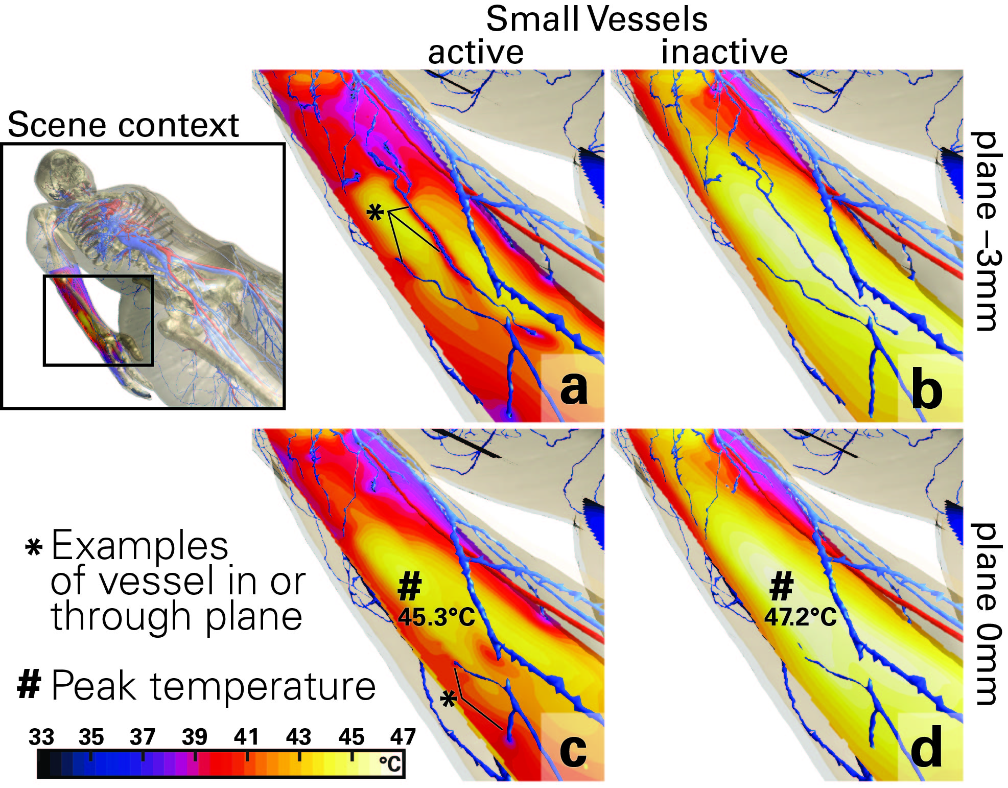
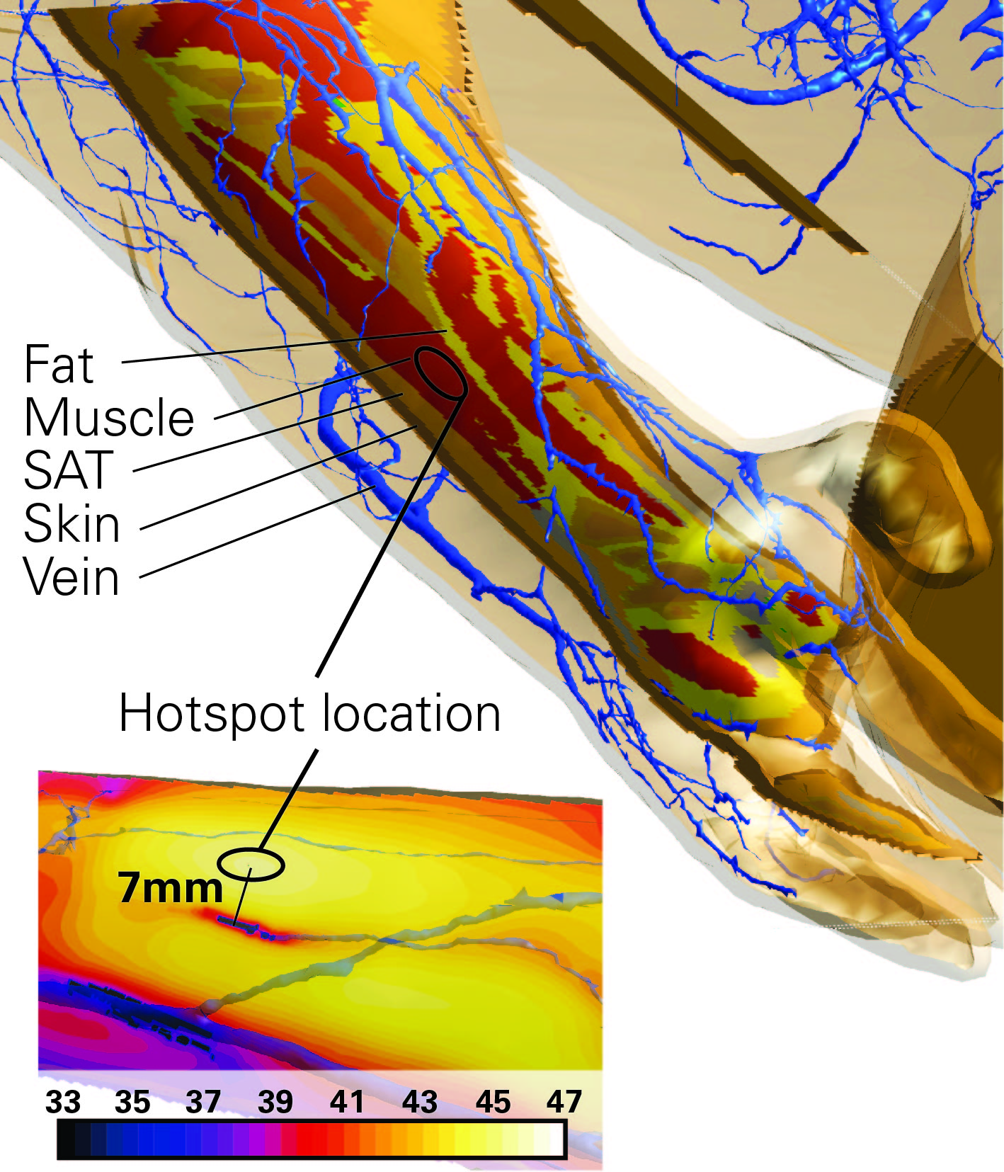
The steady-state temperature distributions for the detailed vessel system and the situation with only large-sized vessels considered are shown in Figure 2. Two slice-views at a vertical distance of 3 mm are provided: panels (a,b) show the situation with a high degree of vasculature detail and panels (c, d) show the location of the actual thermal hotspot. While the simulated medium-sized vessels considerably alter the temperature distribution around their location, the actual peak temperature increase in the thermal hotspot is affected less – a reduction from 47.2°C to 45.2°C, 20% of the temperature increase – when the medium-sized vessels are modeled as boundary conditions. The hotspot location was identical for both simulations (Figure 3).
The real impact of medium-sized vasculature is likely to be between the two simulated scenarios, as medium-sized vessels do not act as perfect coolers – due to Dirichlet boundary conditions – but rather as convective boundary conditions. Also, the heating of the blood in such vessels can no longer be neglected. The situation is further complicated by the active transport of heated blood, which results in less effective cooling or even heating at remote locations.
While a peak temperature increase uncertainty of 20% associated with the modeling of medium-sized vasculature might be smaller than the uncertainty associated with the tissue perfusion parameters and the impact of thermoregulation (on the order of 30%3), it should be noted that the uncertainty in the temperature increase elsewhere – potentially in thermally sensitive tissues – can be much larger, and that only a single, illustrative MRI exposure scenario has currently been studied.
Conclusions
Future investigations of more exposure scenarios and more realistic modeling of the impact of the thermal vasculature (e.g., based on the DIVA model7 that couples 1D convective vascular tree thermal simulations with 3D thermal modelling) must be performed.Acknowledgements
This project is supported by funding from the MRInext and NEUROMAN projects.References
1. Gosselin M-C, Neufeld E, Moser H, et al. Development of a new generation of high-resolution anatomical models for medical device evaluation: the Virtual Population 3.0. Phys Med Biol 2014;59:5287–5303.
2. ViP. Virtual Population. https://itis.swiss/virtual-population/.
3. Murbach M, Neufeld E, Capstick M, Kainz W, Brunner DO, Samaras T, Pruessmann KP, Kuster N. Thermal tissue damage model analyzed for different hole-body SAR and scan durations for standard MR body coils. Magn Reson Med 2014;71:421–431.
4. Murbach M, Neufeld E, Cabot E, Zastrow E, C??rcoles J, Kainz W, Kuster N. Virtual population-based assessment of the impact of 3 Tesla radiofrequency shimming and thermoregulation on safety and B1+ uniformity. Magnetic Resonance in Medicine 2016;76:986–997.
5. Pennes HH. Analysis of tissue and arterial blood temperatures in the resting human forearm. Applied Physiology 1948;1:93–122.
6. Hasgall PA, Di Gennaro F, Baumgartner C, Neufeld E, B L, Gosselin MC, Payne D, Klingenböck A, Kuster N. IT’IS database for thermal and electromagnetic parameters of biological tissues. Version 4.0, May 15, 2018. www.itis.ethz.ch/database 2018.
7. Kotte A, Van Leeuwen G, De Bree J, Van Der Koijk J, Crezee H, Lagendijk J. A description of discrete vessel segments in thermal modelling of tissues. Physics in Medicine and Biology 1996;41:865–884.
Figures