0733
Gadolinium retention in tissues: yttrium as a gadolinium surrogate to investigate the in vivo biodistribution of the retained gadolinium1Athinoula A. Martinos Center for Biomedical Imaging, Department of Radiology, Massachusetts General Hospital and Harvard Medical School, Charlestown, MA, United States
Synopsis
Gadolinium has been found in the brain, skin and bone of patients with normal renal function months to years after the last administration of a gadolinium-based contrast agent (GBCA). Yttrium (Y3+) has similar size and chemical properties to Gd3+. We investigated whether yttrium can be used as a gadolinium surrogate by measuring the biodistribution of Gd-DTPA/Y-DTPA and Gd-DOTA/Y-DOTA (0.6 mmol/kg) in mice. Residual Gd and Y levels 7 days after injection showed a Y:Gd ratio close to 1 in all organs demonstrating that Y can act as a surrogate for Gd.
Introduction
Gadolinium from gadolinium-based contrast agents (GBCAs) is partially retained in the body for long periods after administration.1 First associated with nephrogenic systemic fibrosis in patients with renal insufficiency, gadolinium has also been detected in the skin and central nervous system of patients with normal renal function. Despite increasing scrutiny, the chemical form and the full in vivo biodistribution of the retained gadolinium remain unknown. A strategy to address these questions is to use another metal ion as a surrogate for the gadolinium ion, to take advantage of unique physical properties of the surrogate. Yttrium, a pseudo-lanthanide, appears to be an ideal candidate as Y3+ and Gd3+ ions are of similar size and their complexes have shown similar properties: identical thermodynamic stability constants, and identical structure both in solution and at solid state.2,3 Yttrium also possesses a positron emitting isotope, yttrium-86, that could be used as a PET tracer to investigate the in vivo biodistribution of gadolinium. However, the in vivo behavior of Y(III) and Gd(III) complexes has never been directly compared. Our study aimed to assess if yttrium is a good surrogate for gadolinium by evaluating the in vivo retention of Y(III) and Gd(III) complexes in mice.Method
Complexes synthesis. Y-DTPA, Gd-DTPA, Y-DOTA, and Gd-DOTA were synthesized by mixing 1.1 eq of YCl3 or GdCl3 with H5DTPA or H4DOTA and the pH adjusted to 6. The complexes were purified by cation exchange chromatography and reversed phase HPLC and then freeze-dried. The complexes were solubilized in water and the concentration of the solutions was measured by ICP-MS. The solutions to be injected was prepared by mixing the Y-DTPA and Gd-DTPA solutions or the Y-DOTA and Gd-DOTA solutions in 1:1 ratio. The pH was adjusted to 7 – 7.5, the solutions filtered through a 0.2 mm filter, and the final concentration measured by ICP-MS. The absence of free gadolinium or yttrium was confirmed by HILIC chromatography coupled to ICP-MS. Biodistribution in mice. 12 mice were administered, by IV injection, a dose of Y/Gd-DTPA (n = 6) or Y/Gd-DOTA (n = 6) at a dose of 0.6 mmol/kg of each complex. After 7 days, mice were sacrificed, and 21 organs and tissues were harvested and digested in nitric acid where dysprosium was added as an internal standard. Y and Gd concentration were measured by ICP-MS. The calibration curve consisted in 6 standard solutions in the range 0.1 ppb – 50 ppb with the lowest calibration point corresponding to the LOQ.Results
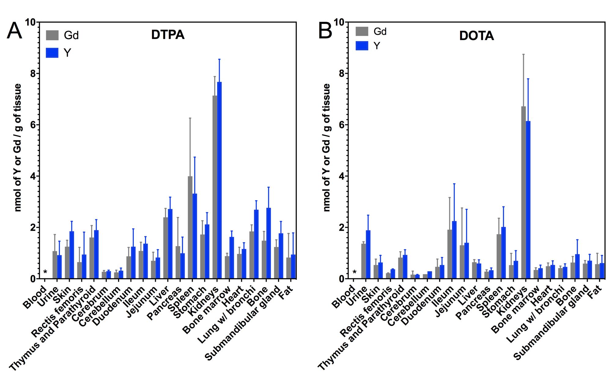
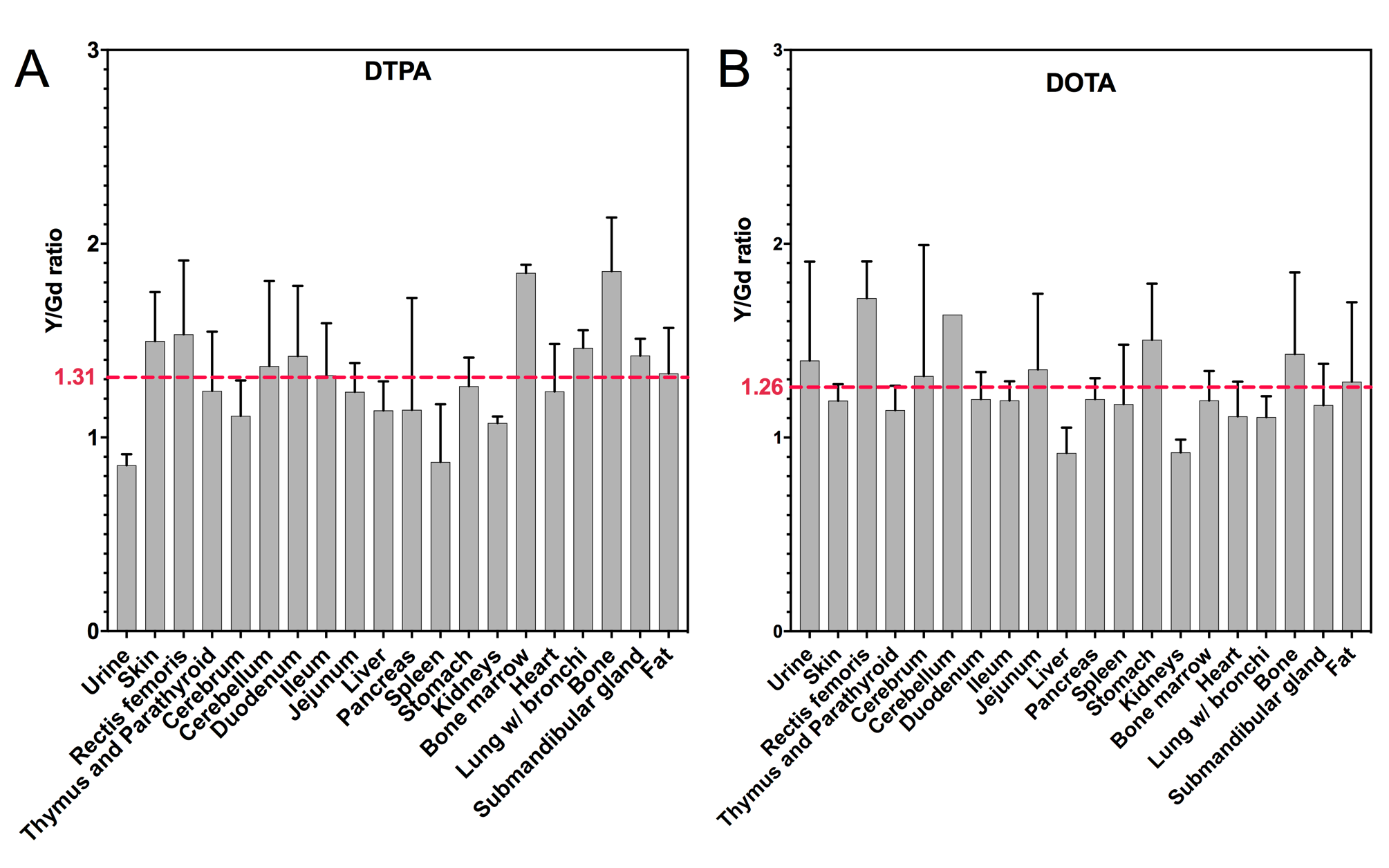
Figure 1 reports the concentration of yttrium and gadolinium in the different organs and tissues 7 days after the injection of either Y/Gd-DTPA or Y/Gd-DOTA. In both cases, yttrium and gadolinium were found in every organ and tissue except in the blood where the detected concentration was below the LOQ. The highest concentration was measured in kidneys. In order to understand if the Y and Gd complexes are similarly retained, the figure 2 shows the Y:Gd ratio in the different organs and tissues, calculated from the Y and Gd concentrations (nmol/g). This ratio was close to 1 in every organ with an average of 1.31 for Y/Gd-DTPA and 1.26 for Y/Gd-DOTA, and there were no statistically significant differences between Y and Gd in any organ.Discussion
The amount of yttrium and gadolinium measured in mice 7 days after injection is consistent with previous studies.4,5 More gadolinium and yttrium were measured in animals exposed to Y/Gd-DTPA than in the Y/Gd-DOTA group, reflecting the lower stability of the linear derivatives. Interestingly, the concentration in the cerebellum and cerebrum are among the lowest in both groups. Y and Gd complexes are retained to the same extent in each organ with a Y/Gd ratio close to 1. In a similar study, Di Gregorio et al. evaluated the in vivo retention of La-DTPA and Gd-DTPA in mice, and La/Gd ratios fom 1 to 15 were found after 96h due to the lower stability of La-DTPA.6 Thus, the same retention of Y and Gd complexes can be attributed to their similar stability constants and Y can be used as a gadolinium surrogate to further investigate the issue of gadolinium retention.Conclusion
The amount of Y and Gd retained in vivo after the concomitant injection of Y-DTPA/Gd-DTPA or Y-DOTA/Gd-DOTA are about the same. Thus, yttrium appears to be a good gadolinium surrogate to investigate the biodistribution of the retained gadolinium in vivo. Future work will utilize 86Y-DTPA and 86Y-DOTA as PET tracers in animals to quantify whole body pharmacokinetics.Acknowledgements
P.C. acknowledges support (EB009062) from the National Institute for Biomedical Imaging and Bioengineering.References
1. McDonald R J, Levine D, Weinreb J, et al. Gadolinium Retention: A Research Roadmap from the 2018 NIH/ACR/RSNA Workshop on Gadolinium Chelates. Radiology, 2018; 289, 517–534.
2. Caravan P, Ellison J J, McMurry T J, Lauffer R B. Gadolinium(III) Chelates as MRI Contrast Agents: Structure, Dynamics, and Applications. Chemical Reviews, 1999; 99(9), 2293–2352.
3. Wadas T J, Wong E H, Weisman G R, Anderson C J. Coordinating radiometals of copper, gallium, indium, yttrium, and zirconium for PET and SPECT imaging of disease. Chemical Reviews, 2010; 110(5), 2858–2902.
4. Bussi S, Coppo A, Botteron C, et al. Differences in gadolinium retention after repeated injections of macrocyclic MR contrast agents to rats. Journal of Magnetic Resonance Imaging, 2018; 47(3), 746–752.
5. Di Gregorio E, Iani R, Ferrauto G, et al. Gd accumulation in tissues of healthy mice upon repeated administrations of Gadodiamide and Gadoteridol. Journal of Trace Elements in Medicine and Biology, 2018; 48(2010), 239–245.
6. Di Gregorio E, Ferrauto G, Furlan C, et al. The Issue of Gadolinium Retained in Tissues: Insights on the role of metal complex stability by comparing metal uptake in Murine Tissues Upon the Concomitant Administration of Lanthanum- and gadolinium-Diethylentriamminopentaacetate. Investigative Radiology, 2018; 53(3), 167–172.
Figures