0726
The PNS oracle: a modified neural activation function metric for rapid assessment of Peripheral Nerve Stimulation (PNS)1Computer Assisted Clinical Medicine, Heidelberg University, Mannheim, Germany, 2A.A. Martinos Center for Biomedical Imaging, Charlestown, MA, United States, 3Harvard Medical School, Boston, MA, United States, 4Harvard-MIT Division of Health Sciences Technology, Cambridge, MA, United States
Synopsis
As gradient engineering advances, Peripheral Nerve Stimulation (PNS) increasingly limits MRI gradient coil use. The ability to predict a winding pattern’s PNS threshold could be useful during design iteration, but recently introduced methods for simulating the threshold in full body models including nerve atlases and neurodynamic simulations are computationally slow. Here we present a simple PNS oracle which is linear (with respect to the E-field) that allows prediction of the gradient-induced stimulation threshold without relying on full neurodynamic modeling. We validated the fast oracle against the full neurodynamic model in multiple gradient coils and two body models.
Target Audience:
Gradient coil designers, those interested in Electromagnetics in MR or MR safetyPurpose:
The steady improvements in gradient coil and power amplifier design in the last 20 years have made Peripheral Nerve Stimulation (PNS) a major limitation in the application of gradient switching speed and strength. We recently proposed and validated a PNS modeling framework to predict PNS thresholds and locations based on coupled electromagnetic and neurodynamic simulations [1,2]. However, full modeling of the nerve dynamics is computationally slow and therefore not useful within advanced numerical coil optimization which must evaluate cost-functions and constraints thousands of times to generate an optimal coil winding pattern. In this work, we present a simple PNS oracle which is linear in the E-field (and thus coil currents) and allows prediction of the gradient-induced stimulation threshold without relying on solving the full neurodynamic differential equations. We validated the fast oracle against the full neurodynamic model.Methods:
Our previously described PNS simulation framework uses electromagnetic body models with co-registered nerve atlases and a neurodynamic model of peripheral nerves [1,2]. After calculating the E-field in the body model, we evaluate the MRG nerve dynamics model [3,4] and look for induced action potentials. The Neural Activation Function (NAF) is a common metric of stimulation, defined as the second spatial derivative of the electric potential along the nerve [5]:
$$\text{NAF}(r)=\frac{\partial^2 V}{\partial r^2}\approx\frac{V(r-h)-2V(r)+V(r+h)}{h^2}$$
where $$$V(r)$$$ is the electric potential at position $$$r$$$ along the nerve and $$$h$$$ is the spatial step (e.g., 0.1 mm). Although the NAF is useful for identifying nerve segments likely to be stimulated, this metric is an imperfect estimator of the quantitative PNS thresholds for three reasons: First, the NAF does not account for the non-myelinated section (nodes of Ranvier), and second it does not account for the myelin thickness (axon diameter). Third, it ignores crosstalk between neighboring nodes of Ranvier (depolarization of a node of Ranvier spreads to neighboring nodes).
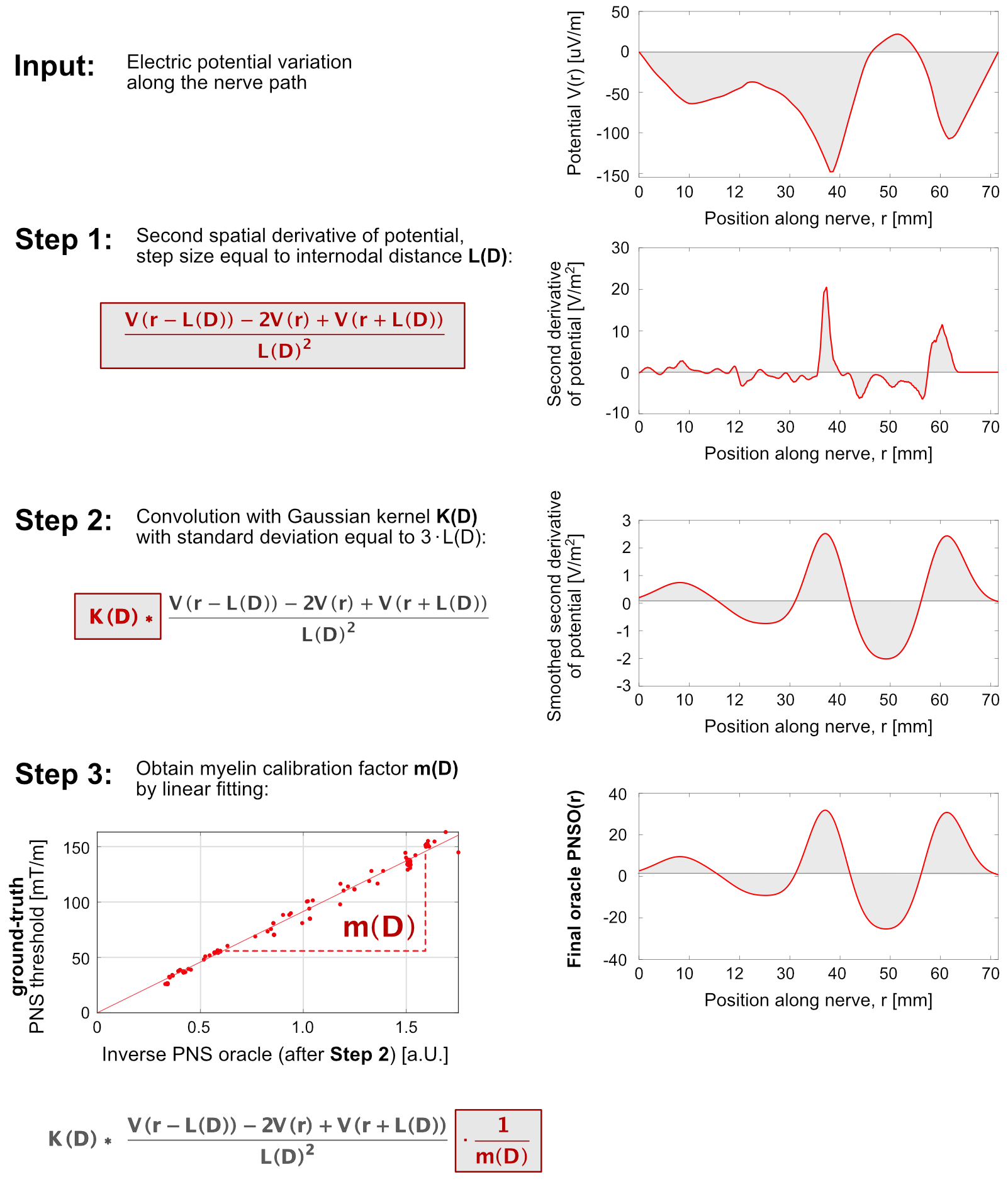
PNS oracle: To overcome these limitations, we propose a new metric that we call the “PNS oracle”:
$$\text{PNSO}(r,D)=K(D)\ast\frac{V(r-L(D))-2V(r)+V(r+L(D))}{L(D)^2}\cdot\frac{1}{m(D)}$$
Here $$$K(D)$$$ is a Gaussian smoothing kernel, $$$(\ast)$$$ denotes the convolution operator, $$$L(D)$$$ is the node spacing (a function of nerve diameter, $$$D$$$) and $$$m(D)$$$ is a calibration factor for myelin thickness (which is also a function of $$$D$$$). The PNS oracle differs from the NAF in three aspects: 1) it uses a step size equal to the distance between nodes of Ranvier (which is a function of the axon diameter [3,4]) for evaluation of the finite derivative. This improves the quantification of the net trans-membrane currents since almost all the voltage drop is across the node of Ranvier. 2) The oracle smooths the result by a Gaussian kernel with SD equal to $$$3\cdot L(D)$$$ to account for crosstalk between neighboring nodes of Ranvier. 3) The PNS oracle is weighted by a factor that models the increased excitability of large nerves. The process is outlined in Fig. 1.
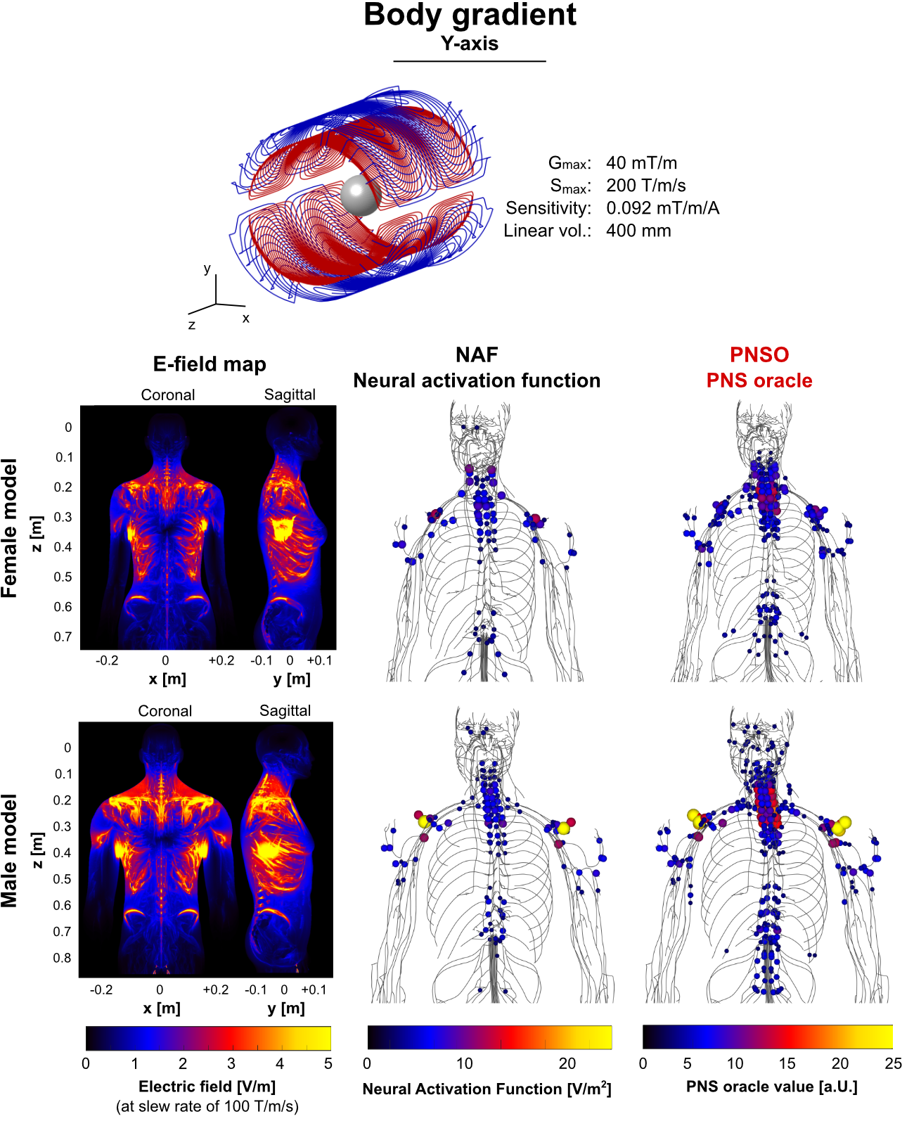
Calibration: We simulated PNS thresholds created in the female body model by an actively-shielded body gradient coil. We solved the neurodynamic model assigning axon diameters of 8 μm, 10 μm, 12 μm, 16 μm, or 20 μm to all nerves. This exhaustive simulation provided the “ground truth” against which the oracle was compared and to empirically determine the myelination calibration factor $$$m(D)$$$.
Results:
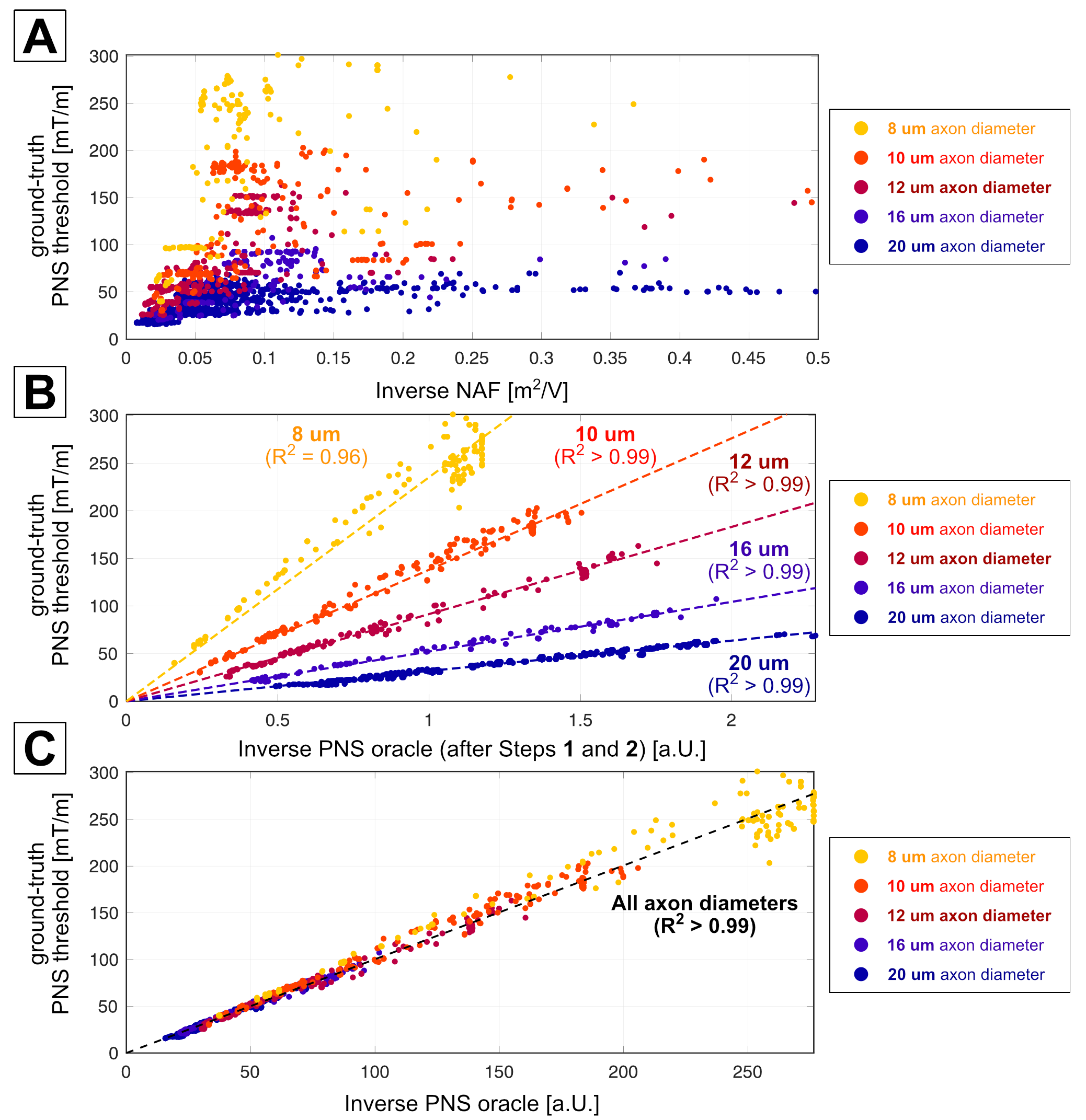
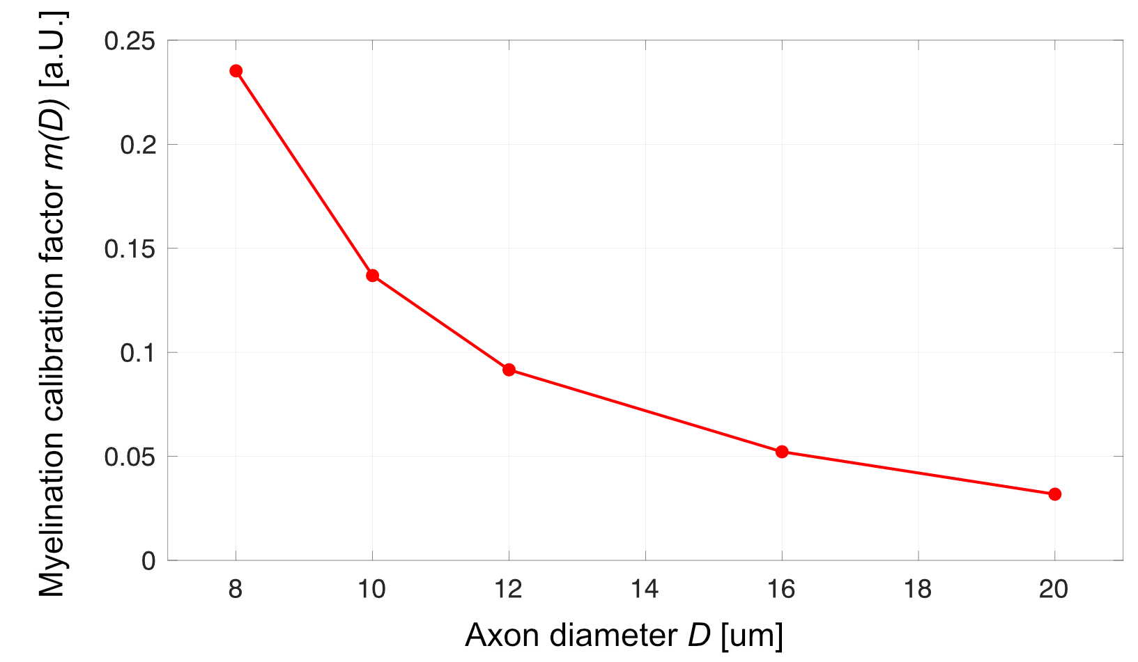
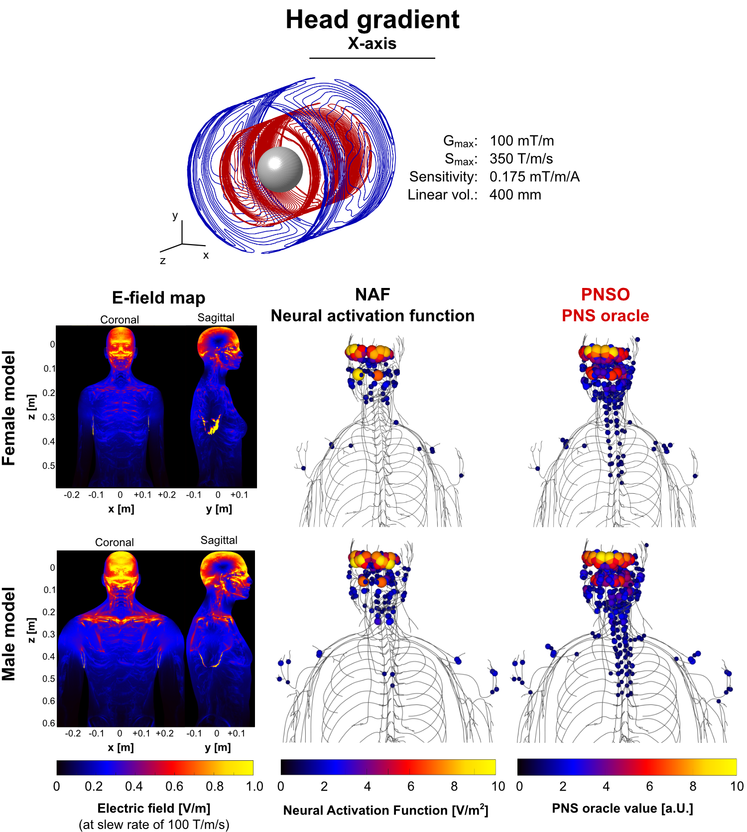
Figure 2A shows the poor correlation between the 1/NAF value and the neurodynamic determined threshold. Figure 2B shows the improvement after accounting for nodal crosstalk ($$$K(D)$$$) and the internodal-distance ($$$L(D)$$$). Now, the correlation is improved but clearly has a different slope for each axon diameter. The linear fit determines $$$m(D)$$$ and application of this yields the final PNS oracle shown in Fig. 2C. The correlation with the full modeling is good ($$$R^2$$$ > 0.99) with a slope of 1, so that the PNS threshold is simply given by 1/PNSO for the gradient efficiency under examination. Figure 3 show the plot of $$$m(D)$$$ determined from the simulations. Figures 4 and 5 show that the oracle generalizes well to the male body model and to other gradient coils.Conclusion:
We present a novel approach to quickly and accurately estimate PNS thresholds without the need to simulate the complex neurodynamics of the nerve. Our PNS oracle is derived from the potential changes along the nerve paths using only linear operations, which dramatically speeds up computation (factor x1000) and allows simple addition of pre-computed oracles associated with wire segments to formulate the total oracle for a gradient winding pattern. By design, the PNS threshold for the test waveform and given gradient efficiency is the inverse of the oracle.Acknowledgements
No acknowledgement found.References
[1] Davids et al., “Predicting magnetostimulation thresholds in the peripheral nervous system using realistic body models”, Sci. Rep. 7:5316, 2017
[2] Davids et al., “Prediction of peripheral nerve stimulation thresholds of MRI gradient coils using coupled electromagnetic and neurodynamic simulations”. Magn. Reson. Med., 2018
[3] McIntyre et al., “Modeling the excitability of mammalian nerve fibers: Influence of afterpotentials on the recovery cycle”. J Neurophysiol. 87(2), 2002
[4] Richardson et al., “Modelling the effects of electric fields on nerve fibres: Influence of the myelin sheath”. IEEE Trans. Bio. Eng. 38(4), 2000
[5] Basser et al., “The activating function for magnetic stimulation derived from a three-dimensional volume conductor model”. Medical and Biological Engineering and Computing. 39(11), 1992
Figures