0720
Mildronate Modulates In-Vivo Metabolism and Improves Ex-Vivo Functional Recovery Post-ischemia in the Diabetic Heart1University of Oxford, Oxford, United Kingdom, 2University of Cambridge, Cambridge, United Kingdom
Synopsis
Carnitine acts as a buffer of acetyl-CoA units in the mitochondria, as well as facilitating
Introduction
One of the leading causes of mortality in type I diabetic patients is cardiovascular disease1. Improving our understanding of the metabolic changes that occur in the diabetic heart may be key to understanding the pathophysiology of the cardiac effects of the disease. One key metabolic effect observed in the diabetic heart is an increase in fatty acid oxidation. The anti-ischemic drug, Mildronate has been shown to reduce fatty acid oxidation through inhibition of the mitochondrial fatty acid transporter, CPT1. Mildronate inhibits CPT1 by reducing both the biosynthesis of L-carnitine and its reabsorption in the kidneys2,3. In this study, the metabolic and functional effects of Mildronate treatment were investigated in the healthy and diabetic heart to observe if it could result in a metabolic shift away from fatty acid oxidation towards glucose oxidation, with potential functional benefits.Methods
36 male Wistar rats (~250g) were split into two groups. In one group, rats were injected with streptozotocin (STZ, 55mg/Kg) to induce a model of type 1 diabetes and in the other group, rats were injected with citrate buffer (CTR) to act as controls. Two weeks after STZ/citrate injection, daily intraperitoneal injections of either saline or Mildronate (100mg/kg/day) were initiated and continued for three weeks. Following treatment, the animals were anaesthetized with isoflurane and ECG gated, 13C MR pulse-acquire cardiac spectra were acquired over 60s following injections of hyperpolarised [1-13C]pyruvate and [2-13C]pyruvate (Injections separated by one hour; repetition time 1s; excitation flip angle 15°; sweep width 13,021Hz; acquired points 2,048; frequency centred on the C1 pyruvate resonance). Acquired spectra were summed over 30s from the first appearance of pyruvate and analysed with jMRUI for metabolic assessment of the heart4. In addition, eight-ten short-axis slices (slice thickness:1.6mm, matrix size:128×128, TE/TR:1.67/4.6ms, flip angle:15°, number of averages: 4) were acquired with a CINE-FLASH sequence and analysed with ImageJ for assessment of cardiac function. Following imaging, the hearts were excised and Langendorff perfused with an initial normal-flow for 20min, followed by 30min of low-flow (0.4ml/min) ischemia and re-perfused again with normal-flow for 20min as described by Heather et al.5Results
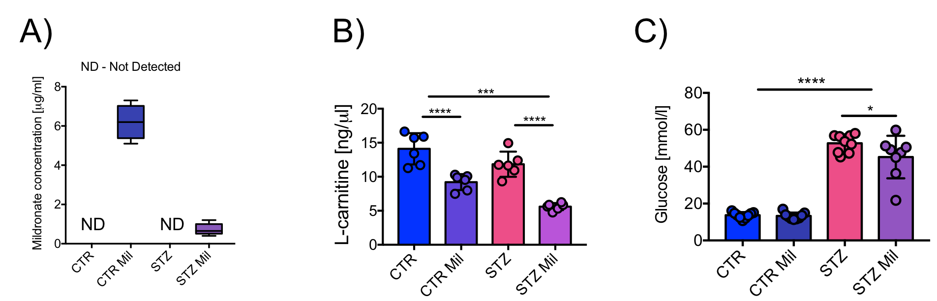
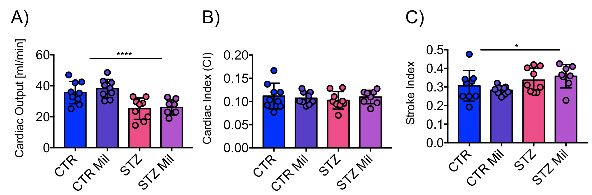
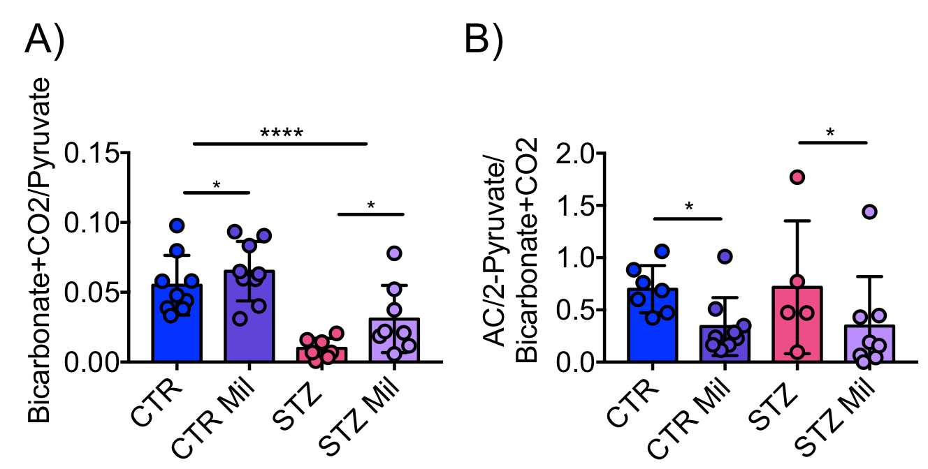
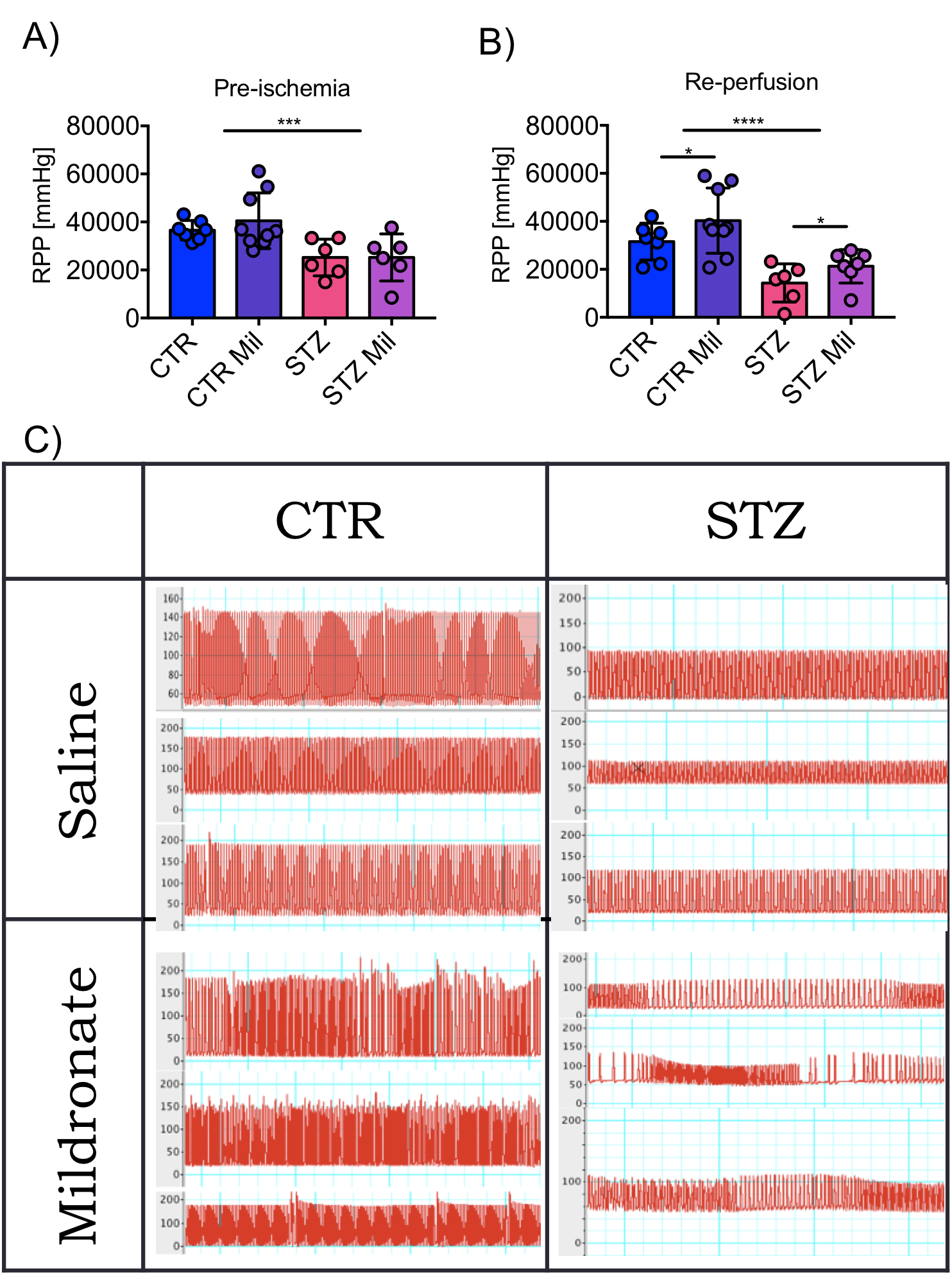
Mildronate was detected in the plasma of the control and diabetic rats treated with Mildronate but not in the saline-treated animals (Fig.1A). However, Mildronate was detected at much lower concentrations in the diabetic group compared to the control group. L-carnitine levels were significantly lower in the Mildronate treated control and diabetic groups (Fig.1B). Glucose levels were elevated in the diabetic groups, however, Mildronate reduced glucose levels slightly (p < 0.05) (Fig.1C).Cardiac output was decreased in both diabetic groups compared to the two control groups (Fig.2A), but when normalized to body weight there was no difference in cardiac index (Fig.2B), however stroke volume normalized to body weight showed an elevated stroke index in the two diabetic groups compared to the control groups with no significant differences observed with Mildronate treatment. Flux through pyruvate dehydrogenase (PDH), as indicated by the bicarbonate+CO2 to pyruvate ratio, was reduced in the two diabetic groups compared to the control animals. However, despite this reduction, PDH-flux was significantly higher with Mildronate treatment in both groups (Fig.3A). The incorporation of [2-13C]pyruvate into acetylcarnitine normalised to PDH flux reduced with Mildronate treatment in both the control and the diabetic groups (Fig.3B). In the Langendorff ischemia-reperfused protocol, the diabetic groups had lower rate pressure product (RPP) at both pre- and post-ischemia. However, Mildronate elevated RPP post-ischemia (p<0.05) in both the diabetic and control groups (Fig.4AB). Multiple irregular heartbeats were observed with Mildronate treatment post-ischemia compared to the saline-treated groups post-ischemia (Fig.4C).
Discussion and Conclusion
Mildronate was detected in higher concentrations in the control groups compared to the diabetic groups, likely due to elevated Mildronate excretion through the urine in the diabetic animals. Blood glucose levels were reduced with Mildronate treatment, but only in the diabetic group, which could have been related to lower food intake. No beneficial effect was observed with Mildronate on in-vivo cardiac function. However, despite a higher excretion of Mildronate in the diabetic group, an elevated PDH-flux was still observed, that was higher than the elevation of PDH-flux in the controls. This suggests that possibly these benefits could have been elevated even further if this group could have retained a higher Mildronate concentration. Incorporation of the 13C label into acetylcarnitine was reduced in the Mildronate treated groups, confirming the L-carnitine lowering effect of Mildronate as observed previously2,3. Finally, when perfused in a Langendorff ischemia-reperfusion protocol, the Mildronate treated groups recovered better post-ischemia, however despite this recovery, there were irregular heartbeats observed in the groups treated with Mildronate, that requires further investigation.Acknowledgements
We would like to thank our funding body; The Danish Research Council for Strategic Research (DNP-LIFE).References
1. Aaron M Secrest et al. "Mortality in Type 1 Diabetes" Diabetes Care, 33,2573-2579, 2010.
2. Maija Dambrova et al. "Mildronate: Cardioprotective Action Through Carnitine-Lowering Effect" TCM Vol 12, No. 6, 2002.
3. Edgars Liepinsh et al " Mildronate treatment alters ϒ-butyrobetaine and L-carnitine concentrations in healthy volunteers" JPP, 63, 1195-1201, 2011.
4. Vanhamme L, et al. "Improved method for accurate and efficient quantification of MRS data with use of prior knowledge." J Magn Reson 1997;129:35-43
5. Heather L et al. " Differential translocation of the fatty acid transport, FAT/CD36, and the glucose transporter, GLUT4, coordinates changes in cardiac substrate metabolism during ischemia and reperfusion" Circ. 6, 2013,1058-66.
Figures