0718
Hyperpolarized [1-13C]Pyruvate MRSI at 48 hrs post anti-VEGF Treatment Predicts Response in C6 Glioma Implanted Rats1Radiology, Stanford University, Stanford, CA, United States, 2Electrical Engineering, Stanford University, Stanford, CA, United States, 3Neurology, Stanford University, Stanford, CA, United States
Synopsis
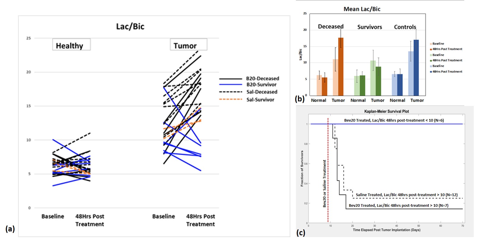
Using a bolus injection of hyperpolarized [1-13C]Pyruvate, we show that measurement of the resulting 13C-Lactate to 13C-Bicarbonate ratio 48-hours post-treatment with anti-angiogenic drug Bevacizumab (Bev20) predicts survival in a C6-Glioma rat model. A positive correlation of Lac/Bic with tumor growth rate further supports our hypothesis that the effect of the drug in survivors is to reverse the tumor Warburg metabolic phenotype necessary to support rapid proliferation.
Introduction
Tumor metabolism shifts from Oxidative Phosphorylation (OXPHOS) towards Glycolysis (Gly), even in the presence of oxygen, to utilize Glucose as biomass for proliferation (Warburg-Folkman effect1). Hyperpolarized [1-13C] Pyruvate (Pyr) imaging is an established technique for probing the underlying cancer metabolic processes via the measurement of surrogate bio-markers [1-13C]Lactate (Lac) and [1-13C]Bicarbonate (Bic) for Gly and OXPHOS respectively2. In this longitudinal study, we investigated whether a single time-point measurement of 13C Lac/Bic 48 hours after treatment with the anti-angiogenic drug Bevacizumab (Bev20) can be used to predict survival in a C6 Glioma rat model.Methods
Animal model: ~1x10⁶ N-methyl-N-nitrosourea (MNU) induced C6 glioma cells were injected into the right striatum of male Wistar rats (N=13, 246-290g) and imaged with hyperpolarized [1-13C]Pyruvate on day 10 to assess baseline metabolism. Subsequently, rats were treated with the anti-vascular endothelial growth factor (VEGF) monoclonal antibody B20-4.1.1 (Roche/Genentech, South San Francisco, CA, USA) administered intra-peritoneally (5mg/kg). To assess early treatment response, animals were again imaged using hyperpolarized [1-13C]Pyruvate 48-hours post-treatment. The animals were then monitored for up to 70 days after which rats were classified as “survivors” for those still alive versus “deceased” for those animals that had to be euthanized at an early time-point due to worsening of symptoms from the tumor. Saline treated male Wistar rats (N=12, 245-300g) were used as controls.
Substrate preparation: 54ul of 15.5M [1-13C] Pyruvic acid (Signa Aldrich) mixed with 15mM trityl radical AH11141(GE Medical systems) was polarized in SPINLab (GE) and dissolved with 16g of 40mM tris(hydroxymethyl)aminomethane, 125mM NaOH, 100mg/L ethylenediaminetetraacetic acid (EDTA) and 50mM NaCl, resulting in a 125mM pyruvate solution. 3ml of this solution was injected into the animal through a tail vein catheter and imaged in a clinical 3T scanner (GE Systems) 30s post injection to maximize the signal from Lac and Bic.
Proton & metabolic imaging: A custom-built birdcage coil tuned to the 1H resonance was used to acquire anatomical references and T2-weighted images (256x256, 2mm thick, 0.5mm in place resolution) for tumor localization. For the metabolic imaging of pyruvate and its metabolites, a custom-made 13C surface coil (dia=4cm) was used to acquire data from an axial 5mm slice of the brain centered on the tumor, using a 16x16 phase encoded free-induction decay sequence (fidcsi) resulting in a 4mm in-plane resolution metabolic image (5000Hz spectral bandwidth, N=256). Metabolite maps were generated by integrating the corresponding peaks in absorption mode after applying 15Hz line broadening and interpolating by a factor of 4 in both spectral and spatial dimensions. Contrast-enhanced T1-weighted proton images were also collected post-hyperpolarized imaging to confirm the tumor location.
Results and Discussion
The tumor Lac/Bic ratio measured 48-hours-post treatment was highly predictive of survival in this C6 glioma model (Fig. 1). Using a Kaplan-Meier survival analysis (Fig. 1c), tumor Lac/Bic ratios below 10 were highly predictive of survival (p=0.03), with 100% survival of Bev20 treated animals with 48-hrs Lac/Bic <10. No correlation was observed between survival and tumor volume (Fig. 2), and the Lac/Bic ratio did not correlate with tumor size at either the baseline or 48-hr time points. However, there was a significant correlation (r=0.7) between the 48-hr Lac/Bic ratios and the rate of change in tumor volume (as measured by the differential volume observed on Gd-enhanced MRI at baseline verses 48-hrs). This positive correlation of 48-hr tumor 13C Lac/Bic ratios with tumor growth rate supports our hypothesis that the effect of the drug in survivors is to reverse the tumor Warburg metabolic phenotype necessary to support rapid proliferation1.Conclusion
In this longitudinal survival study of C6-Glioma implanted rats treated with anti-VEGF Bev20, we show that a single hyperpolarized MRSI measurement of the 13C Lac/Bic ratio at 48-hours post-treatment is predictive of survival. If the same trend is observed in humans, hyperpolarized [1-13C]Pyr imaging could provide a vital tool for evaluating disease progression and treatment response in brain tumor patients.Acknowledgements
The Lucas Foundation, GE Healthcare, and NIH grants R01 CA17683603, R01 EB01901802, and P41 EB01589121. We would like to thank Dr Jarrett Rosenberg and Dr Tie Liang for their help with statistical analysis.References
1. Piia Thomas, Daniel Spielman and Lawrence Recht (2011). The Bevacizumab “Pseudoresponse” in Glioma: Disappointment or Opportunity?, Brain Tumors - Current and Emerging Therapeutic Strategies, Dr. Ana Lucia Abujamra (Ed.), ISBN: 978-953-307-588-4, InTech, Available from: http://www.intechopen.com/books/braintumors-current-and-emerging-therapeutic-strategies/the-bevacizumab-pseudoresponse-in-gliomadisappointment-or-opportunity
2. Park JM, et al. Hyperpolarized 13C-lactate to 13C-bicarbonate ratio as a biomarker for monitoring the acute response of anti-vascular endothelial growth factor (anti-VEGF) treatment. NMR Biomed. 2016 May;29(5):650-9.
Figures