0716
First in cellulo and in vivo metabolic studies using ParaHydrogen Hyperpolarized [1-13C]pyruvate1Torino University, Torino, Italy
Synopsis
Hyperpolarized (HP) [1-13C]pyruvate [1] has been widely exploited for the metabolic processes. The development of this agent has enabled the in vitro and in vivo real-time detection of pyruvate-lactate metabolic conversion. The possibility of obtaining HP pyruvate using the cost effective and fast PHIP (ParaHydrogen Induced Polarization) method, instead of the dissolution Dynamic Nuclear Polarization (d-DNP), would allow a widespread application of this powerful diagnostic tool, that so far has been hampered by cost, technical complexity and intrinsically low polarization time. Here we report an in vitro and in vivo study carried out on different cancer cell lines and in vivo using PHIP-SAH HP-[1-13C]pyruvate.
Introduction
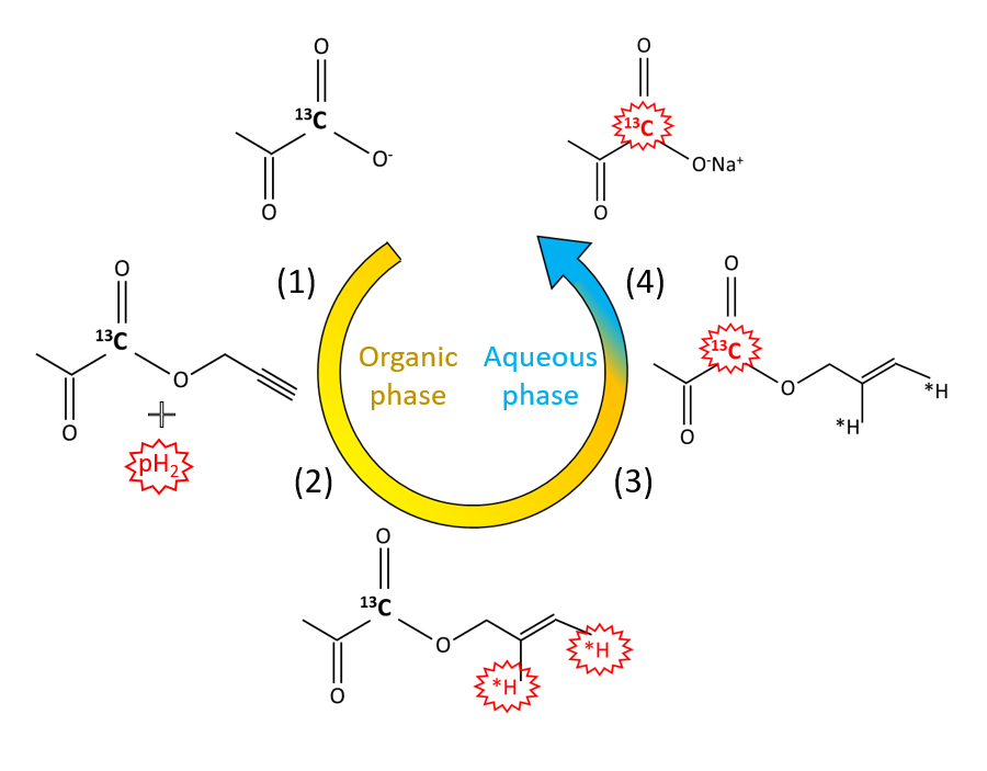
PHIP is a method for the generation of HP molecules that requires much more affordable equipment than d-DNP and it owns the great advantage that the polarization cycle takes only few seconds. The recently introduced PHIP-SAH strategy (PHIP by means of Side Arm Hydrogenation) [2] allowed to hyperpolarize pyruvate, and other metabolites, that, previously, could be obtained only by d-DNP.Methods
The [1-13C] pyruvate was hyperpolarized by means of PHIP-SAH and added into cells suspension (at the concentration of 5.0 ± 0.2 mM), or injected through the tail vein (at the concentration of 48.1 ± 6.9 mM) of healthy and diseased mice.
MRS acquisition allowed the investigation of the kinetics of the metabolic exchange of the 13C HP label with lactate.
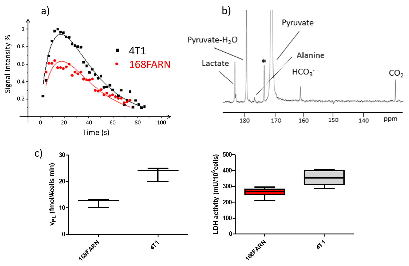
In the in vitro studies, a series of 13C-NMR spectra were acquired after addition of the HP dose to the suspension of cancer cells (at 600 MHz). By using a new perfusion set-up, less than 107 cells were sufficient for each experiment.
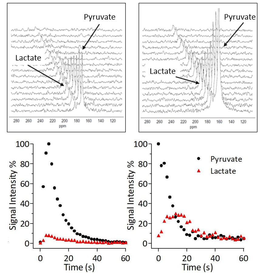
13C dynamic studies were performed and space-selective 13C-MR spectra acquired on voxel centred on the tissue of interest. Mice were anesthetized, catheterized through the tail vein and placed in 1 T (Aspect) or 3 T (Bruker Biospec) MRI scanner. The buffered solution containing HP pyruvate was injected in about 4-5 seconds and series of 13C-MR spectra were acquired, using small flip angle pulses and a tailored space-selective sequence.
Results
The 13C signal enhancement observed for the carboxylate moiety of [1-13C]pyruvate resulted to be 5x10⁴ times higher with respect to thermal polarization at 1 T, corresponding to a polarization level of 4.5 ± 0.5%. 13C hyperpolarization were back-calculated at time zero, its nascent value would have been 10.6 ± 1.5 %. In order to obtain a bio-compatible aqueous solution of the HP metabolite, we succeeded to avoid the hydrogenation co-solvent (ethanol), by modifying the hydrogenation procedure.
The improvement of the quality the used aqueous solution allowed to enhance the accuracy of our metabolic investigation, compared to previously reported results [3].
Different cancer cells lines were compared, characterized by a different degree of aggressiveness and ability to induce metastasis (Breast cancer cells: 4T1-168FARN, Prostate cancer cells: LnCap, DU145 and PC3).
In the in vivo experiments the 13C-MR spectra were acquired on a voxel centred on the tissue of interest to assess the metabolic build-up of lactate. Kinetic analysis of the metabolic exchange of the 13C label between [1-13C]pyruvate and [1-13C]lactate were carried out by monitoring the time courses of the signals of hyperpolarized lactate and pyruvate. The metabolic response of a control healthy mouse was compared to the one obtained in analogous experiments with tumour-bearing animals. As expected the 13C HP label exchange rate between pyruvate and lactate is markedly higher in the tumour bearing mice.
Discussion
Cancer cells exhibit a unique metabolic program characterized by an increased pyruvate-lactate conversion and at the same time a reduction in mitochondrial metabolism. The observed boost in the pyruvate-lactate conversion can be accounted for in terms of the disease progression and provides useful insights on the relative of the investigated tumour cells.Conclusion
This study has shown that in vitro and in vivo investigations of cancer metabolism, based on the administration of a dose of HP [1-13C]pyruvate obtained from the PHIP-SAH procedure is possible. The results obtained from PHIP-SAH hyperpolarized pyruvate are consistent with those already reported in the literature. Consequently, one may expect that the easy access to HP pyruvate by PHIP-SAH methodology will prompt new comers to the use of this powerful metabolic imaging tool that was previously available only using d-DNP technique.Acknowledgements
Airc (Italian Association for Cancer Research, TRIDEO call 2015)and Compagnia di San Paolo (Athenaeum Research 2016, n.CSTO164550) are gratefully acknowledged for financial support.References
[1] Ardenkjaer-Larsen, J. H. et al. Increase in signal-to-noise ratio of > 10,000 times in liquid-state NMR. Proc. Natl. Acad. Sci. U. S. A. 100, 10158–63 (2003)
[2] Reineri, F., Boi, T. & Aime, S. ParaHydrogen Induced Polarization of 13C carboxylate resonance in acetate and pyruvate. Nat. Commun. 6, 5858 (2015).1] Gallagher, F. A., Kettunen, M. I. & Brindle, K. M. Biomedical applications of hyperpolarized 13C magnetic resonance imaging. Prog. Nucl. Magn. Reson. Spectrosc. 55, 285–295 (2009)
[3] Cavallari E., Carrera C., Sorge M., Bonne G., Muchir A., Aime S., Reineri F., “The 13C hyperpolarized pyruvate generated by ParaHydrogen detects the response of the heart to altered metabolism in real-time”, Scientific Reports, May 2018.
Figures