0711
Surface-based diffusion MRI analysis of the superficial white matter in autism: association with functional networks and symptom severity1Multimodal Imaging and Connectome Analysis Lab, Montreal Neurological Institute, Montreal, Quebec, QC, Canada, 2Center for the Developing Brain, Child Mind Institute, New York, NY, United States
Synopsis
Our study focused on a region that remains relatively unexplored by autism neuroimaging: the superficial white matter (SWM) immediately beneath the cortical interface, a compartment known to play a key role in corticogenesis and connectivity. A surface-based diffusion-MRI analysis revealed SWM anomalies in a multicentric ASD cohort compared to neurotypical controls in medial parietal and
Introduction
Autism spectrum disorders (ASD) are neurodevelopmental conditions characterized by atypical social cognition, communication, as well as repetitive behaviors and interests 1. Multiple neuroimaging studies have shown alterations in cortical morphology in ASD 2. White matter changes remain less well understood, particularly their association to cortical structure and function. The current study focused on a compartment that has gained only little attention in ASD neuroimaging so far: the superficial white matter (SWM) immediately beneath the cortical interface. In addition to its role in genesis and maturation of the folded cortex 3, 4, its spatial proximity ensures intrinsic correspondence to the cortical ribbon, making it an ideal candidate for integrative studies on cortical grey matter morphology, function, and white matter organization in ASD. Notably, the SWM harbours both short- and long-range fibers mediating cortico-cortical connectivity 5-8, likely contributing to functional network alterations and behavioral symptomatology in ASD.Methods
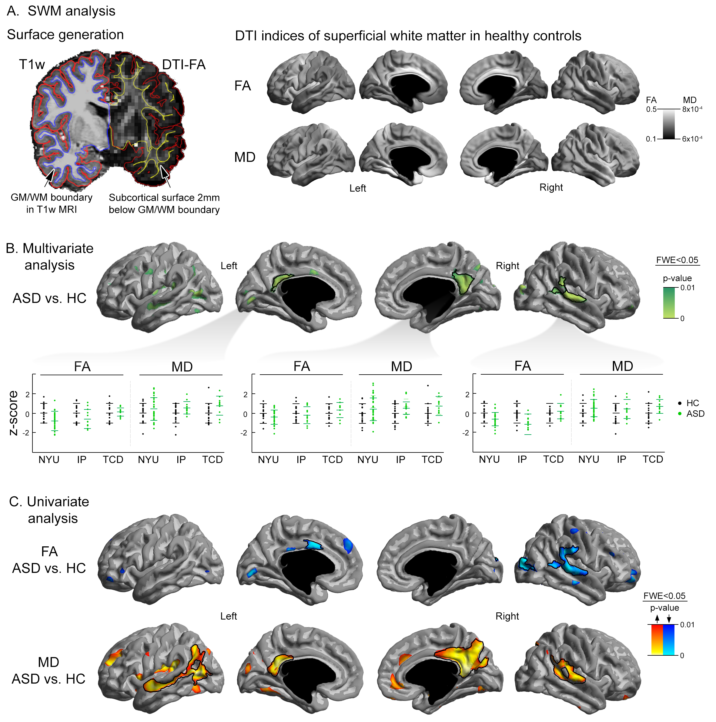
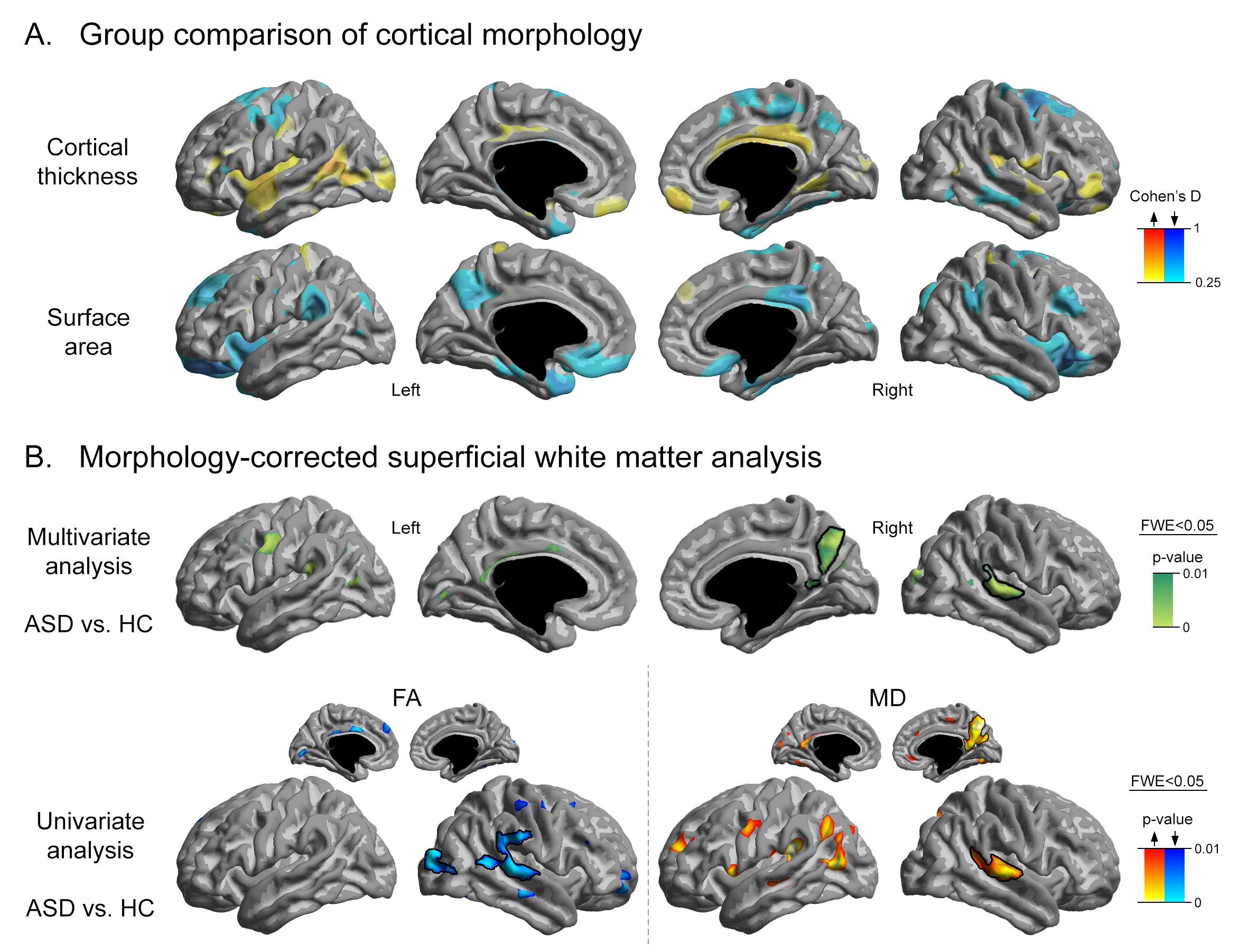
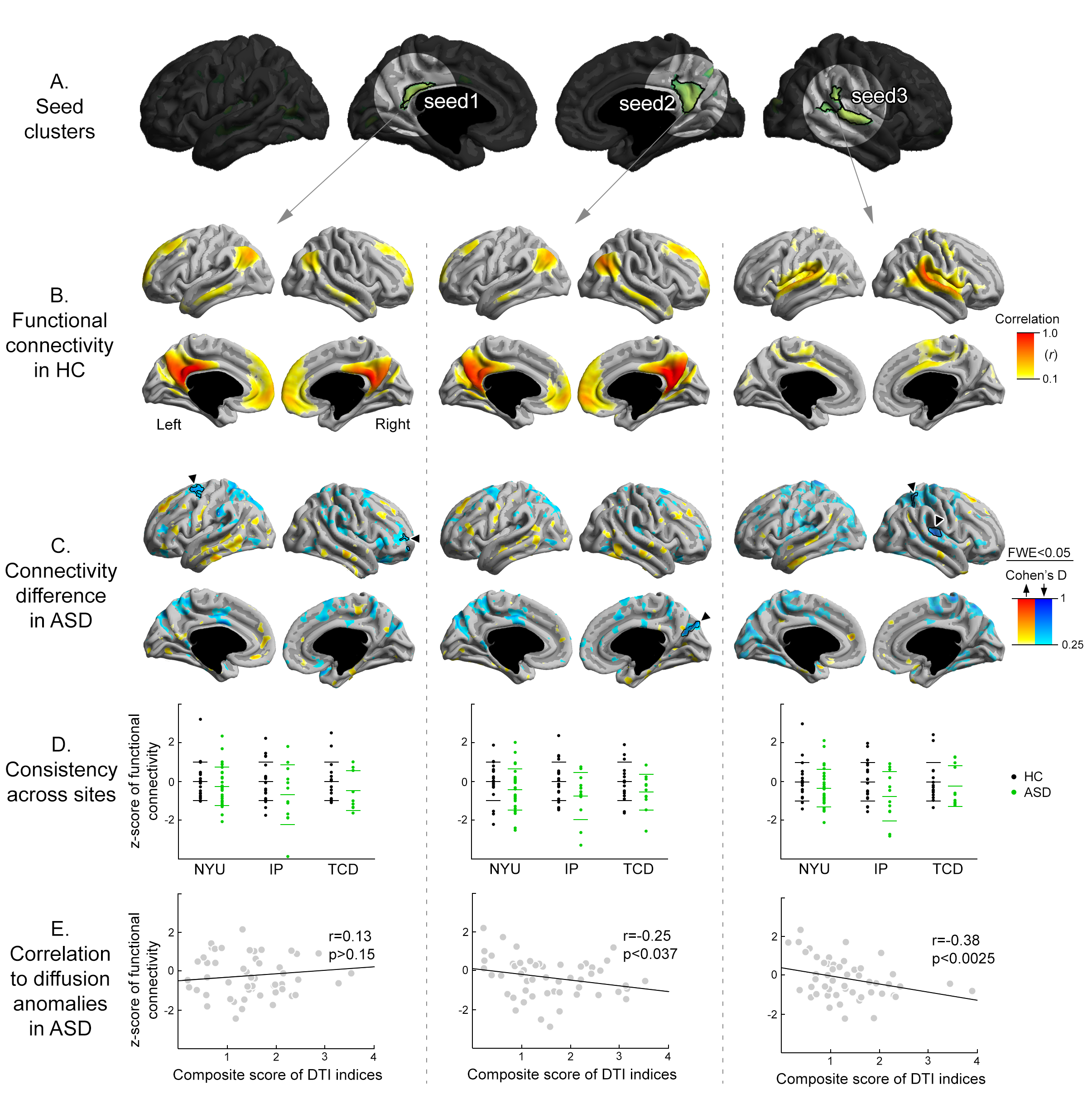
From the Autism Brain Imaging Data Exchange repository, we selected three sites (i.e., IP, NYU, TCD) that contained: i) males and females, ii) children and adults, and iii) diffusion MRI and T1-weighted MRI data. Following cortical surface extraction and QC, our sample included 53 ASD and 57 controls. To examine the SWM, we generated a surface running 2mm below the cortex by following a Laplacian potential field towards the ventricles (Fig 1A). Surface-wide statistics mapped alterations in fractional anisotropy (FA), mean diffusivity (MD), and a multivariate aggregate of both in ASD compared to controls, controlling for age, sex, and site. To rule out confounds of cortical morphological variations, we corrected for cortical thickness at each cortical point and carried out seed-based functional connectivity analyses from clusters of significant diffusion findings. To examine associations to behavioral symptoms of ASD, we correlated inter-individual differences in SWM measures with ADOS scores in ASD and finally built path analytical models, evaluating the role of functional connectivity on the relation between diffusion anomalies and ADOS scores. Surface-based analyses were corrected using random-field theory for non-isotropic images 9 with a threshold of family-wise error of pFWE<0.05 (cluster defining threshold, CDT=0.01).Results
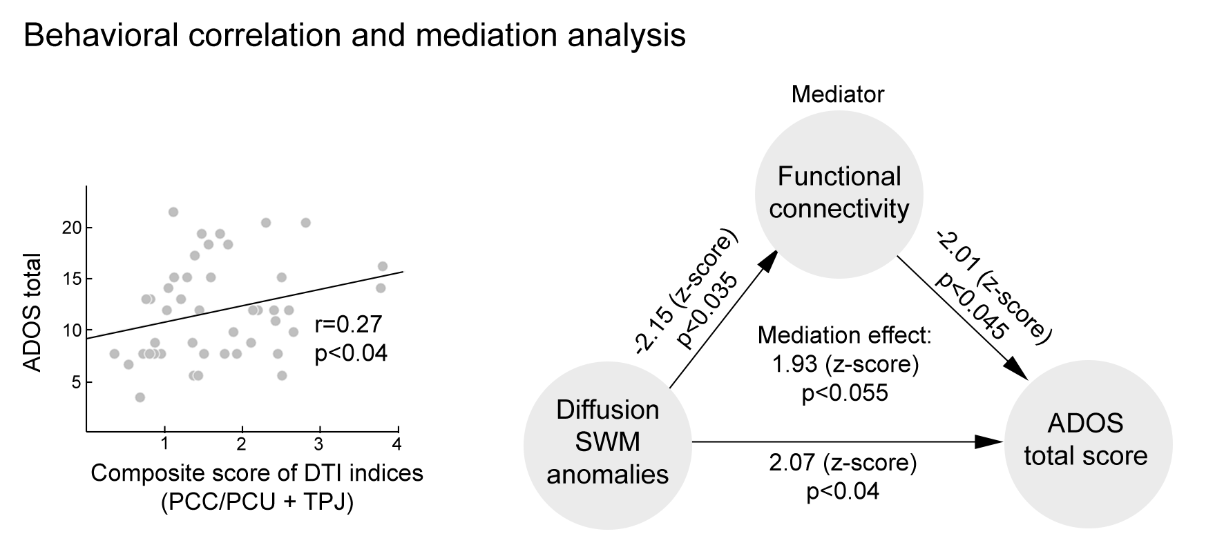
Multivariate analysis mapped SWM diffusion anomalies in ASD compared to controls (Fig 1B) in bilateral medial parietal cortices (i.e. precuneus and posterior cingulate [PCU/PCC]) and the right temporo-parietal cortex). Effects were similar in children and adults, and findings were consistent across sites. Importantly, although the extent of SWM anomalies in ASD was reduced after controlling for cortical thickness and surface area at each cortical vertex (Fig 2), effects in right hemisphere clusters persisted after correcting for these morphological variations, specifically in the right PCC/PCU and TPJ clusters. Notably, functional connectivity analysis from these regions revealed decreased intrinsic connectivity in ASD compared to controls, with right PCC/PCU being disconnected from adjacent cuneus (Fig 3A-C). Right temporoparietal cortex also showed reduced connectivity to rather proximal regions, including insular and superior parietal cortex. Again, connectivity reductions were similar across sites (Fig 3D). Notably, in the ASD group, inter-individual differences in SWM anomalies in both right hemispheric clusters correlated with overall degrees of functional connectivity reductions to those target areas (i.e., adjacent cuneus, and the posterior insula, and superior parietal cortex). In other words, individuals with ASD who showed more marked SWM anomalies (defined by multivariate composite scores of FA and MD) also displayed more marked reductions in functional connectivity (SWMTPJ: r=-0.38, p<0.0025; SWMPCC/PCU: r=-0.25, p<0.037, Fig 3E). Overall SWM profile (averaged score of SWMTPJ and SWMPCC/PCU) was also associated to more severe ASD symptoms, as indexed by total ADOS (r=0.27, p<0.04). Separate univariate analyses suggested that results were mainly driven by FA (r=-0.24, p=0.054), not MD (p>0.4). Finally, correlations between SWM anomalies and total ADOS scores were found to be partially mediated by reduced functional connectivity (z=1.93, p<0.06), suggesting a disease-related path between SWM alterations, functional connectivity reductions, and behavioral symptoms (Fig 4). Analysis of specific ADOS subscales revealed that the observed mediation was mainly attributable to social cognition (mediation effects z=1.84, p<0.07 for social cognition; p>0.1 for communication and repeated behavior domains).Discussion
Our study targeting the SWM in ASD offers a novel perspective on the interplay between white matter anomalies and atypical functional networks, providing potentially important evidence to better understand the complex biological factors contributing to its diverse behavioral symptoms.Acknowledgements
No acknowledgement found.References
1. Robertson CE, Baron-Cohen S. Sensory perception in autism. Nat Rev Neurosci 2017; 18(11): 671-684.
2. Courchesne E, Mouton PR, Calhoun ME, Semendeferi K, Ahrens-Barbeau C, Hallet MJ et al. Neuron number and size in prefrontal cortex of children with autism. JAMA: the journal of the American Medical Association 2011; 306(18): 2001-2010.
3. Toro R, Burnod Y. A Morphogenetic Model for the Development of Cortical Convolutions. Cerebral Cortex 2005; 15(12): 1900-1913.
4. Herculano-Houzel S, Mota B, Wong P, Kaas JH. Connectivity-driven white matter scaling and folding in primate cerebral cortex. Proc Natl Acad Sci U S A 2010; 107(44): 19008-19013.
5. Schüz A, Braitenberg V. The Human Cortical White Matter: Quantitative Aspects of Cortico-Cortical Long-Range Connectivity, Cortical areas: unity and diversity. Taylor & Francis: London; New York, 2002.
6. Parent A, Carpenter MB. Carpenter's human neuroanatomy. 9 edn. Williams & Wilkins: Baltimore, MD, 1996.
7. Oishi K, Zilles K, Amunts K, Faria A, Jiang H, Li X et al. Human brain white matter atlas: identification and assignment of common anatomical structures in superficial white matter. NeuroImage 2008; 43(3): 447-457.
8. Oishi K, Huang H, Yoshioka T, Ying SH, Zee DS, Zilles K et al. Superficially located white matter structures commonly seen in the human and the macaque brain with diffusion tensor imaging. Brain connectivity 2011; 1(1): 37-47.
9. Worsley K, Andermann M, Koulis T, MacDonald D, Evans A. Detecting changes in nonisotropic images. Human brain mapping 1999; 8(2-3): 98-101.
Figures