0690
Water/fat separation for distortion-free EPI with point spread function encoding1Center for Biomedical Imaging Research, Department of Biomedical Engineering, School of Medicine, Tsinghua University, Beijing, China, 2Philips Healthcare, Beijing, China
Synopsis
Effective removal of chemical-shift artifacts in EPI is a challenging problem especially with severe field inhomogeneity. In this study, water/fat separation thus fat suppression is achieved using the intrinsic signals of point spread function (PSF) encoded EPI (PSF-EPI), in which chemical-shift encoding is realized in the intermediate images with different time shifts. The results from three imaging regions show that this PSF-EPI based method can separate water/fat robustly. Thus fat signals can be effectively removed from EPI even with severe field inhomogeneity.
Introduction
Echo-planar imaging (EPI), especially single-shot EPI, suffers from T2* blurring and geometric distortions. These problems can be addressed by the PSF-EPI technique 1,2, which has the potential for practice use via tilted-CAIPI 3. With the distortion problem being well addressed, the large chemical-shift of fat along the phase encoding (PE) direction is another challenge in EPI, which may obstruct important structures and affect quantification analysis. Commonly used fat suppression techniques, such as SPIR 4, SPAIR 5, STIR 6 may fail in regions with severe field inhomogeneities. Water/fat separation is an effective fat suppression strategy, and water- and fat-only images can provide additional valuable information to biomedical research and clinical diagnosis of diseases. In EPI, water/fat separation is complicated by the large fat shift, and additionally by physiological motion in EPI-DWI. Current studies on water/fat separation for EPI-DWI still suffer from the distortion problem and the long acquisition time resulted from multiple acquisitions with echo shifts, especially for multi-shot EPI 7,8. In this study, water/fat separation thus fat suppression is achieved in PSF-EPI using the intrinsic multi-echo signals of PSF-EPI without extra scans.Methods
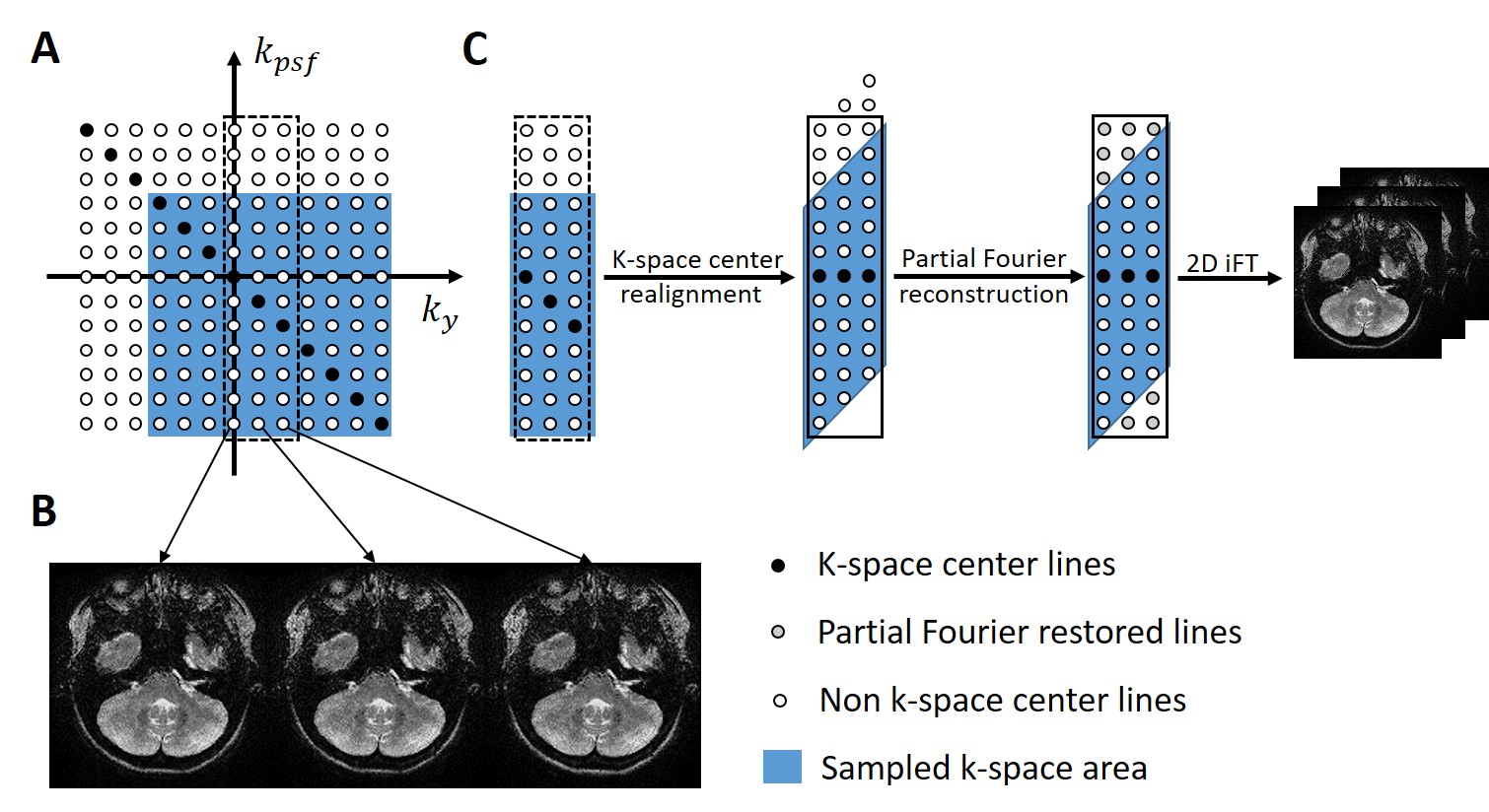
(1) Echo-shifts in PSF-EPI: The 3D k-space data in PSF-EPI consists of frequency encoding ($$$k_x$$$), phase encoding ($$$k_y$$$) and PSF encoding ($$$k_{psf}$$$) directions. Taking a gradient-echo PSF-EPI as an example, when considering the chemical-shift encoding, the distortion-free 2D image $$$I_m$$$ at $$$k_y = k_m$$$ can be expressed as: $$I_m=(ρ_w+ρ_Fe^{-i2πf_F(t_0+m∆t)})e^{-(t_0+m∆t)⁄T_2^*}e^{-i\overrightarrow{k}_m\overrightarrow{y}}e^{-iγ∆B(t_0+m∆t)}.$$Here $$$m$$$ is the index of $$$k_y$$$ which starts at 0 for $$$k_y = -k_{ymax}$$$; $$$ρ_w$$$ is the intensity of the water component; $$$ρ_F$$$ is the intensity of the fat component with a frequency shift $$$f_F$$$ (in Hz); $$$t_0$$$ is the echo time of the first echo; $$$∆t$$$ is the echo spacing (ESP) along $$$k_y$$$; $$$e^{-i2πf_F(t_0+m∆t)}$$$ is the chemical-shift encoding term; $$$e^{-(t_0+m∆t)⁄T_2^*}$$$ is the signal decay term; $$$e^{-i\overrightarrow{k}_m\overrightarrow{y}}$$$ is the phase modulation term resulted from PSF encoding gradient; $$$e^{-iγ∆B(t_0+m∆t)}$$$ is the susceptibility-induced phase accumulation, where $$$∆B$$$ is the B0 inhomogeneity. The phase modulation term can be eliminated by shifting the k-space along $$$k_{psf}$$$. Then the remaining phase contains only chemical-shift encoding and B0 inhomogeneity information. Thus 2D images at different $$$k_y$$$ positions can be used for water/fat separation after preprocessing. Taking the three echoes at $$$k_y = 0, 1, 2$$$ as examples (Fig. 1A), the preprocessing includes three steps (Fig. 1C): firstly the k-space center lines are realigned to remove the phase modulation term; secondly partial Fourier reconstruction, such as POCS 9, is conducted to restore the missing lines; then echo-shifted images can be calculated by 2D inverse Fourier transform (iFT).
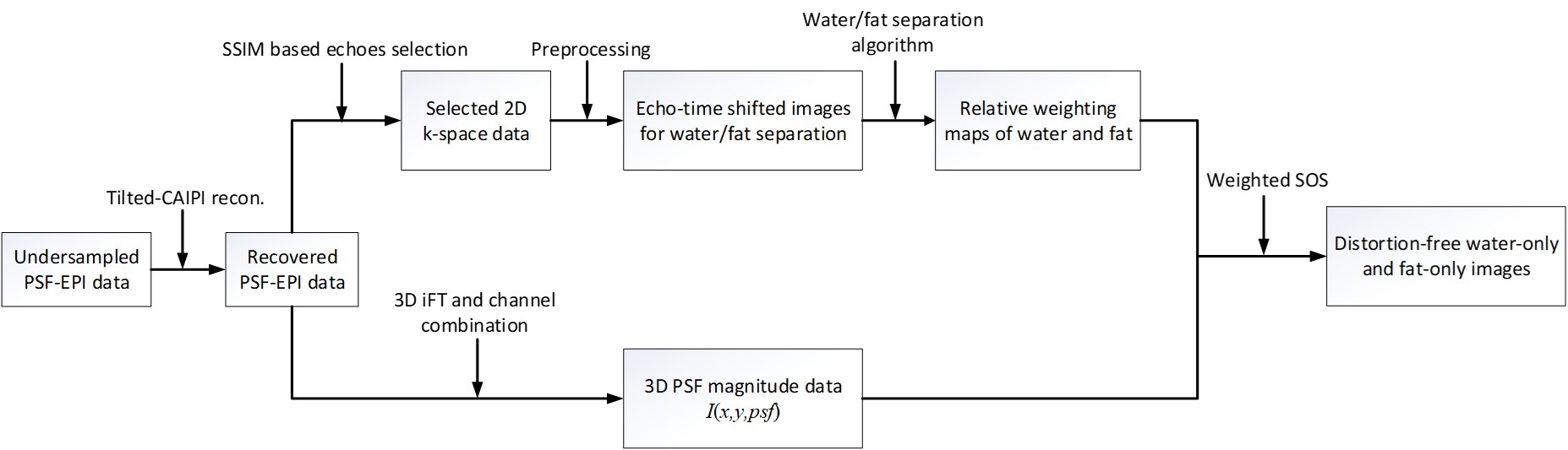
(2) PSF-EPI based water/fat separation: The flowchart of the proposed PSF-EPI based water/fat separation is shown in Fig. 2. After the tilted-CAIPI reconstruction of undersampled PSF-EPI data, multi-echo 2D k-space (three echoes or more) data are selected based on structural similarity index (SSIM) 10 to choose echoes with high SNR and fewer aliasing artifacts. After the preprocessing of these echoes, commonly used algorithms, such as QPBO 11, can be adopted to get the relative weight of water and fat in each voxel. Then the weights are used to combine the 3D image data along $$$k_y$$$ direction via weighted sum-of-square to achieve distortion-free water- and fat-only images.
(3) Experiments: Data for evaluation were acquired on a Philips 3T scanner (Philips Healthcare, Best, The Netherlands), including T1-, T2-weighted brain images, DW head-neck images and cervical spine DWI using PSF-EPI. The PSF-EPI was accelerated in both PE and PSF encoding directions, and the acceleration factors are denoted by $$$R_{PE}$$$ and $$$R_{PSF}$$$, respectively. This study was approved by the Institutional Review Board and written informed consent was obtained from all the participants.
Results and Discussion
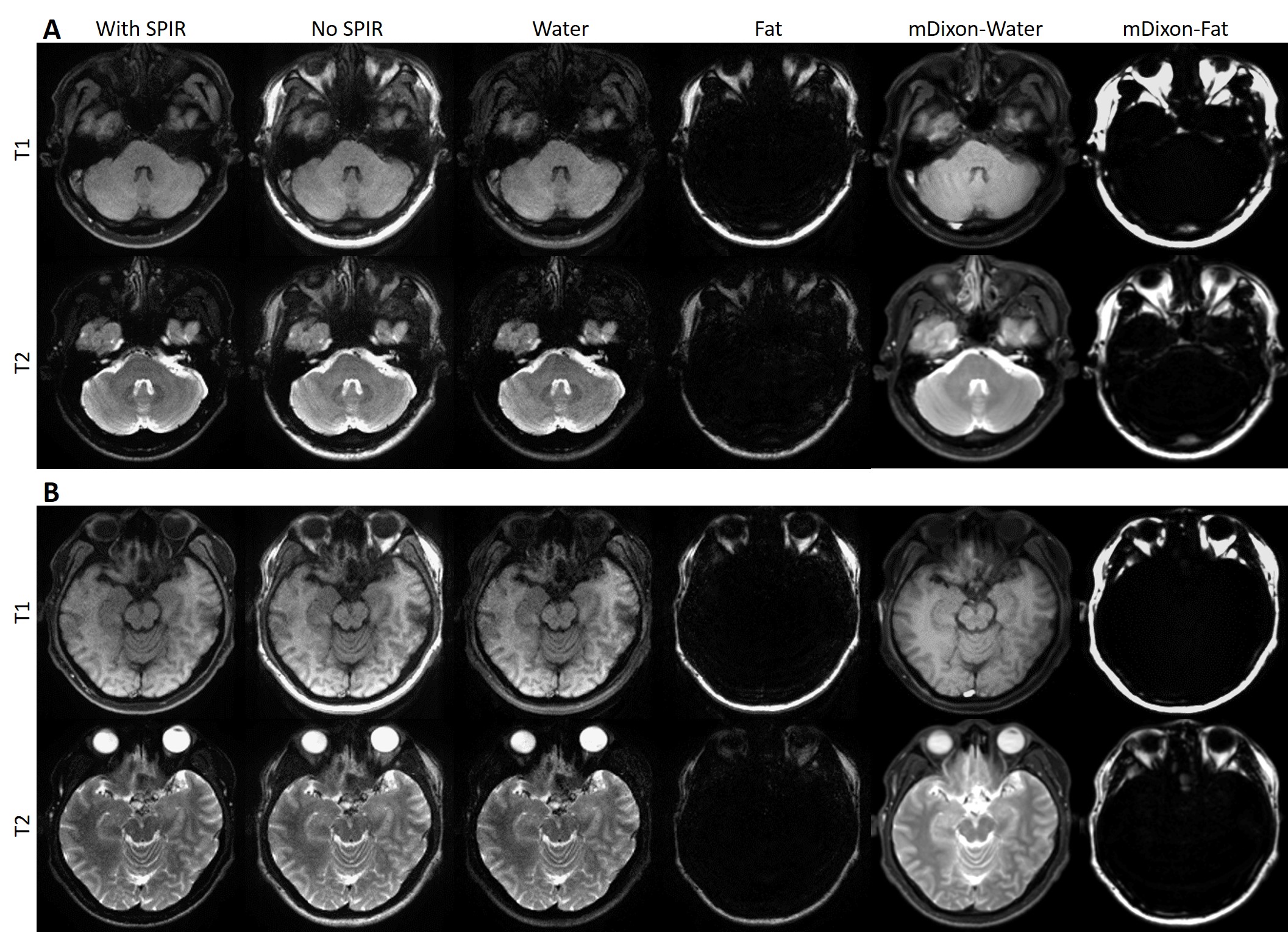
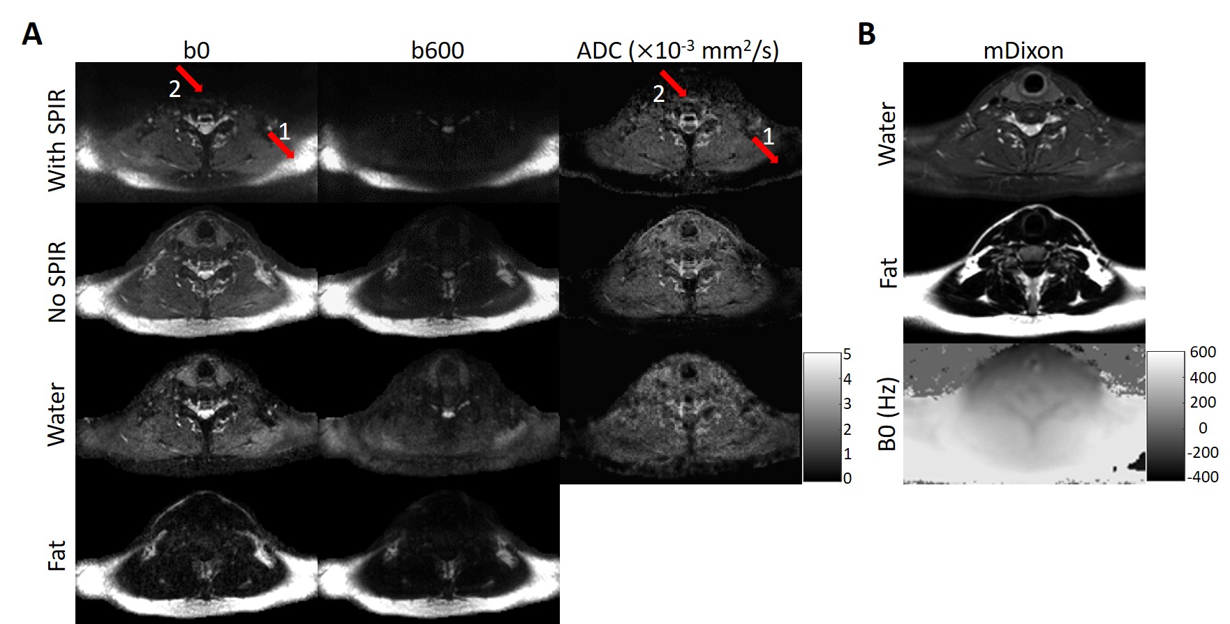
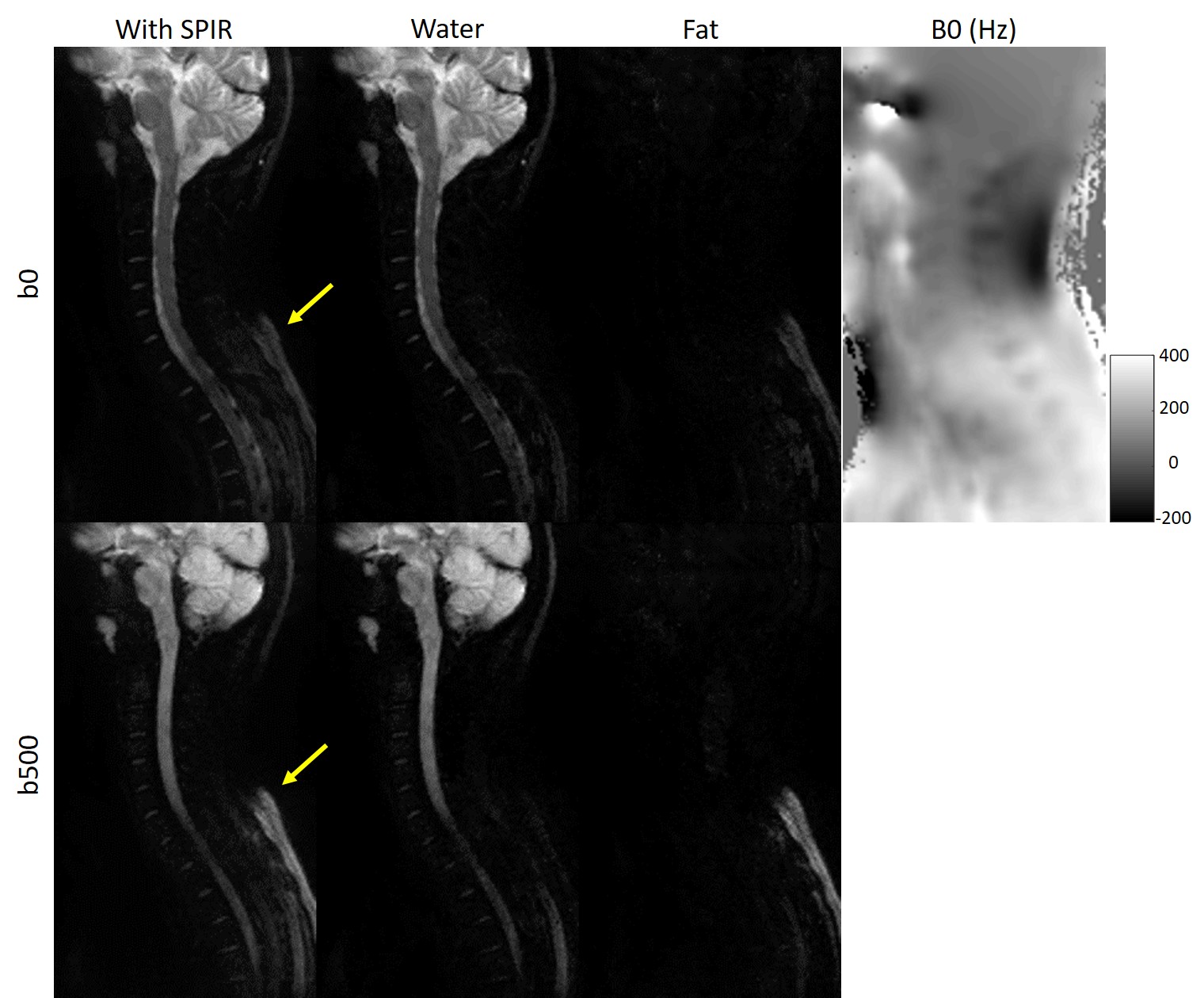
Fig. 3 shows the water/fat separation results of T1- and T2-weighted images using proposed method. Compared with the images with SPIR and mDixon results, no water/fat swapping is observed. Fig. 4 shows the results of head-neck DWI, where imaging suffers severe field inhomogeneity and fat-suppression is very challenging. The unsaturated fat can be successfully separated and the ADC maps show more details in these regions. Fig. 5 shows the application of proposed method in cervical spine DWI, where residual fat can also be successfully removed. These results all indicate that the proposed method can separate fast water/fat and remove fat residues robustly using the intrinsic multi-echo acquisitions of PSF-EPI without extra scans.Conclusion
This PSF-EPI based method can separate water/fat robustly, thus fat signals can be effectively suppressed in EPI even with severe field inhomogeneity. It can be extended to more image contrasts. The distortion-free PSF-EPI technique has the potential to provide anatomical and functional images with high-fidelity and practical acquisition efficiency.Acknowledgements
No acknowledgement found.References
1. Robson MD, Gore JC, Constable RT. Measurement of the point spread function in MRI using constant time imaging. Magnetic resonance in medicine 1997;38(5):733-740.
2. In MH, Posnansky O, Speck O. High-resolution distortion-free diffusion imaging using hybrid spin-warp and echo-planar PSF-encoding approach. NeuroImage 2017;148:20-30.
3. Dong Z, Wang F, Reese TG, Manhard MK, Bilgic B, Wald LL, Guo H, Setsompop K. Tilted-CAIPI for highly accelerated distortion-free EPI with point spread function (PSF) encoding. Magnetic resonance in medicine 2018.
4. Oh C. Selective partial inversion recovery (SPIR) in steady state for selective saturation magnetic resonance imaging (MRI). Abstr of Society of Magnetic Resonanc in Medicine, San Francisco 1988;1042.
5. Kaldoudi E, Williams SC, Barker GJ, Tofts PS. A chemical shift selective inversion recovery sequence for fat-suppressed MRI: theory and experimental validation. Magnetic resonance imaging 1993;11(3):341-355.
6. Krinsky G, Rofsky NM, Weinreb JC. Nonspecificity of short inversion time inversion recovery (STIR) as a technique of fat suppression: pitfalls in image interpretation. AJR American journal of roentgenology 1996;166(3):523-526.
7. Hernando D, Karampinos DC, King KF, Haldar JP, Majumdar S, Georgiadis JG, Liang ZP. Removal of olefinic fat chemical shift artifact in diffusion MRI. Magnetic resonance in medicine 2011;65(3):692-701.
8. Burakiewicz J, Charles‐Edwards GD, Goh V, Schaeffter T. Water–fat separation in diffusion‐weighted EPI using an IDEAL approach with image navigator. Magnetic resonance in medicine 2015;73(3):964-972.
9. Haacke E, Lindskogj E, Lin W. A fast, iterative, partial-Fourier technique capable of local phase recovery. Journal of Magnetic Resonance (1969) 1991;92(1):126-145.
10. Wang Z, Bovik AC, Sheikh HR, Simoncelli EP. Image Quality Assessment: From Error Visibility to Structural Similarity. IEEE Transactions on Image Processing 2004;13(4):600-612.
11. Berglund J, Kullberg J. Three-dimensional water/fat separation and T2* estimation based on whole-image optimization--application in breathhold liver imaging at 1.5 T. Magnetic resonance in medicine 2012;67(6):1684-1693.
Figures