0689
Dynamic Water/Fat Separation and Field Inhomogeneity Mapping at a Temporal Resolution of 40 ms1Biomedizinische NMR, Max-Planck-Institute for Biophysical Chemistry, Göttingen, Germany, 2Department of Diagnostic and Interventional Radiology, University Medical Center Göttingen, Göttingen, Germany, 3DZHK (German Center for Cardiovascular Research), Göttingen, Germany
Synopsis
To achieve dynamic water/fat separation even in the presence of rapid physiological motions and large magnetic field inhomogeneities, this work presents a multi-echo multi-spoke radial FLASH sequence and a model-based non-linear inverse reconstruction. Asymmetric echoes are integrated into the sequence to shorten echo times. A spatial-smoothness constraint on field inhomogeneity maps is developed to counteract local minima in the non-convex inverse problem.
Introduction
Water/fat separation based on proton water/fat chemical shift in multi-gradient-echo acquisitions1,2,3 has been of great interest in scientific research and clinical diagnostics. Due to the use of multiple echoes, however, it suffers from poor temporal resolution. Secondly, the successful separation of water and fat requires an accurate estimation of the B0 field inhomogeneity. Existing joint estimation techniques4,5 rely on proper initialization via region growing6, which is not applicable to dynamic imaging. To overcome these limitations, this work aims at developing: (1) an undersampled asymmetric-echo multi-echo radial FLASH sequence and (2) a model-based reconstruction technique with a spatial-smoothness regularization on B0 field map.Methods
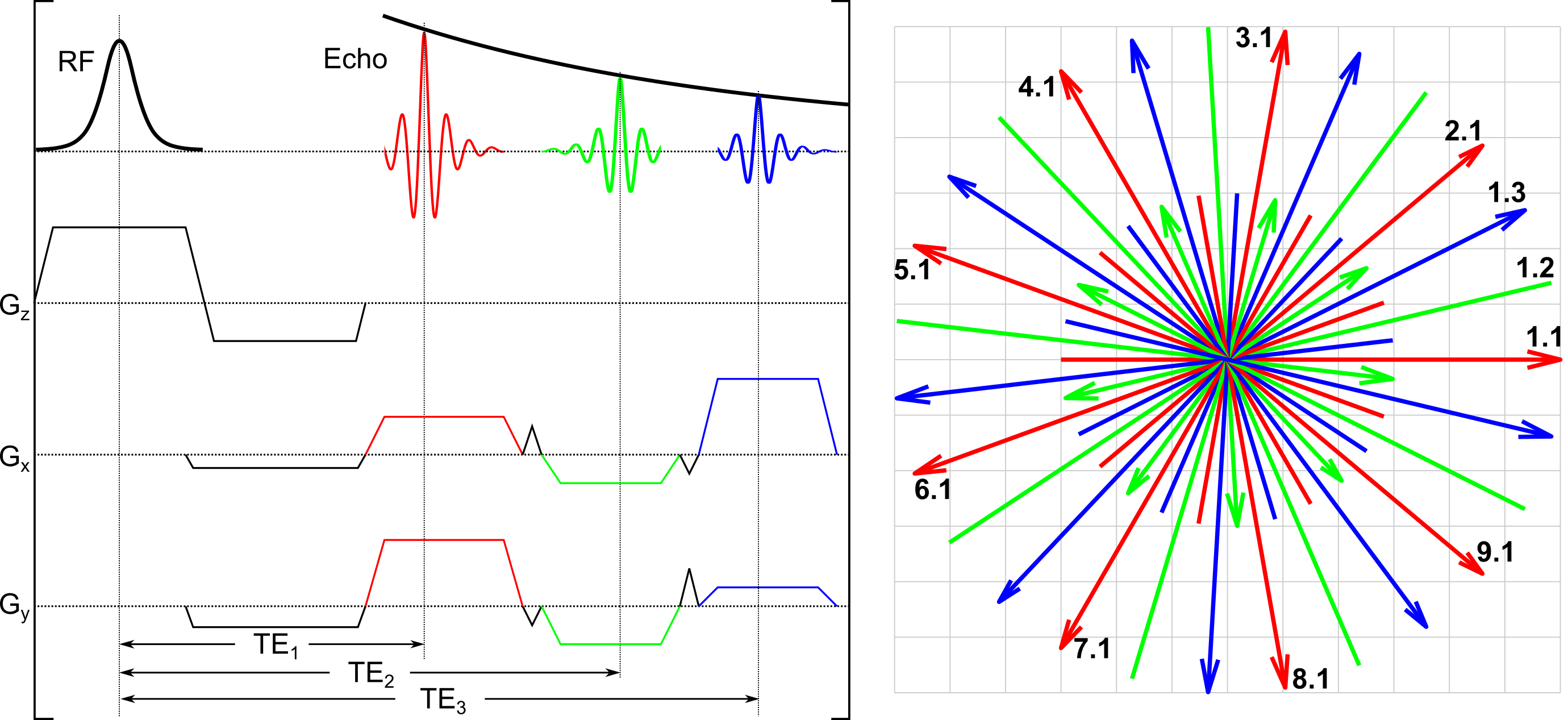
Figure 1 illustrates the multi-echo multi-spoke radial FLASH sequence and its corresponding k-space trajectory, where an echo asymmetry 75% is integrated to shorten TE and TR7. To achieve optimal coverage of k-space, radial spokes with the same TE are uniformly distributed in k-space, and the incremental angle between frames is empirically chosen as Golden angle (68.75o).
The signal model is:
$$F_{j,l} (x) = P_l \mathcal{F} \{ (W + F \cdot z_l) \cdot e^{i 2\pi f_{B0} \text{TE}_l \cdot c_j} \} \; \text{with} \; x = (W, F, f_{B0}, c_1, ... c_N)^T$$
Here, $$$P_l$$$ and $$$\mathcal{F}$$$ are the sampling pattern for the $$$l$$$th echo and 2D FFT, respectively. The unknown $$$x$$$ consists of water ($$$W$$$), fat ($$$F$$$), B0 field inhomogeneity (off-resonance) frequency ($$$f_{B0}$$$), and coil sensitivity maps ($$$c_j$$$). The fat modulation8 follows $$$z_l = \sum_{p=1}^{6} a_p \cdot e^{i 2\pi f_p \text{TE}_l}$$$. To jointly estimate all unknowns, the cost function is
$$\Phi(\hat{x}) = \text{argmin}_{\hat{x}} \left \| y - F(T\hat{x}) \right \|_2^2 + \alpha \left \| \hat{x} \right \|_2^2 \; \text{with} \; x = T\hat{x} $$
$$$y$$$ is the gridded k-space data without roll-off corrections. The weighting matrix is $$$T = \mathcal{F}^{-1} \Big(1 + w \cdot \left \| \vec{k} \right \| \Big)^{-h}$$$, with $$$w=11$$$ and $$$h=18$$$ for the B0 field map, $$$w=880$$$ and $$$h=16$$$ for coil sensitivity maps, while an identity matrix ($$$T=I$$$) is applied onto water and fat maps. $$$\vec{k}$$$ is a 2D Cartesian grid matrix. Although weaker than coil sensitivity maps, the weighting on the B0 field map enforces spatial smoothness and assures accuracy. This cost function is minimized by the iteratively regularized Gauss-Newton method9 with automatic scaling10 for water and fat, while the scaling for the B0 field map is kept as 1.5 to warrant convergence. The reconstruction starts with $$$W=F=1$$$, $$$f_{B0}=0$$$, and $$$c_j=0$$$, while the initialization for the following frames is set as the estimate from the preceding frame damped by 0.9 to enforce temporal continuity.
Data acquisitions were performed on a 3T scanner (Magnetom Prisma, Siemens Healthineers, Erlangen, Germany) with an 18-channel body matrix coil. Acquisition parameters were: 8o FA, standard shimming, 1560 Hz/Px bandwidth, 320 x 320 mm2 FoV, 200 x 200 matrix size, 1.6 x 1.6 x 6 mm3 spatial resolution, and 40 ms temporal resolution with 9 RF excitations per frame and TR = 4.43 ms, TE = 1.26/2.66/3.69 ms.
In addition, a whole-body scan with the built-in 2-channel body receiver coil was conducted. The volunteer was pulled through the isocenter from the lower leg to the head. Acquisition parameters were: 16o FA, tune-up shimming, 1200 Hz/Px bandwidth, 448 x 448 mm2 FoV, 320 x 320 matrix size, 1.4 x 1.4 x 10 mm3 spatial resolution, and 50 ms temporal resolution with 9 RF excitations per frame and TR = 5.54 ms, TE = 1.54/3.12/4.70 ms. This scan covers the whole body within only 40 s. To accompany with rapid change of anatomies from slice to slice, a real non-negative constraint is applied on water and fat maps during image reconstructions.
Results
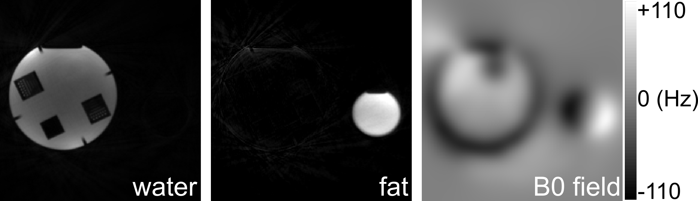
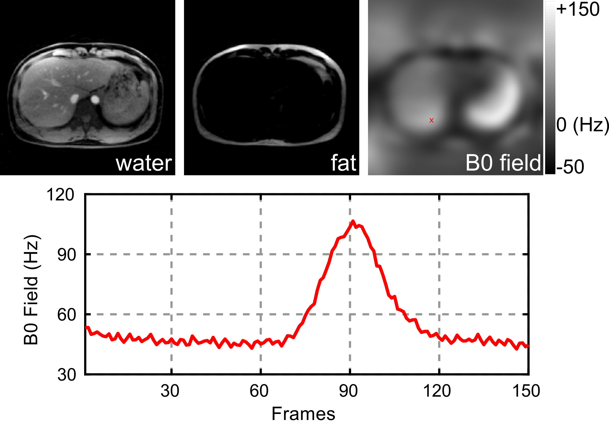
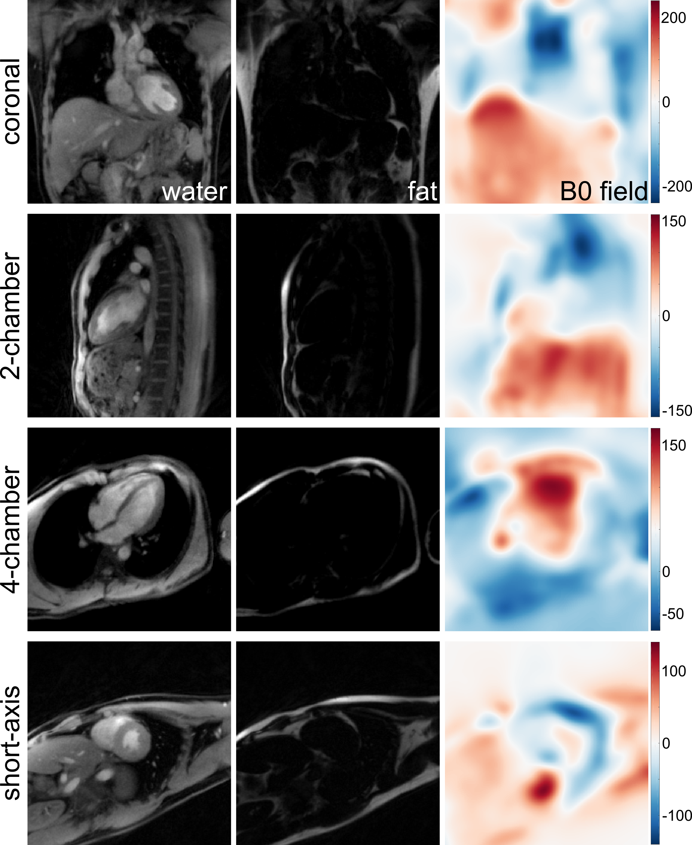
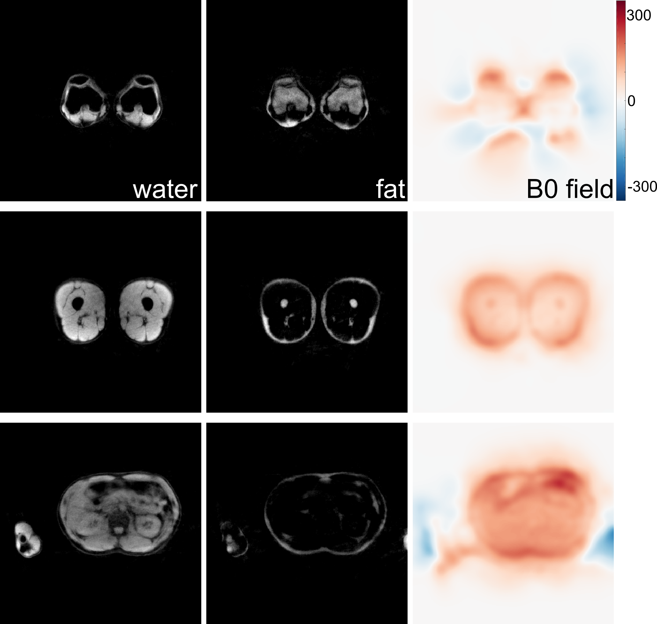
Figure 2 shows results of a static phantom using the proposed acquisition and joint estimation techniques. The reconstruction is able to capture fast varying B0 field inhomogeneity across the FoV. Moreover, temporal B0 field inhomogeneity variations in the presence of abdominal breathing are well resolved (see Figure 3). When imaging the beating heart, the proposed method again accurately separates water and fat in different sections and for all cardiac phases (see Figure 4). In the experiment where the volunteer was pulled through the isocenter, the field inhomogeneity changes rapids along with anatomy, as depicted by the selected slices in Figure 5.Discussion and Conclusion
B0 field homogeneity is affected by various factors, e.g., shimming, tissue composition, and motions. Therefore, accurate estimation of dynamic field inhomogeneity maps is crucial for the successful separation of water and fat. The proposed sequence and joint estimation reconstruction provide a practical solution to time-resolved water/fat separation at a temporal resolution of 40 ms.Acknowledgements
The authors would like to thank Dr. Arun Joseph, Olkesandr Kalentev, Klaus-Dietmar Merboldt, and Thomas Michaelis for their help on the experiments.References
- Dixon WT. Simple proton spectroscopic imaging. Radiology 1984;153:189-194.
- Glover GH. Multipoint Dixon technique for water and fat proton and susceptibility imaging. J Magn Reson Imaging 1991;1:521-530.
- Reeder SB, Wen Z, Yu H, et al. Multicoil Dixon chemical species separation with an iterative least-squares estimation method. Magn Reson Med 2004;51:35-45.
- Doneva M, Börnert P, Eggers H, et al. Compressed sensing for chemical shift-based water-fat separation. Magn Reson Med 2010;64:1749-1759.
- Benkert T, Feng L, Sodickson DK, et al. Free-breathing volumetric fat/water separation by combining radial sampling, compressed sensing, and parallel imaging. Magn Reson Med 2017;78:565-576.
- Berglund J, Johansson L, Ahlström H, et al. Three-point Dixon method enables whole-body water and fat imaging of obese subjects. Magn Reson Med 2010;63:1659-1668.
- Untenberger M, Tan Z, Voit D, et al. Advances in real-time phase-contrast flow MRI using asymmetric radial gradient echoes. Magn Reson Med 2016;75:1901-1908.
- Hu HH, Börnert P, Hernando D, et al. ISMRM workshop on fat-water separation: Insights, applications and progress in MRI. Magn Reson Med 2012;68:378-388.
- Uecker M, Hohage T, Block KT, et al. Image reconstruction by regularized nonlinear inversion – Joint estimation of coil sensitivities and image content. Magn Reson Med 2008;60:674-682.
- Tan Z, Hohage T, Kalentev O, et al. An eigenvalue approach for the automatic scaling of unknowns in model-based reconstructions: Application to real-time phase-contrast flow MRI. NMR Biomed 2017;30:e3835.
Figures