0687
Regional Variation of Water Permeability at the Blood-Brain Interface in the Mouse Brain using Multi-TE ASL: The Role of Aquaporin-41UCL Centre for Advanced Biomedical Imaging, University College London, London, United Kingdom, 2Office of the President, Karolinska Institutet, Stockholm, Sweden, 3GliaLab and Letten Centre, University of Oslo, Oslo, Norway, 4Neuroradiological Academic Unit, UCL Institute of Neurology, University College London, London, United Kingdom, 5Leonard Wolfson Experimental Neurology Centre, UCL Institute of Neurology, University College London, London, United Kingdom
Synopsis
We apply a multi-TE ASL technique to the mouse brain to assess the regional variation of water permeability at the blood brain interface, and measure the expression of brain AQP4 water channels as a marker of water transport. We report a significant decrease in the intravascular fraction of the ASL signal in the cerebellum compared to the cortex, 0.61 (± 0.22) and 0.90 (± 0.08) respectively, which is consistent with a marked increase (~400%) in Aqp4 expression in the cerebellum. This technique is a promising tool to better understand the dynamic role of AQP4 in pathological conditions.
Introduction
Changes in water permeability at the blood brain interface (BBI) may be an upstream indicator of neurodegenerative processes. Whole brain MRI can capture the signature patterns of regional pathology that define different neurodegenerative conditions, such as Alzheimer’s disease. However, to date, there has been very limited evaluation of regional difference to water permeability in healthy and pathological brain tissue using non-invasive MRI techniques. Brain aquaporin-4 (AQP4) water channels are central to the transfer of water across the BBI [1]. We have recently demonstrated that our measures of water permeability using multiple echo-time (multi-TE) ASL are sensitive to AQP4 polarisation at the BBI [2]. Marked regional differences in AQP4 expression within the healthy mouse brain have also been noted [3]. To investigate the possible application of multi-TE ASL in different brain regions, we build on our previous work and compare the cortical and cerebellum brain regions. We hypothesise that the cerebellum may have increased capacity to exchange water across the BBI compared to the cortex, given the markedly higher expression of AQP4 within this region [3]. Measuring regional difference in water transport across the healthy mouse brain may represent a novel approach for characterising changes in BBI water permeability within different neurodegenerative conditions.Method
Images were acquired using a 9.4T Bruker BioSpec imaging system with a four-channel array surface coil (BioSpec) in 6 month old female C57/Bl6 WT mice (n = 5). A multi-TE ASL protocol, based on flow-alternating inversion recovery (FAIR) sequence with single shot SE-EPI readout, was implemented at inflow times (TI) = 800ms and 1500ms. Sequence parameters were: TE = 8, 10, 12 15, 18, 23, 30, 40, 50, 65ms; TR = 5000ms; FOV = 25x25mm; matrix size = 64x64; slice thickness = 2mm; repetitions = 10. Mice were induced with 2% and maintained at ~ 1.5% isoflurane anesthetic in a mixture of 1.0L/min medical air. Multi-TE ASL data was evaluated with Matlab R2015a (Mathworks) using the analysis pipeline shown in Figure 1, which assumes a two compartment, intravascular (IV) and extravascular (EV), model for ASL signal: $$$ \small \triangle M = \triangle M_{IV}\exp(- \frac{TE}{T2_{IV}}) + \triangle M_{EV}\exp(- \frac{TE}{T2_{EV}}) $$$ (Equation 1) to calculate the intravascular fraction at each inflow time. The IV and EV signal contributions were used to calculate the exchange time ($$$\small T_{ex}^w$$$) as a surrogate index of BBI permeability to water.
Aqp4 mRNA expression was quantified in the caudal cortex and cerebellum brain regions of female C57/Bl6 mice (n = 6). Total RNA was extracted and converted to cDNA using RNeasy® Plus Microkit and QuantiTect® Reverse Transcription Kit (Qiagen). TaqMan® Gene Expression assays were used for Aqp4 and reference housekeeper (ACTB and GAPDH) gene quantification using an Eppendorf Mastercycler with Realplex software (v1.5, Eppendorf). Final Aqp4 expression levels were quantified by the 2–∆∆Ct method [4]. All data are reported as the mean and associated error (± std), with statistical analysis performed using GraphPad Prism6 (GraphPad Software).
Results
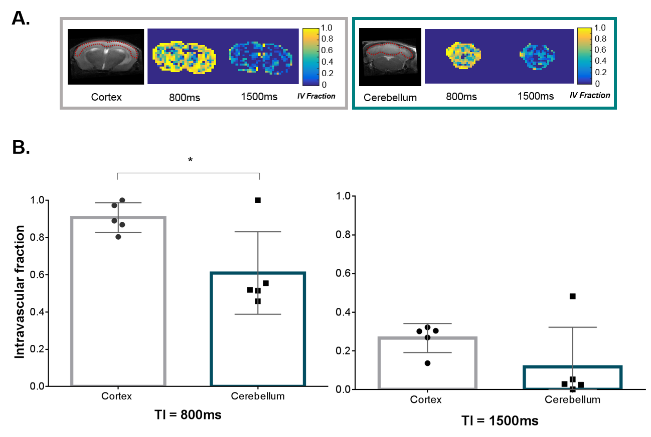
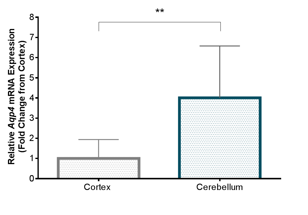
We observed a significant reduction in the intravascular fraction in the cerebellum compared to the cortical brain region at TI = 800ms: 0.61 ± 0.22 and 0.90 ± 0.08 respectively (p = 0.012) (Figure 2B). A similar decreasing trend between the two brain regions occurred at 1500ms: 0.11 ± 0.20 and 0.26 ± 0.08 (p = 0.084). The mean exchange times at TI = 1500ms were measured at 335 ± 97ms (cortex) and 150 ± 266ms (cerebellum). A large reduction of the intravascular fraction with increasing inflow time in both cortical and cerebral brain regions was observed (p < 0.0001 both regions; see Figure 2A). A 4-fold increase in Aqp4 mRNA expression was found in the cerebellum compared to the cortex (p = 0.0098) (Figure 3).Discussion
Multi-TE ASL is able to detect regional changes in BBI water permeability in the mouse brain. Our results suggest that labelled blood water is transferred into the brain tissue of the cerebellum more rapidly than in the cortex. This is consistent with the marked increase in AQP4 expression (~400%) measured within the cerebellum relative to the cortex (Figure 3), and supports our previous results which suggested that BBI water permeability is highly associated with the expression of AQP4 water channels [2]. Of note, the exchange time values from the cortical region acquired here using a 9.4T Bruker system with a similar (though not identical) acquisition protocol are also very consistent with our previous measurements using a 9.4T Agilent scanner [2].Conclusion
Regional differences in BBI water permeability in the mouse brain are highly associated with the expression of AQP4. Multi-TE ASL is a promising non-invasive technique for better understanding the dynamic role of AQP4 in pathological conditions.Acknowledgements
This work is supported by the Medical Research Council (MR/K501268/1), the EPSRC-funded UCL Centre for Doctoral Training in Medical Imaging (EP/L016478/1) and the UCL Leonard Wolfson Experimental Neurology Centre (PR/YLR/18575), together with the Wellcome Trust and Royal Society.References
1. Nagelhus, E.A. and O.P. Ottersen, Physiological Roles of Aquaporin-4 in Brain. Physiological Reviews, 2013. 93(4): p. 1543-1562.
2. Ohene, Y., et al., Non-invasive MRI of Brain Clearance Pathways using Multiple Echo Time Arterial Spin Labelling: An Aquaporin-4 Study NeuroImage, 2018. In press.
3. Hubbard, J.A., et al., Expression of the Astrocyte Water Channel Aquaporin-4 in the Mouse Brain. ASN Neuro, 2015. 7(5)
4. Livak, K.J. and T.D. Schmittgen, Analysis of relative gene expression data using real-time quantitative PCR and the 2(-Delta Delta C(T)) Method. Methods, 2001. 25(4): p. 402-8.
Figures
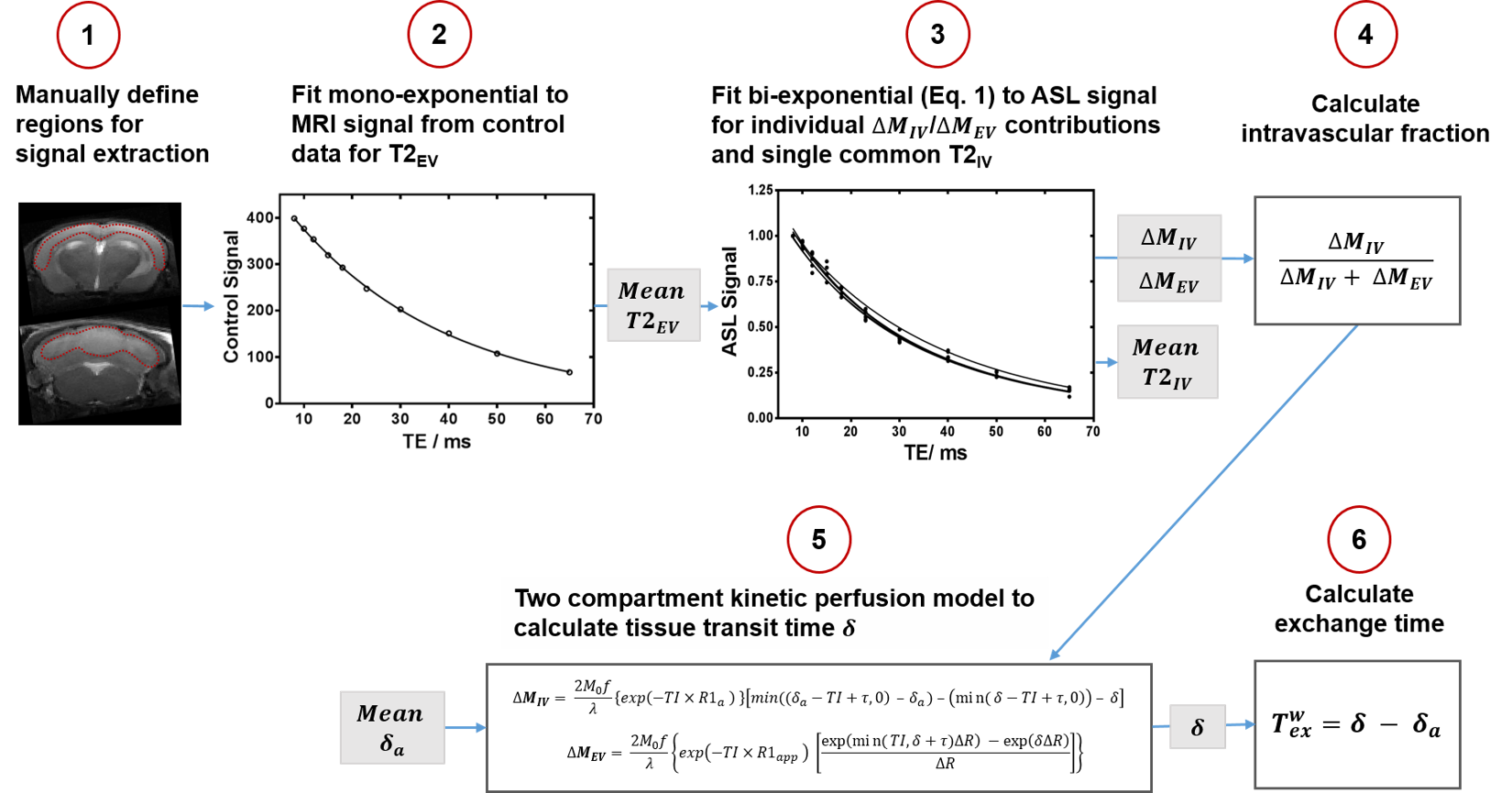
Figure 1: Analysis pipeline to calculate the intravascular fraction (ΔMIV / ΔMEV +ΔMIV) and exchange time (Texw) from the multi-TE ASL data, measuring transverse relaxation of extravascular tissue (T2EV), intravascular transverse relaxation (T2IV), tissue transit time (δ) and assuming arterial transit time (δa) for each brain region.