0684
Motion robust distortion-free Arterial Spin Labeling1Fraunhofer MEVIS, Bremen, Germany
Synopsis
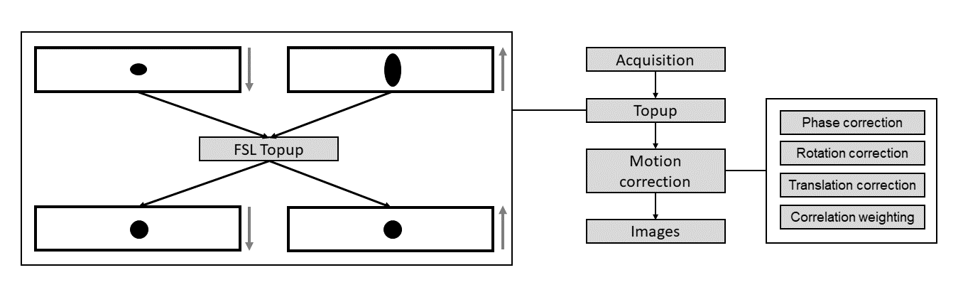
An improved reconstruction pipeline for arterial spin labeling (ASL) by means of a 3D GRASE PROPELLER (3DGP) acquisition is presented. Quality of ASL perfusion images is affected by motion artefacts, which can be mitigated by self-referred motion correction with 3DGP. However, since 3DGP is based on an EPI readout, geometric distortion is introduced in images, especially for long echo train lengths (ETL). The proposed method allows self-referred correction of motion as well as geometric distortion by playing each PROPELLER brick twice with toggled phase-encoded direction. The Topup technique then enables distortion correction followed by PROPELLER motion correction.
Introduction
Arterial spin labeling (ASL) MRI, being a subtractive technique, is sensitive to motion artefacts. However, ASL by means of a 3D GRASE PROPELLER (3DGP) readout allows for in-plane correction of rigid body motion1. A further challenge is that 3DGP, carrying an EPI readout, is sensitive to magnetic fi eld inhomogeneities that lead to geometric distortion, especially in case of a long echo train length (ETL). A constraint in ETL can be particularly detrimental in case of large fi eld of view, high-resolution ASL in organs other than brain. To this aim, we show that 3DGP ASL with label and control played twice with opposite polarity of the phase-encoded blips can allow to correct both geometric distortions and subject's motion.
Methods
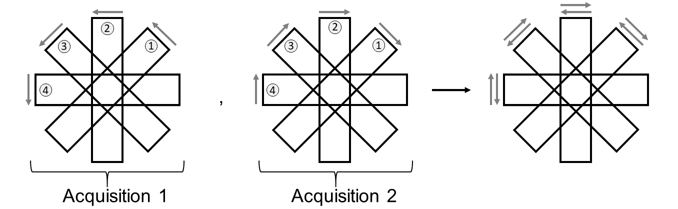
The acquisition scheme for 3DGP data is shown in Fig. 1. Since each brick is acquired twice with toggled polarity of the EPI blips, the Topup technique can be used to correct geometric distortions2. Our Topup-3DGP (T3DGP) reconstruction pipeline is shown in Fig. 2. The reconstruction pipeline is implemented in Matlab (The MathWorks, Natick, MA) with integration of Topup from the FSL toolbox3.
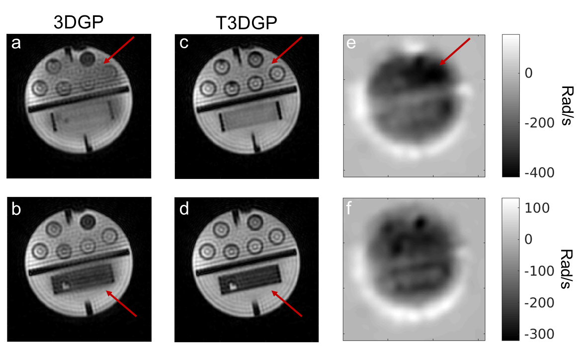
Measurements were performed on a 3T Siemens Skyra Magnetom with a twenty-channel headcoil. Phantom data were acquired using the following parameters: 18 label/control bricks of size 128x32x12 with an ETL of 32, TR/TE = 4190ms/14.7ms, cartesian resolution after gridding = 2.97x2.97x5mm3, bandwidth= 2298Hz/Px, echo-spacing = 0.5ms. In-vivo data of a healthy volunteer were acquired using the following parameters: 26 label/control bricks of size 192x48x12 with an ETL of 48, TR/TE = 5000ms/35ms, cartesian resolution after gridding = 2.5x2.5x5mm3, bandwidth = 1860Hz/Px, echo-spacing = 0.58ms. Background suppression was provided by two FOCI pulses. ASL(pCASL) was used with a post-labeling delay of 1780ms and a bolus length of 1800ms. The number of bricks arises from merging both acquisitions in Fig. 1. The subject was told to remain still during the measurements. Shimming was performed during the pre-scan of the fi rst acquistion of phantom/in-vivo data. 3DGP and T3DGP images are reconstructed by applying the standard PROPELLER reconstruction1 and our proposed reconstruction from Fig. 2 respectively. Image quality is assessed by comparing both reconstructions of original and perfusion images that were zero padded by a factor of two.
Results and Discussion
Fig. 3a and 3b show distortions in the phantoms geometry with 3DGP which are corrected by our proposed T3DGP reconstruction (cf. 3c and 3d).
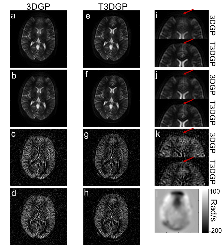
Reconstructed in-vivo images are shown in Fig. 4. The 3DGP images show distortions in the frontal area in the control as well as the perfusion images as indicated by the arrows. With our T3DGP reconstruction, the quality of these areas is signi cantly improved in both cases. The estimated fi eld map (cf. Fig. 4l) shows that the fi eld strength in the frontal area is smaller as expected which agrees with the position of artefacts in the 3DGP images.
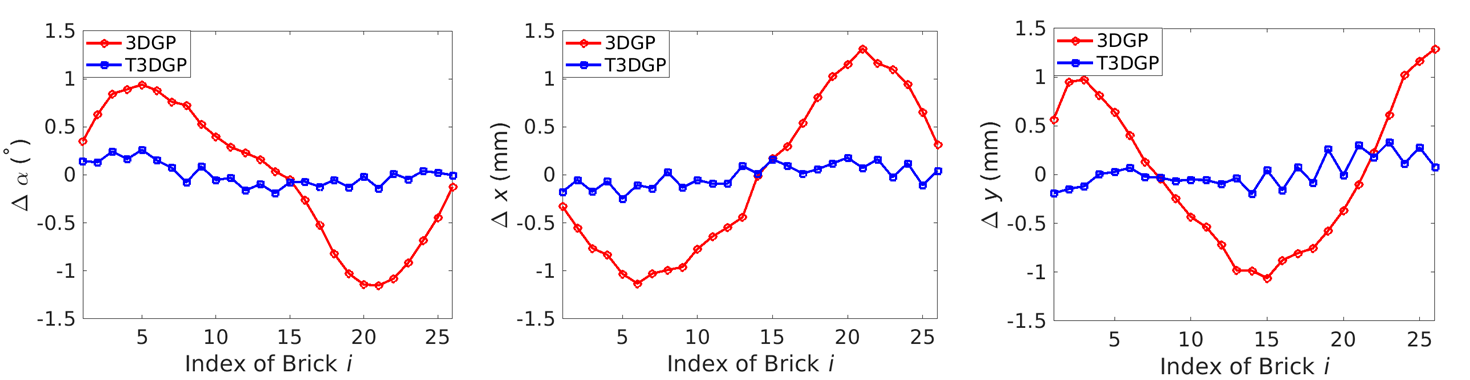
The calculated motion in Fig. 5 shows low frequency oscillation for 3DGP which is the result of the variation of distortion in every brick due to rotation of the phase-encoded direction. However, after rigid motion correction, it shows that there are slight improvements in control image quality for 3DGP (cf. Fig. 4i and Fig. 4j), indicating that the rigid motion correction is able to compensate for the distortions by some extend. Certainly this does not lead to signi cant improvements in the perfusion images (cf. Fig. 4c and 4d) and detection of fictious motion may even lead to artefacts with other sequence protocols4. With T3DGP this modulation is removed, since distortions are corrected and no signifi cant change can be seen in the T3DGP images after motion correction.
Conclusion
We have shown that a double acquisition of label and control with toggled polarities of the phase-encoded blips allows for integration of self-referred off-resonance correction through Topup. Therefore, as a future perspective, we will analyse the following acquisition scheme: Brick 1(Label, Blip+), Brick 1(Label, Blip-), Brick 1(Control, Blip+), Brick 1(Control, Blip-), repeated for all other bricks. So doing the repeated bricks to be used for the Topup field inhomogeneity correction are very close in time and, then, much more robust against motion. This will allow a more robust motion correction as next step of the reconstruction pipeline (cf. Fig. 2). Moreover, the application of Topup by using a label brick and a toggled version of the corresponding control brick will be investigated, since it would avoid to acquire the same brick twice (cf. Fig 1). Finally, our proposed method could be a good candidate for ASL perfusion imaging in organs other than brain like kidneys, where field inhomogeneities may arise from different breathing states during the acquisition.Acknowledgements
No acknowledgement found.References
1. Tan H, Hoge S, Hamilton C, et al. 3D GRASE PROPELLER: Improved Image Acquisition Technique for Arterial Spin Labeling Perfusion Imaging. Magnetic Resonance in Medicine 2011;66:168-173.
2. Andersson J, Skare S, Ashburner J. How to correct susceptibility distortions in spin-echo echo-planar images: application to diffusion tensor imaging. Neuroimage 2003;20:870-888.
3. Smith S, Jenkinson M, Woolrich M, et al. Advances in functional and structural MR image analysis and implementation as FSL. NeuroImage 2004;23:208-219.
4. Huber J, Vicari M, Günther M. Improving Robustness of Motion Correction in 3D GRASE PROPELLER Arterial Spin Labeling. Proceedings of the 34rd Annual Scienti c Meeting ESMRMB 2017; 567.
Figures