0674
Robust Region-Growing Fat-Water Imaging Using MT-Based B0 Field Priors1Radiology, University of Wisconsin, Madison, WI, United States, 2Medical Physics, University of Wisconsin, Madison, WI, United States
Synopsis
Region-growing (RG) is one of most computationally efficient fat-water separation methods, which exploits B0 smoothness assumption for separation with improved accuracy. However, practical robustness of RG method is majorly limited by low accuracy of initial seeded B0 values, which are problematic to select due to competing off-resonance from fat. Recently, it was demonstrated that insensitivity of fat to magnetization transfer (MT) preparation can be utilized to produce fat-insensitive B0 field priors. Here, we present a modified RG method that exploits this phenomenon to solve problems of seeding and stability of the original method and to attain robust F/W separation.
Introduction
Chemical-shift encoded fat/water (F/W) imaging may suffer from errors due to F/W estimation ambiguity caused by competing off-resonance sources (fat chemical shift and B0 field inhomogeneity). The ambiguity can be alleviated by prior knowledge about the B0 field, e.g. the reasonable B0 map estimate and/or smoothness (1,2). Region-growing (RG) is one of the most computationally efficient ways to exploit B0 smoothness. RG propagates initial B0 values from seed locations to each voxel in recursive fashion for initialization of F/W methods such as IDEAL (3). Robustness of RG-IDEAL is majorly limited by low accuracy of seeded B0 values, which are selected heuristically from a B0 field estimate affected by fat (4). Further, RG-IDEAL can lose stability over long range from the seeding area, especially for spatially disjoint tissues. Recently, it was demonstrated that insensitivity of fat to magnetization transfer (MT) preparation (5) can be utilized to produce fat-insensitive B0 field priors (2). Here, we present a modified RG-IDEAL that exploits this phenomenon to solve problems of seeding and stability of the original method and to attain robust F/W separation.Theory
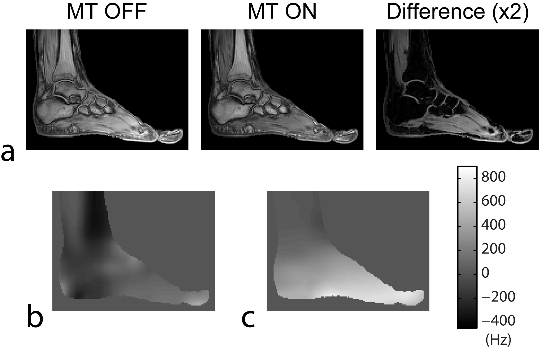
MT-Based B0 Field Estimation: Off-resonance MT pulse does not have a detectable effect on the fat signal due to absence of efficient mechanisms to transfer magnetization from fat protons (5). Simultaneously, it attenuates water via MT between saturated tissue macromolecules and water protons. If all sequence parameters affecting fat (i.e., repetition time TR, excitation angle) are kept the same, the subtraction of images acquired at nth echo time tn without ( $$$S_n^{off}$$$) and with ( $$$S_n^{on}$$$ ) MT saturation creates images without fat signal (Fig. 1a):
$$\Delta{S_n}=S_n^{off}-S_n^{on}=(1-A_{mt})We^{i\psi{t_n}},n=1,...,N_E$$
Here, $$$A_{mt}$$$ is MT attenuation, $$$W$$$ is water signal, and $$$\psi$$$ is the field map. As the fat signal is eliminated, $$$\psi$$$ can now be calculated from $$$\Delta{S_n}$$$ using standard inter-echo phase difference method (6).
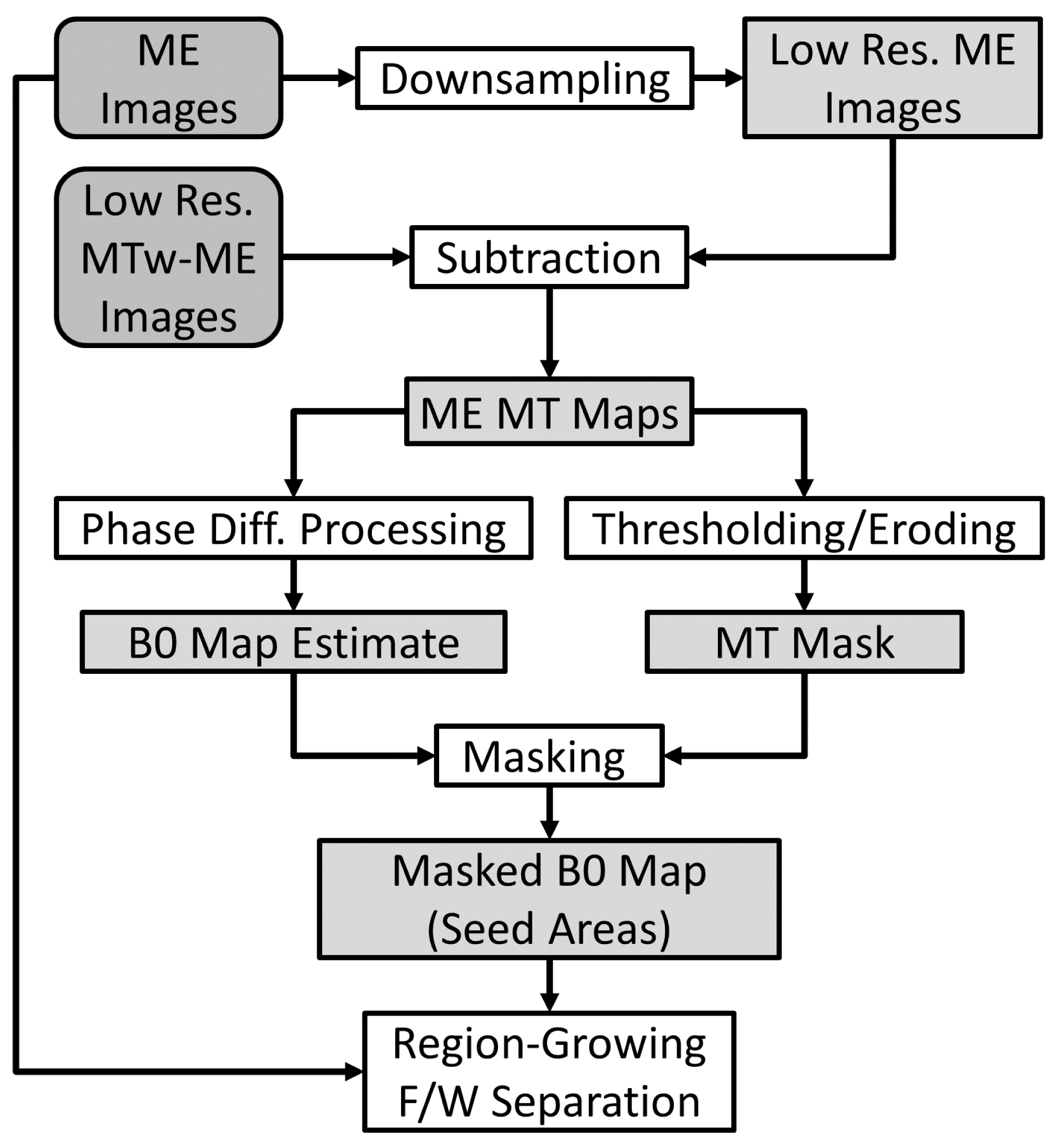
MT-RG-IDEAL: The original RG-IDEAL starts with a seeding area found heuristically from a low-resolution field map obtained by IDEAL, which does not guarantee proper RG initialization, because fat can still affect B0 values (Fig. 1b). In MT-RG-IDEAL (Fig. 2), we find seed areas using MT-based B0 prior not affected by fat. We identify the seed areas from the MT-based B0 map as regions with significant MT effect by thresholding the low-resolution MT image $$$\Delta{S_1}$$$, and morphologically eroding the mask. We note that our scheme allows selection of multiple seeds as opposed to the single region seeding in RG-IDEAL.
Methods
All experiments were performed on a 3.0T GE MR750 (Waukesha, WI). Main multi-echo (ME) SPGR scans were acquired with three-echo readout. MT-weighted ME (MTw-ME) data were acquired using the same sequence but with lower resolution (fewer phase encodes), and with the third echo replaced by MT pulse (Sinc shape/3kHz/170°) of the same duration (1.7ms). The MTw-ME acquisition time did not exceed 6% of main ME datasets. B0 map was estimated from $$$\Delta{S_1}$$$ and $$$\Delta{S_2}$$$ as illustrated in Fig. 2.Results
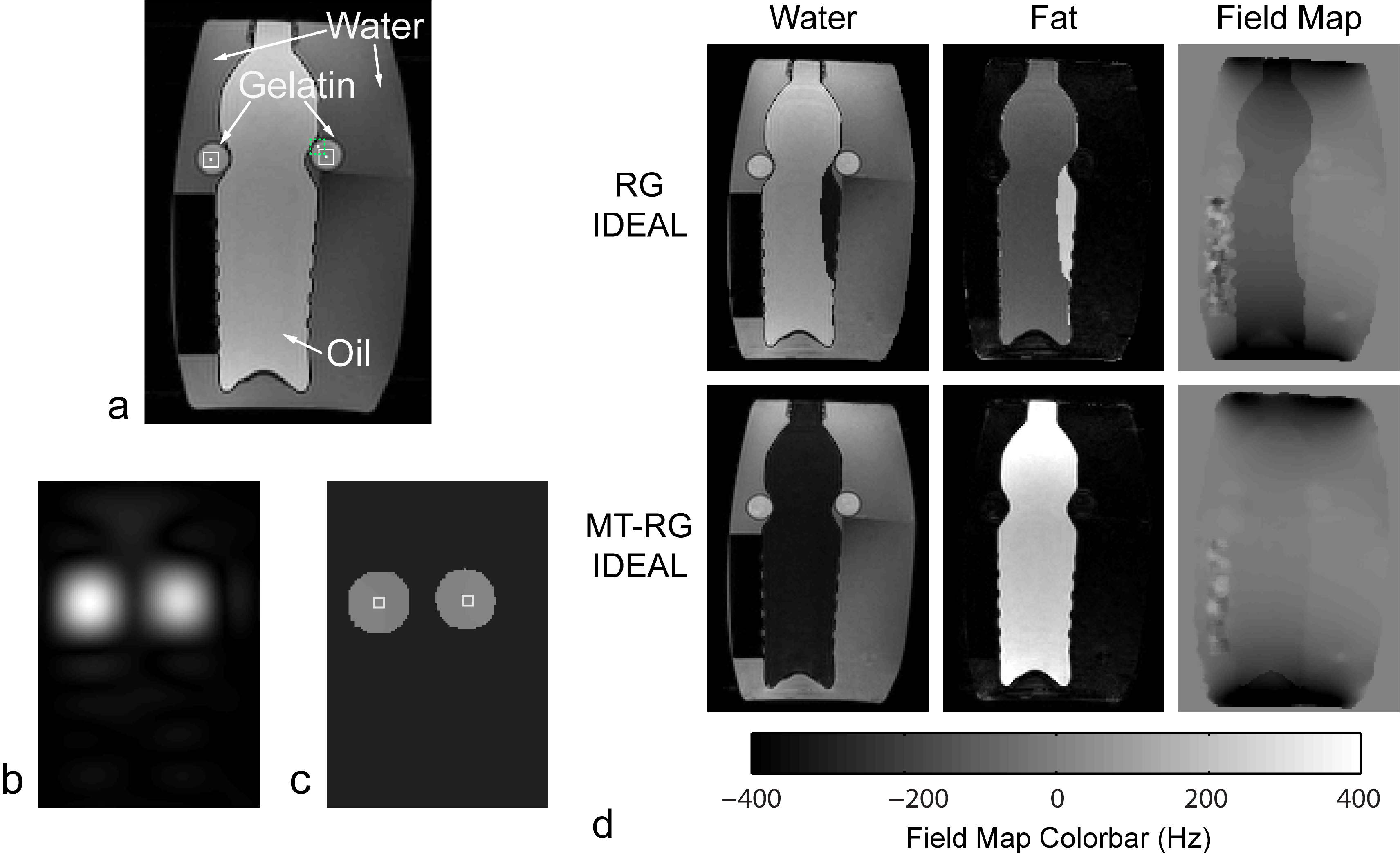
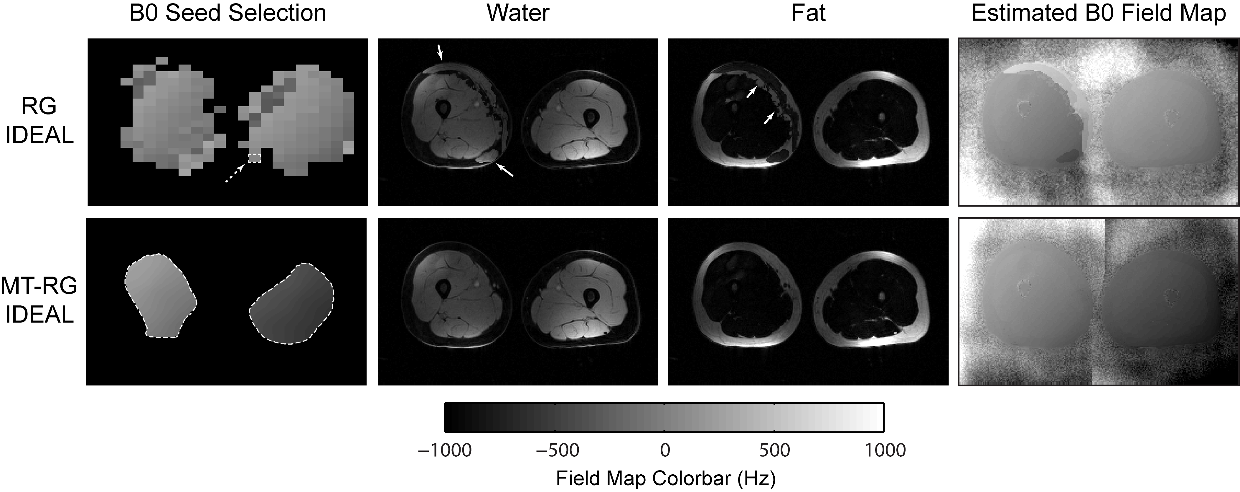
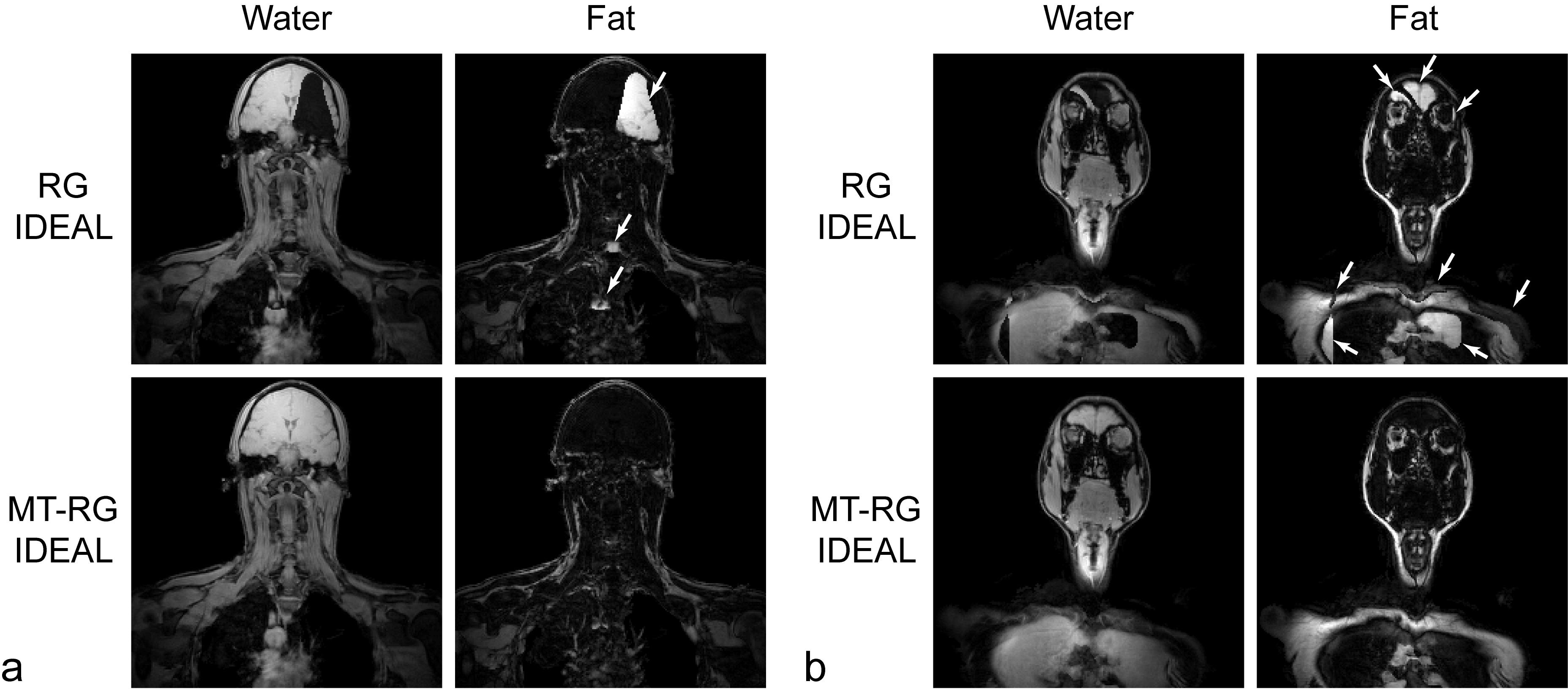
Figure 3 compares RG methods in a phantom object. Standard RG-IDEAL places the seed on the F/W boundary, which causes significant errors. Figure 4 illustrates performance of RG methods for anatomy with spatially disjoint tissues. For RG-IDEAL, F/W errors exist in left thigh, which is disconnected from tissues containing the seed (right thigh). Simultaneously, MT-RG-IDEAL seeding areas are present in both left and right thighs and lead to accurate separation. Figure 5 shows instability of RG with standard seeding, yielding results with sporadic errors in multiple locations. In MT-RG-IDEAL, the greater spatial coverage of seed areas corresponding to MT-sensitive tissues stabilizes separation.Discussion
Our method significantly improves performance of RG-based F/W separation by solving two main problems of the approach: robust seeding and stabilization of RG process. Unlike RG-IDEAL, MT-RG-IDEAL relies on fat-insensitive B0 field pre-estimation enabled by MT effect, thereby exploiting natural abundance of macromolecules in most tissues. We note that the way our B0 field map is utilized does not require it to be defined in non-MT regions (e.g, pure fat), which makes it particularly well suited for the use with RG algorithms. Given proliferation of RG algorithms on clinical scanners, our method may impact quality of clinical exams by improving F/W estimation in suboptimal imaging conditions such as significant B0 inhomogeneities and disjoint tissues. These improvements come at expense of moderate scan time increase (~5-6%) needed to acquire MTw-ME data. This overhead may be reduced in protocols using GRAPPA (7) if the extra MTw-ME acquisition is utilized for GRAPPA calibration thereby avoiding acquisition of fully sampled k-space center for main ME dataset.Acknowledgements
The work was supported by NIH (R21EB018483, R01EB027087) and GE Healthcare.References
1. Sharma SD, Artz NS, Hernando D, Horng DE, Reeder SB. Improving chemical shift encoded water-fat separation using object-based information of the magnetic field inhomogeneity. Magn Reson Med 2015;73(2):597-604.
2. Samsonov AA. Resolving Uncertainties of IDEAL Fat-Water Imaging Using Magnetization Transfer Effect. In: Proc of ISMRM; 2016; Singapore. p 567.
3. Reeder SB, Pineda AR, Wen Z, Shimakawa A, Yu H, Brittain JH, Gold GE, Beaulieu CH, Pelc NJ. Iterative decomposition of water and fat with echo asymmetry and least-squares estimation (IDEAL): application with fast spin-echo imaging. Magn Reson Med 2005;54(3):636-644.
4. Yu H, Reeder SB, Shimakawa A, Brittain JH, Pelc NJ. Field map estimation with a region growing scheme for iterative 3-point water-fat decomposition. Magn Reson Med 2005;54(4):1032-1039.
5. Chen JH, Sambol EB, Decarolis P, O'Connor R, Geha RC, Wu YV, Singer S. High-resolution MAS NMR spectroscopy detection of the spin magnetization exchange by cross-relaxation and chemical exchange in intact cell lines and human tissue specimens. Magn Reson Med 2006;55(6):1246-1256.
6. Jezzard P, Balaban RS. Correction for geometric distortion in echo planar images from B0 field variations. Magn Reson Med 1995;34(1):65-73.
7. Griswold MA, Jakob PM, Heidemann RM, Nittka M, Jellus V, Wang J, Kiefer B, Haase A. Generalized autocalibrating partially parallel acquisitions (GRAPPA). Magn Reson Med 2002;47(6):1202-1210.
Figures