0669
Simultaneous Denoising of Multi-contrast MR Images Using a Novel Weighted Nuclear Norm Minimization Approach1Laboratory of Biomedical Imaging and Signal Processing, The University of Hong Kong, Hong Kong, China, 2Electrical and Electronic Engineering, The University of Hong Kong, Hong Kong, China, 3Diagnostic Radiology, The University of Hong Kong, Hong Kong, China
Synopsis
A typical clinical MRI scanning session produces image sets with same geometries but different contrasts. These multi-contrast images often share strong structural similarities or correlations despite their contrast differences. Most existing MRI denoising methods deal with single-contrast images independently, and fail to explore and utilize such correlations across contrasts. In this study, we present a simultaneous denoising method for multi-contrast images based on low rank multi-contrast patch matrix completion. This denoising method exploits the structural similarities across contrasts, and outperforms the traditional method. Further, it does not compromise the image fidelity in absence of any structural similarities across contrasts.
Introduction
A typical clinical MRI scanning session offers image sets with same geometries but different contrasts. These multi-contrast images often share strong structural similarities or correlations across contrasts. Most existing MRI denoising methods only deal with single-contrast images independently. Thus they fail to exploit the rich structural correlations among contrasts. Inspired by the recent success of multi-spectral image denoising1, this study aims to simultaneously denoise multi-contrast MR images by utilizing the structural similarities across contrasts through a low rank multi-contrast patch matrix completion strategy.Methods
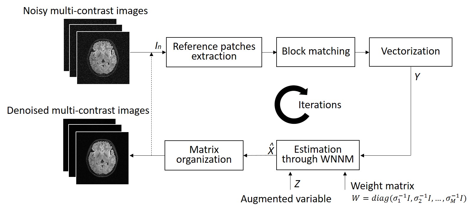
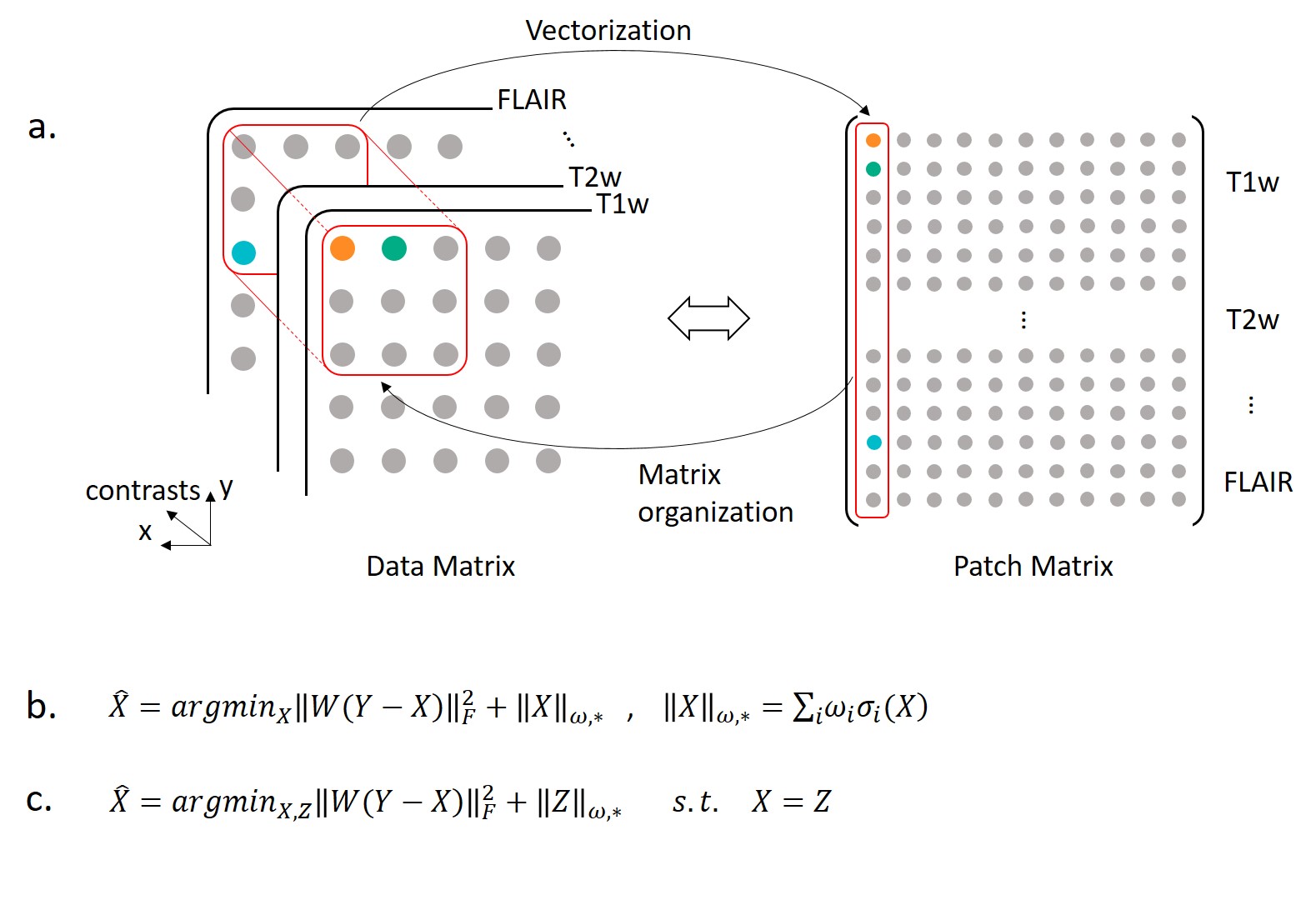
For each set of multi-contrast images at a particular slice location, the proposed denoising procedure is shown in Fig. 1. A data matrix $$$I_n\in R^{N_{x}\times N_{y} \times M}$$$ is structured from a noisy multi-contrast image set, where $$$N_{x}\times N_{y}$$$ stands for the matrix size of each image, and M stands for the contrast number. By sliding a window with size p×p×M across the entire data matrix, 3D reference patches are extracted. For each reference patch, K similar patches (including the reference patch itself) are searched based on Euclidean distance around the reference patch. As shown in Fig. 2a, by stretching K similar patches to patch vectors and stacking them into a matrix, a patch matrix $$$Y\in R^{Mp^{2}\times K}$$$ is then formed. Multi-contrast image denoising process can be modeled as recovering clean patch matrix X from the noisy patch matrix Y=X+N, where N stands for noise. Considering Y is composed of K similar patch vectors, this patch matrix should be a low rank matrix and could be recovered by low rank matrix completion method. Hence, we apply the multi-spectral weighted nuclear norm minimization (WNNM) model1 to describe the denoising process (Fig. 2b). The weight matrix W is a diagonal matrix and determined by the inverse of the noise standard deviation in each contrast. The stronger the noise in one contrast, the less this contrast will contribute to estimate X. By introducing an augmented variable Z, the multi-spectral WNNM model could be reformulated (Fig. 2c) and then be solved through the alternating direction method of multipliers (ADMM)2 framework.
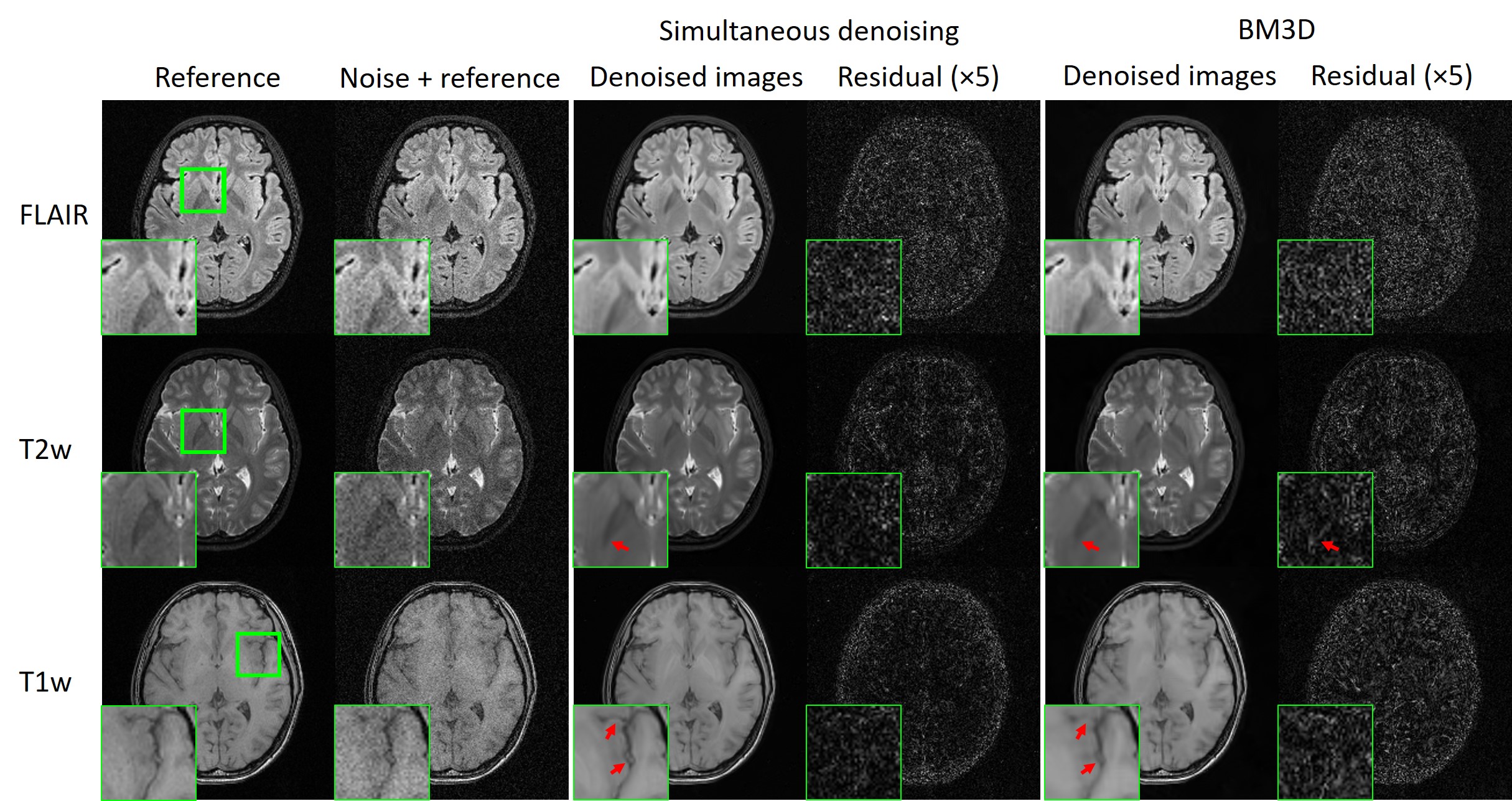
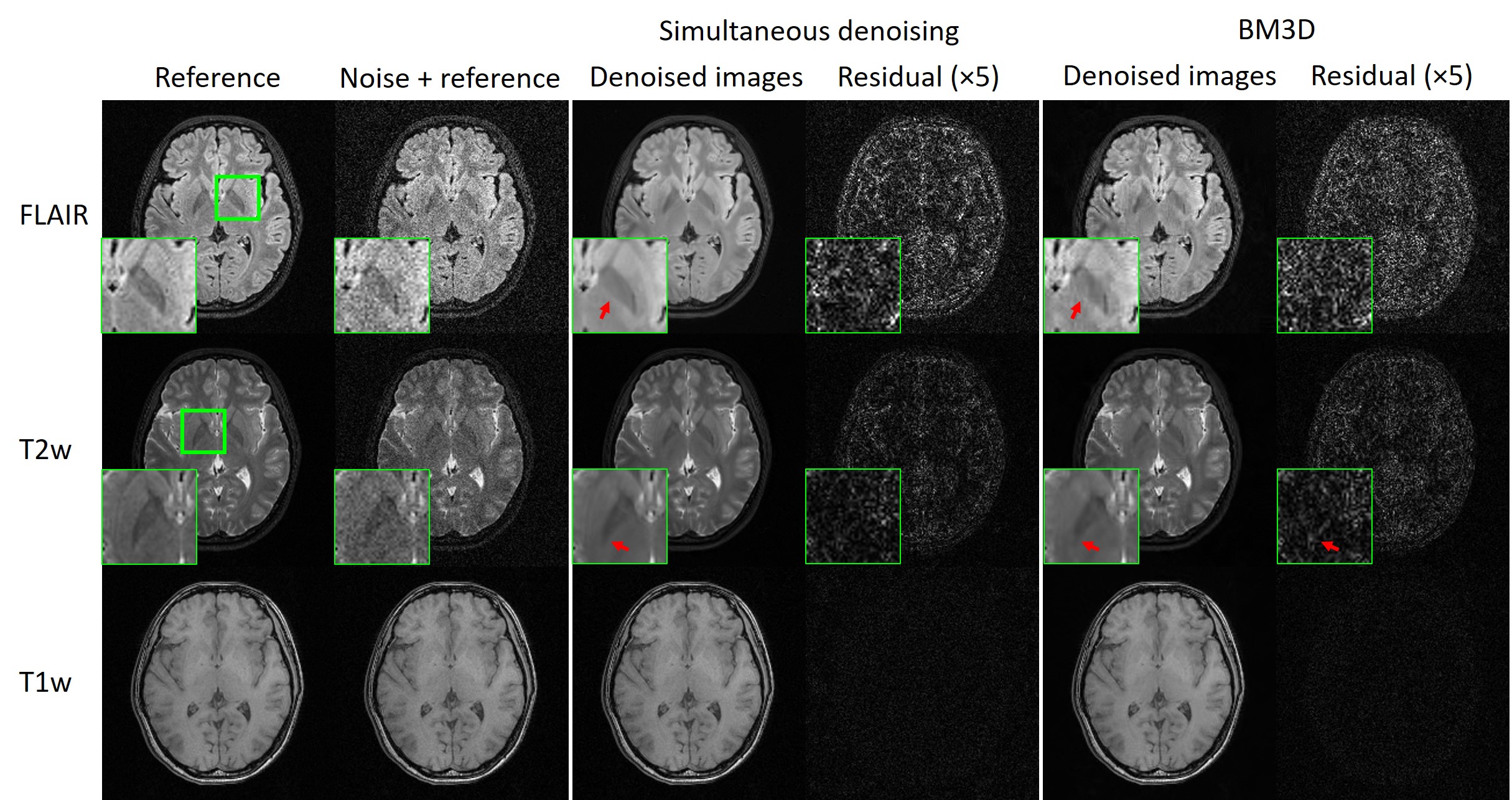
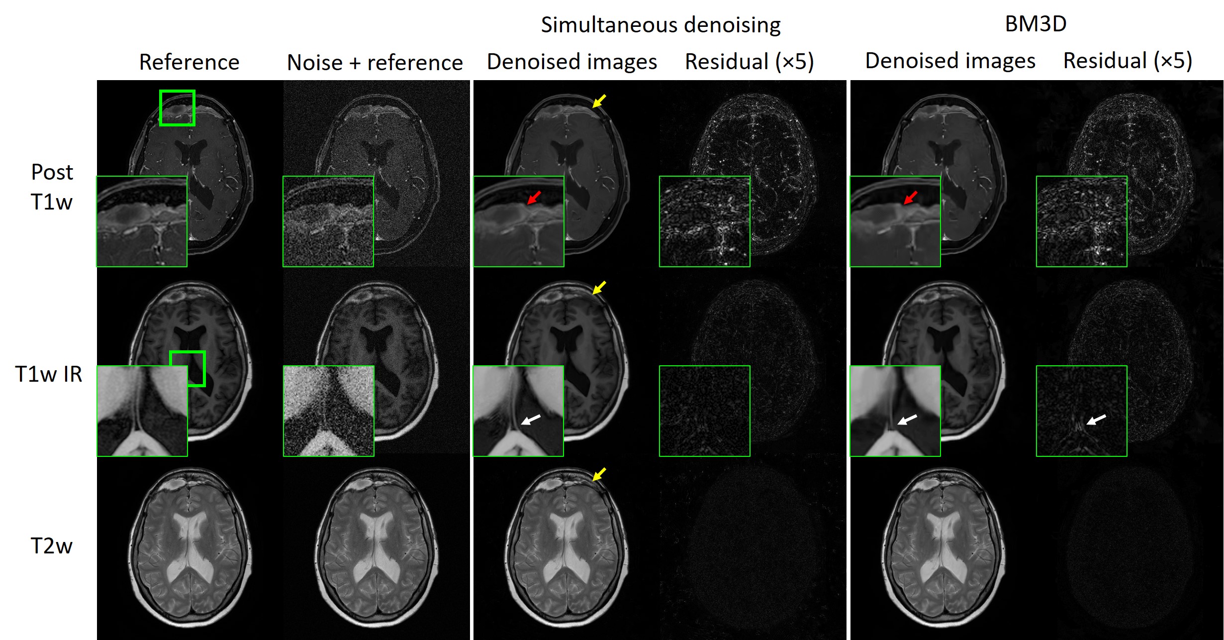
Experiments: Human brain data were acquired on a 3T Philips scanner. MR data from a healthy volunteer were acquired with identical geometries and three contrasts: T1w GE, T2w FSE and FLAIR. The image matrix size for each contrast was 240×240. MR data from a patient with mucosal thickening were also acquired with identical geometries and three contrasts: post-contrast T1w THRIVE, T1w IR and T2w FSE. The image matrix size for each contrast was 400×400. Noisy images were simulated by adding Gaussian noise to reference images. Patch size and similar patch number for WNNM model were p=6 and K=80.
Results
Figs. 3-5 show the performance of our proposed denoising method. As shown in Fig. 3, the same level of noise was added to images with three contrasts. Our method effectively removed noise in all three contrasts while preserving structural details. Fig. 4 shows that our method was also effective in removing noise while preserving details when images with three contrasts had different noise levels. Fig. 5 demonstrates that, in presence of brain pathology that manifests limited structural similarities across contrasts, our method could still reliably preserve pathology details while effectively remove noise. Further, our multi-contrast 2D image denoising method outperforms the popular BM3D method3, 4 that deals with single-contrast 2D images independently.Discussion and Conclusions
Our proposed simultaneous denoising method is based on the rationale that, by stacking similar patch vectors into a matrix, the patch matrix should be a low rank matrix and could be recovered through low rank matrix completion. The superior performance of our method arises from the fact that the structural similarities across contrasts substantially improve the robustness of the block matching process. Further studies can be carried out in two directions. First, the proposed method can be further optimized, e.g., model parameters can be tailored based on different noise level among contrasts. Second, the proposed method can be applied to denoise 3D MR images or multi-channel images by extending the 2D patch matrix to 3D patch matrix. In conclusion, this study presents a new denoising method for multi-contrast MR images based on a low rank patch matrix completion strategy. Our method exploits the rich structural correlations across contrasts and outperforms the traditional single-contrast MR denoising approach.Acknowledgements
This work was supported by the Hong Kong Research Grant Council (C7048-16G and HKU17103015 to E.X.W.).References
1. J. Xu, L. Zhang, D. Zhang, et al, "Multi-channel weighted nuclear norm minimization for real color image denoising," IEEE International Conference on Computer Vision, 2017.
2. S. Boyd, N. Parikh, E. Chu, et al, "Distributed optimization and statistical learning via the alternating direction method of multipliers," Foundations and Trends in Machine Learning, vol. 3, pp.1–122, Jan 2011.
3. P. Elahi, B. Soosan, and H. Masoud, "BM3D MRI denoising equipped with noise invalidation technique," IEEE International Conference on Acoustics, Speech and Signal Processing (ICASSP), 2014.
4. X. Lin, and T. Qiu, "Denoise MRI images using sparse 3D transformation domain collaborative filtering," IEEE International Conference on Biomedical Engineering and Informatics (BMEI), 2011.
Figures