0645
Comparison of reduced field-of-view (rFOV) and full FOV (fFOV) diffusion-weighted imaging (DWI) in the assessment of insulioma: image quality and WHO grading1Department of Radiology, Peking Union Medical College Hospital, Beijing, China, 2Philips Healthcare, Beijing, China, 3Cancer Research UK Cambridge Institute, University of Cambridge, Cambridge, United Kingdom
Synopsis
Reduced-FOV DWI (rFOV-DWI) of the pancreas has been applied in small cohorts and demonstrated improved image quality, but it has not been studied in the detection and characterization of insulioma. In this study, we compared the imaging quality (IQ) of rFOV-DWI and full FOV DWI sequence in insulioma detection. We also explored the correlation between the ADC value and WHO classification.
INTRODUCTION
Previous studies have shown DWI sequence as a powerful tool in the detection of insulinoma, and the apparent diffusion coefficient (ADC) value has a direct correlation with insulioma WHO grading system [1,2]. However, challenges to DWI remains including poor spatial resolution, ghosting, and susceptibility artifacts[3]. Although reduced-FOV DWI (rFOV-DWI) of the pancreas has been applied in small cohorts and demonstrated improved image quality [4,5], it has not been studied in the detection and characterization of insulioma. In this study, we compared the imaging quality (IQ) of rFOV-DWI and full FOV (fFOV) DWI sequence in insulioma detection. We also explored the correlation between the ADC value and WHO classification.METHODS
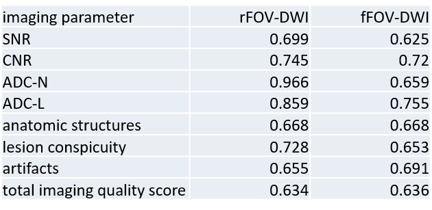
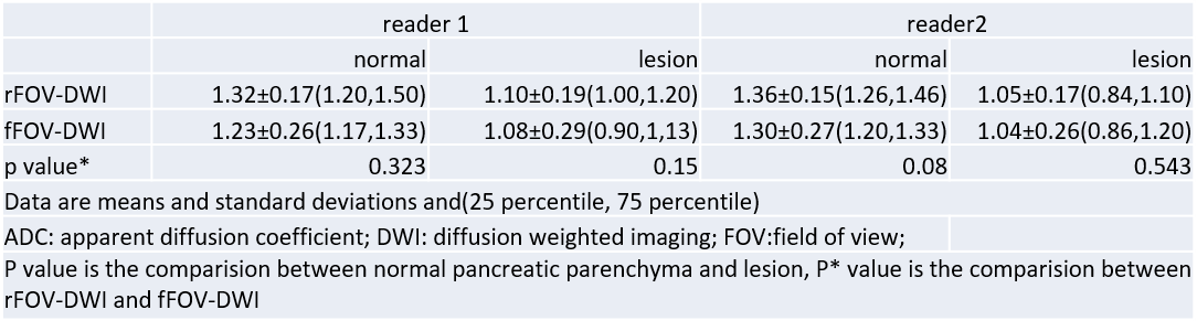
From October 2017 to September 2018, 77 patients (mean age: 46.11±15.31years, age range: 13-78 years) with clinically suspected insuliomas, among which 45 tumor resections and 35 histological results were obtained through surgery (G1, n = 21; G2, n = 14), went through pancreas MRI with two DWI sequences, study approved by the local IRB. Two radiologists independently assessed the imaging quality (IQ) including anatomic details, lesion conspicuity, distortion, and presence of artifacts via visual observation with 4-point scale. The images were also evaluated on signal-to-noise ratio (SNR) and contrast-to-noise ratio (CNR) between the two DWI sequences by the two radiologists. The Wilcoxon signed rank test was used to compare IQ scores, CNR, and SNR. The ADC values of the lesion and normal pancreatic parenchyma were calculated and compared for the two DWI sequences using paired t-test. The Spearman correlation analysis was used to explore the association between the ADC values and the WHO classification. The inter-observer agreement was evaluated using linearly weighted kappa coefficients for IQ and ICC for the ADC values, CNR, and SNR.RESULTS
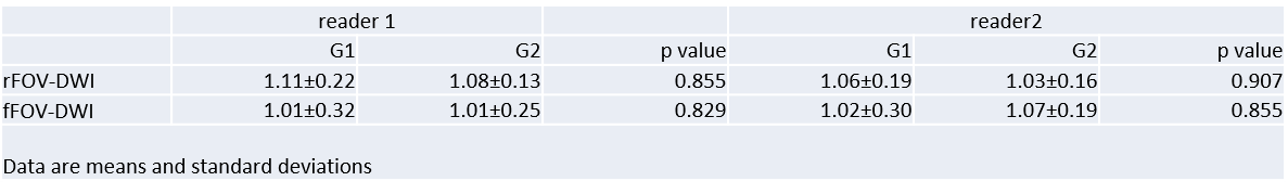
The rFOV-DWI images showed both clearer lesions and more fine structures than the fFOV-DWI images (Figure 1). As shown in Figure 2, the IQ score, SNR, and CNR were significantly higher in rFOV DWI than in fFOV DWI from both reader1 (IQ: 3.46±0.57vs.3.22±0.61,SNR:23.73±10.48vs.13.80±6.66, CNR: 8.72±6.44vs.3.94 ±4.42, all p﹤0.05) and reader 2 (IQ: 3.41±0.57vs.3.13 ±0.54, SNR: 26.50±15.59vs.17.12 ±6.56, CNR: 9.58±10.12vs.6.71±4.62, all p﹤0.05). There were no significant differences between rFOV and fFOV DWI sequences in ADC values of the tumor (reader1:1.10±0.19vs.1.08±0.29 ×10⁻³ mm2/s, reader2: 1.05±0.17vs.1.04±0.26 ×10⁻³ mm2/s) and normal pancreas parenchyma (reader1:1.32±0.17vs.1.23±0.26×10⁻³ mm2/s, reader2: 1.36±0.15vs.1.30±0.27×10⁻³ mm2/s), according to both reader 1 and reader 2 (Figure 3). There were slight differences in the ADC values of G2 insulioma both between the sequences and between the readers (Figure 4), yet not significant (p value was 0.855, 0.829, 0.907, and 0.855 respectively). There were correlations (Figure 5) between the WHO grading and the ADC values for both sequences (rFOV-DWI: r= 0.855, p=0.001; fFOV-DWI: r= 0.908, p=0.001). Agreement between the two readers was good to excellent for both sequence in both qualitative and quantitative assessments (rFOV-DWI:0.634-0.966, fFOV-DWI:0.636-0.755).DISCUSSION AND CONCLUSIONS
rFOV-DWI of the pancreas provides significant improvement in image quality than the fFOV-DWI sequence. The ADC values of the lesion and normal pancreas parenchyma were equivalent between these two sequences. The ADC value of the lesion is correlated with the WHO grading, but the difference of ADC value between G1 and G2 insulioma is not statistically significant, which may due to the relatively small sample size.
Acknowledgements
No acknowledgement found.References
1. De Robertis R, D'Onofrio M, Zamboni G, et al. Pancreatic Neuroendocrine Neoplasms: Clinical Value of Diffusion-Weighted Imaging. Neuroendocrinology. 2016;103(6):758-70.
2. Lotfalizadeh E, Ronot M, Wagner M, et al. Prediction of pancreatic neuroendocrine tumour grade with MR imaging features: added value of diffusion-weighted imaging. Eur Radiol. 2017;27(4):1748-59.
3. Barral M, Taouli B, Guiu B, et al. Diffusion-weighted MR imaging of the pancreas: current status and recommendations. Radiology. 2015;274(1):45-63.
4. Mannelli L, Monti S, Corrias G, et al. Comparison of Navigator Triggering Reduced Field of View and Large Field of View Diffusion-Weighted Imaging of the Pancreas. J Comput Assist Tomogr. 2018.
5. Riffel P, Michaely HJ, Morelli JN, et al. Zoomed EPI-DWI of the pancreas using two-dimensional spatially-selective radiofrequency excitation pulses. PLoS One. 2014;9(3):e89468.
Figures
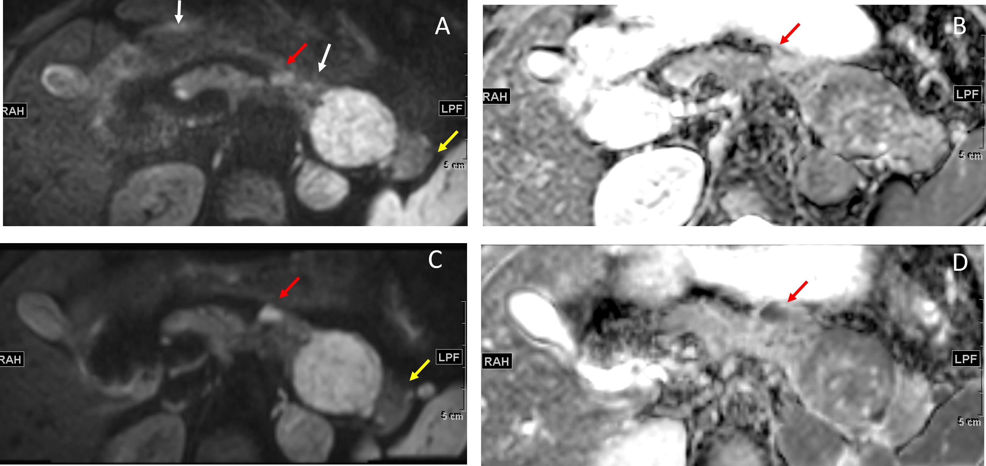
27 years old female, finally diagnosed as MEN-1. fFOV-DWI image (A) and its ADC map (B), and rFOV-DWI image (C) and its ADC map (D). High intensity lesions in the pancreas body and tail were found by using both DWI sequences. There were artifacts and distortion in the fFOV image (white arrow) but not in the rFOV image. The boundary of the pancreas was much clearer in the rFOV image when compared to that in the fFOV image (yellow arrow). The lesion in the body was better displayed in the rFOV compared to that in the fFOV (red head).