0634
T1 and T2 Weighted Image Segmentation from 1.0T Neonatal MRI1King's College London, London, United Kingdom, 2Medical Physics and Biomedical Engineering, University College London, London, United Kingdom, 3Shaare Zedek Medical Centre, Jerusalem, Israel, 4Faculty of Medicine, The Hebrew University, Jerusalem, Israel
Synopsis
In this work we present joint-image segmentation results from a novel 1.0T neonatal-specific MRI scanner. Data from machines such as this represent a new way to observe the growth and development of the preterm brain and can transfer our understanding of neurodevelopment prematurity. Advanced image analysis techniques are required to understand these developmental processes and we show preliminary results showing that good results can be obtained from these data.
Introduction
It is not currently practical to repeatedly scan neonates on high-field MRI due to the repeated transfer of the baby to and from the machine [1]. Low-field, small size MRI machines have the capability to transform our current understanding of neonatal brain development since they can be based in the neonatal intensive care unit. What is not known is how data obtained from 1T machines compares to standard 1.5T or 3T field strengths, nor how they can be used to provide longitudinal data to complement the more detailed imaging obtained from a single high-field strength scan [4].
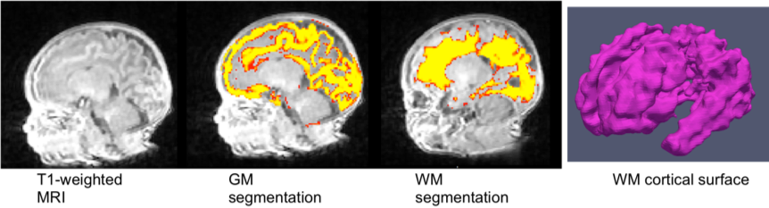
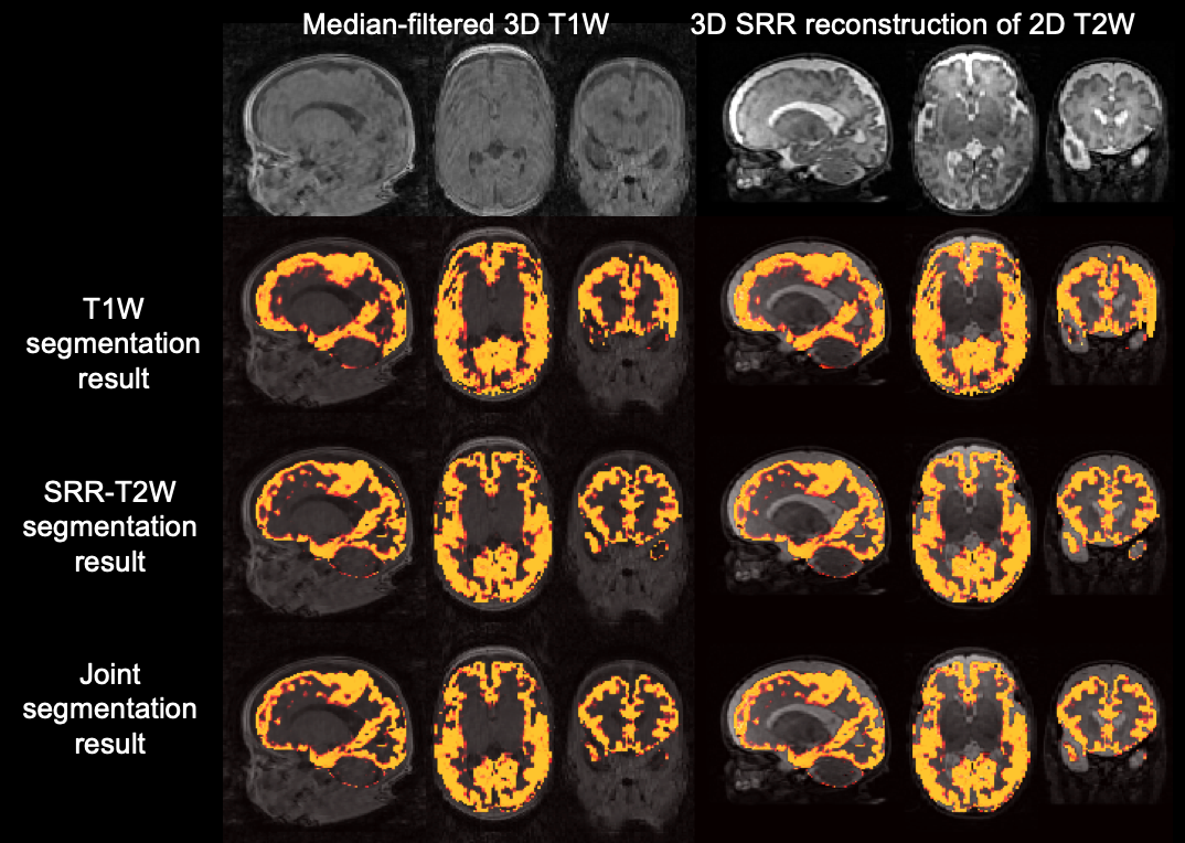
Developments in perinatal care in the last twenty years have resulted in reduced mortality, but neurodevelopmental injury remains a major concern among infants born at low gestational age [2]. Brain imaging has evolved in the last decades from cranial ultrasonography, to more detailed MRI scans, allowing for more detailed injury patterns, correlating with better prediction of neurodevelopmental outcome [3]. State-of-the-art segmentation routines make use of a mix of template matching tissue intensity classification. These algorithms can be adapted to neonates [5,6]. Multiple gestational-age matched existing high-quality segmentations from MRI data can be propagated using image registration and combined based on their local image similarity to form a segmentation prior. This prior is used to initialise a GMM expectation maximisation algorithm to refine the segmentation [6]. Occasionally, imaging contrasts can be slow to acquire and acquisition may be constrained to 2D rather than full-3D imaging. Widely applied in fetal imaging, 3D super-resolution reconstruction from multiple-plane T2-weighted data can be used to generate a higher-resolution full-3D reconstruction [7]. The reconstruction result can be aligned to the space of the other imaging data and this enables joint-intensity segmentation [6]. This has the advantage of making use of improved contrast or weaker magnetic field bias from one image where contrast is poorer in the other. The cortical surface of the brain can then be analysed to monitor cortical folding (see figure 1).
In this work, we apply joint-image segmentation [6] to data from a commercial 1.0T neonatal specific MRI machine. We use this segmentation to measure brain volume and estimate the shape of the cortical surface in several neonates and show the utility of joint image segmentation over segmentation using a single modality.
Methods
In an IRB approved study 55 premature infants, born at a mean gestational age of 28.8 (±2.1) weeks were scanned at term equivalent age (37.3±2.3 post conceptional age), without sedation in the neonatal dedicated MRI scanner. We use 1T Gradient-echo scans (T1W) from 13 neonates (TE/TR=2.2/20, 1.09x1.09x1.5mm). We a make use of additional T2 weighted (T2W) data acquired for each infant during the same scan in three orthogonal planes (TE/TR = 165/6400, 0.55x0.55x3 mm). The three-plane T2w data is reconstructed making use of the algorithm described in [7]. Imaging data is reconstructed in the space of the T1-weighted data, so that imaging data are co-registered. Affine registration could be used to correct for residual motion, but this is not found to be necessary for the infants in this study. We used a state-of-the-art segmentation routine to obtain measurements of white matter volume, grey matter volume, cerebellum volume and the cortical surface shape [4,5,6]. The segmentation method is based on an algorithm that combines template matching from previously segmented individuals with further mathematical modeling to refine the segmentation [5].Results
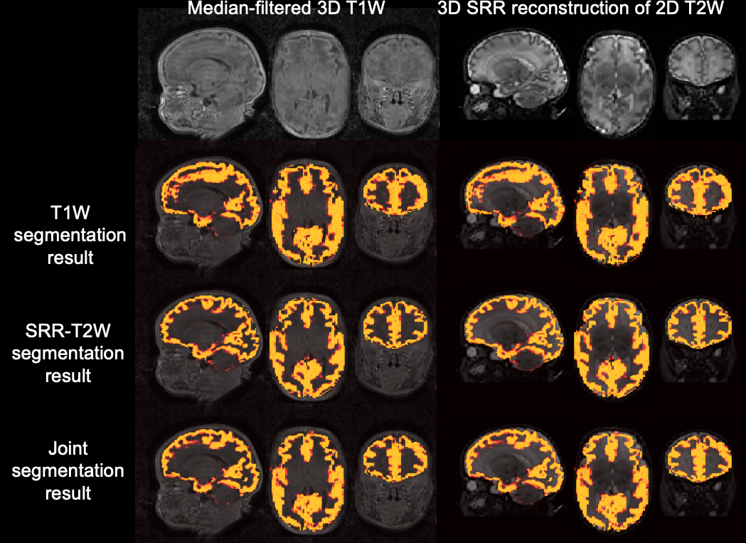
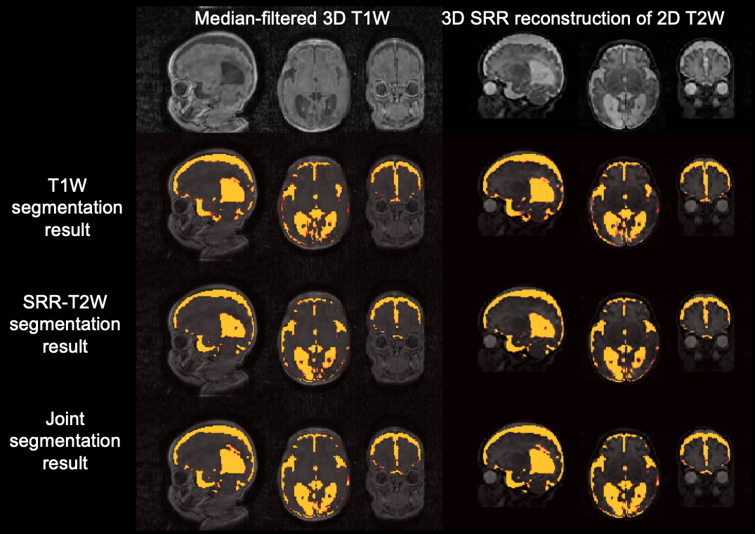
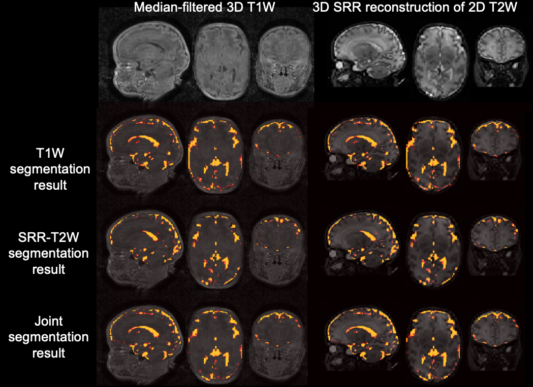
Segmentations obtained from 1T data are compared and contrasted, specifically we compare imaging volumes between segmentation results from only T1w data, only T2w data and joint segmentation data. Figure 2 shows general differences between grey matter segmentations. In this case joint segmentation gives improved lateral grey matter delineation in the absence of good contrast in the T1 data in this region. Figure 3 and 4 show improved segmentation of the ventricles when using T2 weighted data and joint segmentation. Figure 5 shows the effect of joint segmentation in the presence of motion.Conclusion
Our work provides the first evidence of the effectiveness of the dedicated neonatal machines for research into early neurodevelopment and provides the ground-work for future longitudinal studies of a type not currently possible with standard clinical high-field MRI. Existing segmentation software can be applied and used to measure brain volume in this new cohort opening the door for more sophisticated, multi-timepoint acquisitions of the neonatal brain. Future validation will compare and contrast these results with those from gold standard MRI data at normal field strengths of the same individuals. The novelty of the machine and the availability of clinical and research data will help us to begin to answer questions that are currently difficult to study effectively using full-size MRI.Acknowledgements
This work received funding from the Wellcome Trust (210182/Z/18/Z, 101957/Z/13/Z) and the EPSRC (NS/A000027/1, (EP/L016478/1).References
[1] Van Wezel-Meijler G, Leijser LM, de Bruïne FT, Steggerda SJ, van der Grond J, Walther FJ. Magnetic resonance imaging of the brain in newborn infants: practical aspects. Early Hum Dev. Feb; 2009 85(2):85–92.
[2] Adams-Chapman I, Heyne RJ, DeMauro SB, Duncan AF, Hintz SR, Pappas A, Vohr BR, McDonald SA, Das A, Newman JE, Higgins RD; Follow-Up Study of the Eunice Kennedy Shriver National Institute of Child Health and Human Development Neonatal Research Network. Neurodevelopmental Impairment among Extremely Preterm Infants in the Neonatal Research Network. Pediatrics. 2018 May;141(5).
[3] Mirmiran M, Barnes PD, Keller K, Constantinou JC, Fleisher BE, Hintz SR, Ariagno RL. Neonatal brain magnetic resonance imaging before discharge is better than serial cranial ultrasound in predicting cerebral palsy in very low birth weight preterm infants. Pediatrics. 2004 Oct;114(4):992-8.
[4] Eliza Orasanu, Andrew Melbourne, M. Jorge Cardoso, Herve Lombaert, Giles S. Kendall, Nicola J. Robertson, Neil Marlow, and Sebastien Ourselin. Cortical folding of the preterm brain: a longitudinal analysis of extremely-preterm born neonates using spectral matching. Brain and Behavior, 2016 Aug; 6(8): e00488.
[5] Andrew Melbourne, M. Jorge Cardoso, Giles S Kendall, Marc Modat, Nicola J Robertson, Neil Marlow, and Sebastien Ourselin. AdaPT: An adaptive preterm segmentation algorithm for neonatal brain MRI. Neuroimage, 65:97–108, Jan 2013 (Joint first authorship).
[6] M.J. Cardoso, M. Modat, R. Wolz, A. Melbourne, D. Cash, D. Rueckert, S. Ourselin. Geodesic Information Flows: Spatially-Variant Graphs and Their Application to Segmentation and Fusion. IEEE Trans. Medical Imaging, 2015, Volume:PP Issue:99 DOI:10.1109/TMI.2015.2418298.
[7] Ebner, M., Wang, G., Li, W., Aertsen, M., Patel, P. A., Melbourne, A., Doel, T., David, A. L., Deprest, J., Ourselin, S., and Vercauteren, T. (2018). An Automated Localization, Segmentation and Reconstruction Framework for Fetal Brain MRI. In Medical Image Computing and Computer-Assisted Intervention -- MICCAI 2018. Springer (Joint first authorship).
[8] Andrew Melbourne, Giles S Kendall, Manuel J Cardoso, Roxanna Gunny, Nicola J Robertson, Neil Marlow, and Sebastien Ourselin. Preterm birth affects the developmental synergy between cortical folding and cortical connectivity observed on multimodal MRI. NeuroImage 2014 Apr 1;89:23-34. [
Figures