0631
Altered development of structural brain networks in young children with prenatal alcohol exposure1Radiology, University of Calgary, Calgary, AB, Canada, 2Department of Paediatrics, University of Calgary, Calgary, AB, Canada
Synopsis
Prenatal alcohol exposure (PAE) can result in lifelong cognitive and behavioral deficits. Structural brain abnormalities have been shown in older children, but whether they are apparent in younger children is unclear. We investigated structural brain connectivity in 32 children with PAE aged 2-7 years compared to 95 healthy controls using diffusion tensor imaging. Group differences in structural connectivity and correlations with age were examined within and between eight different brain networks. Children with PAE had lower connectivity within and between several networks, but faster development of connectivity than controls, suggesting delayed development.
Introduction
Prenatal alcohol exposure (PAE) can result in lifelong cognitive and behavioral deficits1. Previous studies have shown brain alterations including decreased cortical thickness and white matter anisotropy in adults and adolescents with PAE 2. Early childhood is a critical period of brain development, and a time when many behavioral and cognitive difficulties first emerge 3,4, but it is unclear whether brain differences are apparent in early childhood, and how they change during this critical period of development. In the present study, we aimed to investigate structural brain connectivity and its development in young children with PAE.Methods:
Thirty-two children with PAE (17 males; aged 2.9 to 7.0 years) and ninety-five typically-developing children with no PAE (52 males; aged 2.5 to 6.9 years) were included in the present study. PAE was assessed through medical records, children’s services records, and/or interviews with close family members. Most control participants had multiple scans spaced ~6 months apart (282 total control datasets); PAE participants had one scan each (32 datasets total). All neuroimaging data were collected on a General Electric 3T scanner (GE, Waukesha, WI) at the Alberta Children’s Hospital after obtaining informed written consent from a parent. Diffusion tensor imaging (DTI) data was acquired with spin-echo EPI: voxel size: 0.78 x 0.78 x 2.2 mm3; TR = 6750 ms; TE = 79 ms; 30 directions at b = 750 s/mm2 and 5 b=0 s/mm2 images.
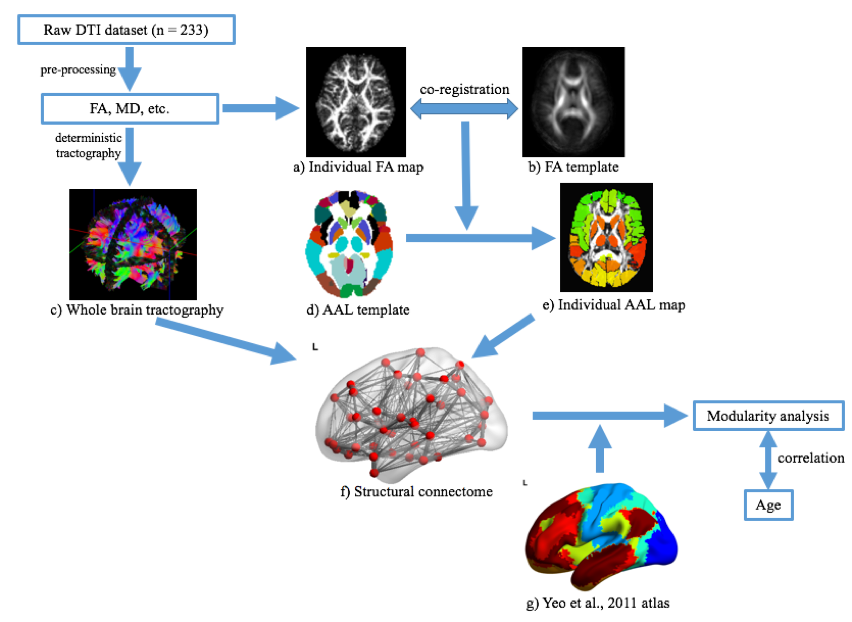
DTI data were preprocessed including correction of signal drift, Gibb’s ringing, motion, and eddy current distortion, calculation of the diffusion tensor parameters, and deterministic tractography of the whole brain fiber tracts by ExploreDTI 5. The Automated Anatomical Labeling (AAL) template was used to subdivide the cerebrum into 90 regions. A connectome matrix was built based on the AAL regions and whole brain tractography, then binarized. Eight networks were defined: visual, somatomotor, dorsal attention, ventral attention, limbic, frontoparietal, default mode, and a deep gray matter network based on Yeo et al6, and Diez et al7 (Fig. 1). Interactions (i.e., total number of edges) within and between each network were calculated, as well as the global participation coefficient, in the GRETNA toolbox 8. Correlation between modularity measures and age were performed in Matlab, including sex as a covariate. Group-level comparisons also included age as a covariate.
Results
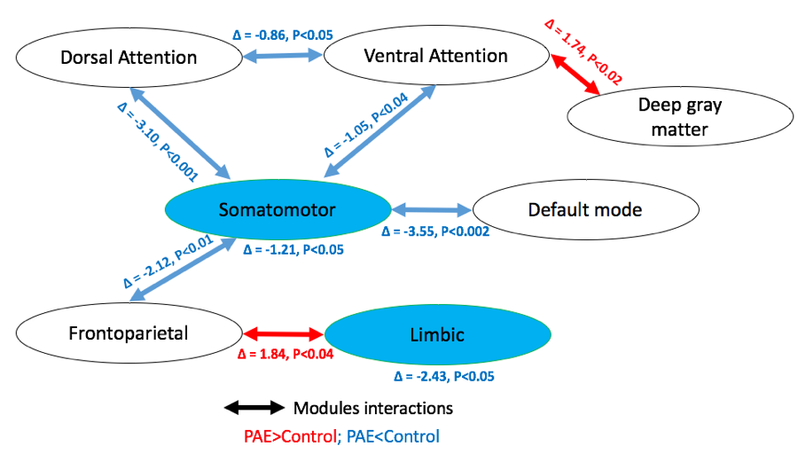
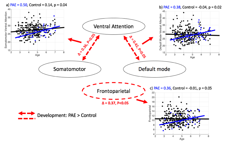
The somatomotor and limbic networks had decreased within-module connectivity in the children with PAE compared to unexposed controls (Fig. 2). Many networks had decreased interactions in the PAE group, while the deep gray matter-ventral attention, and frontoparietal-limbic interactions were higher in the PAE group. The frontoparietal network itself, and the somatomotor and the default mode network interaction with the ventral attention network, showed faster age-related changes in the PAE group than in unexposed controls (Fig. 3). The global participation coefficient was significantly lower in children with PAE than controls (t = -2.09, p = 0.04).Discussion
These results indicate reduced structural connectivity both within and between key brain networks in young children with PAE. In particular, the somatomotor network and its associations with other networks were reduced in children with PAE. However, age-related changes were also observed such that children with PAE appear to be catching up to controls, suggesting delayed development of within and between network connectivity. These results link previous findings of reduced functional connectivity within somatomotor areas in infants with PAE 9, and increased functional connectivity in the somatomotor network in adolescents with PAE 10, suggesting altered development patterns in children with PAE that include a catch-up, and possibly an overshoot in development. The increased connectivity between the deep gray matter and attention network, frontoparietal and limbic network were observed in PAE group, suggest higher network integration, which may be related to reduced volume of these structures11,12.Conclusions
Here, we show decreased within and between network connectivity in children with PAE, as well as altered developmental trajectories. The patterns suggest under-developed networks that appear to catch-up during childhood, especially within motor and attention areas. These network alterations may underlie motor and attention deficits in children with PAE. Future studies linking these age-related changes to behavioral and cognitive development will help clarify the implications of these changes in PAE populations.Acknowledgements
This work was supported by grants from CIHR and the Alberta Children’s Hospital Research Institute (ACHRI). Salary support was provided by University of Calgary I3T program (XL, PK), ACHRI (PK), and CIHR (CL).References
1. Riley, E. P. et al. Fetal alcohol spectrum disorders: An cverview. Neuropsychol. Rev. 21, 73–80 (2011).
2. Donald, K. A. et al. Neuroimaging effects of prenatal alcohol exposure on the developing human brain: A magnetic resonance imaging review. Acta Neuropsychiatr. 27, 251–269 (2015).
3. Brown, T. T. & Jernigan, T. L. Brain development during the preschool years. Neuropsychol. Rev. 22, 313–333 (2012).
4. Paus, T., Keshavan, M. & Giedd, J. N. Why do many psychiatric disorders emerge during adolescence? Nat. Rev. Neurosci. 9, 947–957 (2008).
5. Leemans, A., Jeurissen, B., Sijbers, J. & Jones, D. ExploreDTI: a graphical toolbox for processing, analyzing, and visualizing diffusion MR data. Proc. 17th Sci. Meet. Int. Soc. Magn. Reson. Med. 17, 3537 (2009).
6. Yeo, B. T. T. et al. The organization of the human cerebral cortex estimated by intrinsic functional connectivity. J Neurophysiol 106, 1125–1165 (2011).
7. Diez, I. et al. A novel brain partition highlights the modular skeleton shared by structure and function. Sci. Rep. 5, 1–13 (2015).
8. Wang, J. et al. GRETNA: a graph theoretical network analysis toolbox for imaging connectomics. Front. Hum. Neurosci. 9, 386 (2015).
9. Donald, K. A. et al. Interhemispheric Functional Brain Connectivity in Neonates with Prenatal Alcohol Exposure: Preliminary Findings. Alcohol. Clin. Exp. Res. 40, 113–121 (2016).
10. Long, X., Little, G., Beaulieu, C. & Lebel, C. Sensorimotor network alterations in children and youth with prenatal alcohol exposure. Hum. Brain Mapp. 39, 2258–2268 (2018).
11. Lebel, C., Walker, L., Leemans, A., Phillips, L. & Beaulieu, C. Microstructural maturation of the human brain from childhood to adulthood. Neuroimage 40, 1044–1055 (2008).
12. Nardelli, A., Lebel, C., Rasmussen, C., Andrew, G. & Beaulieu, C. Extensive deep gray matter volume reductions in children and adolescents with fetal alcohol spectrum disorders. Alcohol. Clin. Exp. Res. 35, 1404–1417 (2011).
Figures