0618
In vivo MRI detection of β-amyloid pathologies at early and late stages of Alzheimer’s disease1Laboratory of Biomedical Imaging and Signal Processing, The University of Hong Kong, Hong Kong, China, 2Department of Electrical and Electronic Engineering, The University of Hong Kong, Hong Kong, China, 3Department of Biomedical Engineering, City University of Hong Kong, Hong Kong, China, 4F.M. Kirby Research Center for Functional Brain Imaging, Kennedy Krieger Research Institute, Baltimore, MD, United States, 5Russell H. Morgan Department of Radiology and Radiological Science, The Johns Hopkins University School of Medicine, Baltimore, MD, United States
Synopsis
Early diagnosis of Alzheimer’s disease (AD) is crucial. However, there is a lack of effective diagnostic tools to detect AD at the early stage. Early stage β-amyloid (Aβ) oligomers (AβOs) and
Purpose
Alzheimer’s disease (AD) is a degenerative brain disease and the most common cause of dementia1. Early stage β-amyloid (Aβ) oligomers (AβOs) and late stage Aβ plaques are the pathological hallmarks of AD brains. AβOs are known to be more neurotoxic and contribute to neuronal damage2-5. Most current approaches are focused on detecting Aβ plaques, which occurs at the late stage of AD, and are limited by poor sensitivity and/or contrast agent toxicity6-8. We have recently synthesized a new curcumin-conjugated magnetic nanoparticle (Cur-MNP) to target the Aβ pathologies9. In this study, we investigated the in vivo feasibility of this novel Cur-MNPs to detect Aβ pathologies at both early and late stages of AD in transgenic AD mice. We further performed immunohistochemical examinations to validate our in vivo MRI findings by confirming the specific Cur-MNP targeting of various form of Aβ pathologies.Methods
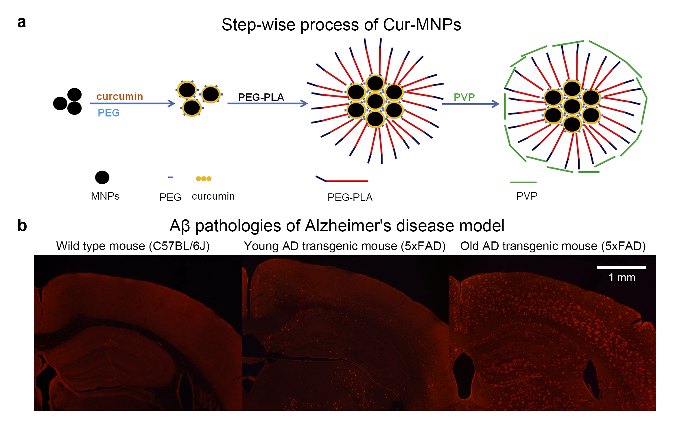
Animal groups and preparation: 6 old (10- to 14-month-old) and 6 young (3- to 4-month-old) transgenic AD mice (5xFAD), and 12 age-matched wild type mice (C57BL/6J) were used in this study. The superparamagnetic iron oxide (SPIO) based contrast agent, Cur-MNPs, were used to detect the Aβ pathologies (Figure 1a). Cur-MNPs suspension was concentrated to 2.7 mg Fe/ml and immediately injected into the animals (10μl/g) intravenously through the tail vein. Mice were examined in MRI 4 hours after injection. Longitudinal measurements were performed on old transgenic AD mice (n=4) 8 hours, 1 day, 2 days, 4 days and 8 days after injection. During MRI experiments, animals were anesthetized under 1.0-1.5% isoflurane mixed with 99% oxygen.
Data acquisition: MRI data was acquired on a Bruker 7T preclinical scanner using a 2D GRE sequence with TR/TE=3000/12.3ms, FA=80.1o, bandwidth=25kHz, FOV=25.6mm×25.6mm, matrix size=512×512, in-plane resolution=50μm×50μm, slice thickness=0.38mm with 0.02mm gap, 35 slices, two repetitions, and acquisition time of 51 mins.
Immunohistology: Animals were immediately perfused with saline after MRI experiments. The brain sections were either double-stained with anti-beta Amyloid 1-42 antibody and Prussian blue or stained with oligomer A11 Polyclonal antibody only. Curcumin was examined under fluorescent view (excitation/emission = 488/510-590nm).
Results
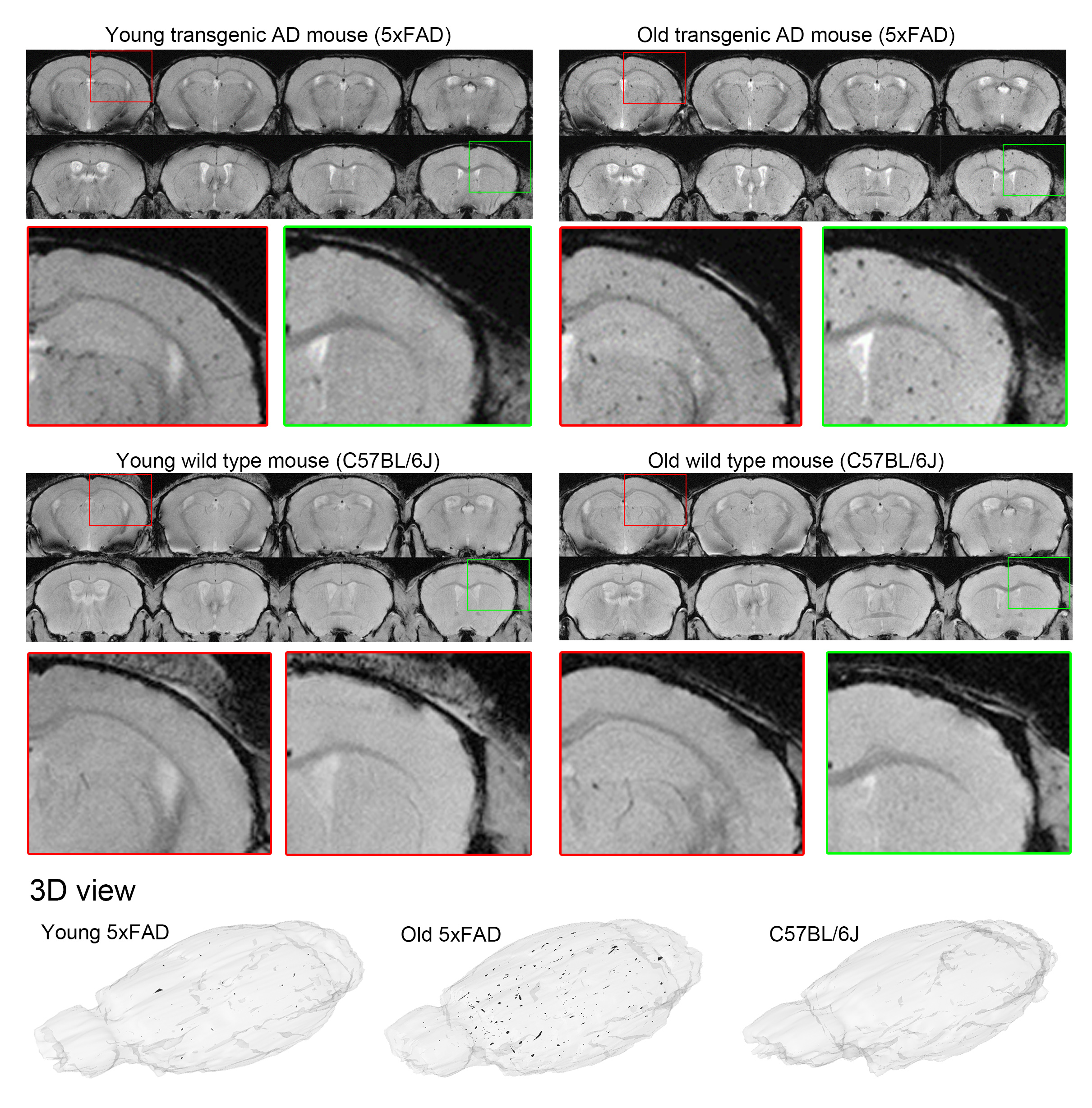
Figure 2 presents the serial in vivo T2*-weighted brain images of representative young and old transgenic AD (5xFAD) mice and their age-matched wild type (C57BL/6J) controls 4 hours after intravenous Cur-MNPs injection. Iron-induced hypointense spots were observed in both young and old 5xFAD mice but not in controls. The old 5xFAD mouse showed denser and larger hypointense spots, likely due to the significant increase of Aβ pathologies in old 5xFAD mouse brain.
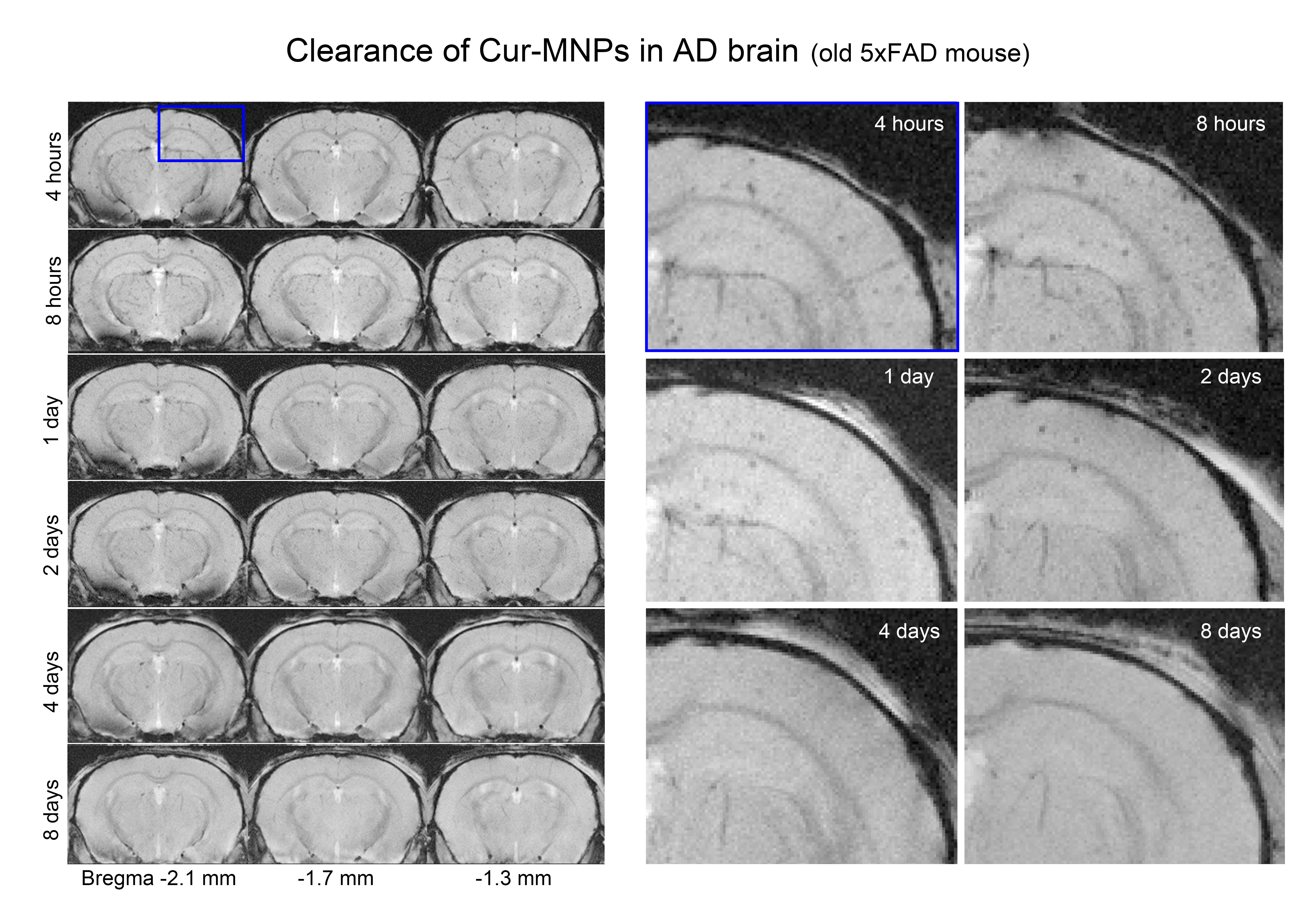
Figure 3 shows the longitudinal MRI measurements of Cur-MNPs at 4 hours, 8 hours, 1 day, 2 days, 4 days and 8 days after injection in a representative old transgenic AD mouse. In general, hypointense spots in old 5xFAD mice had similar density at 4 hours and 8 hours after injection, but were barely observable 1 day after injection. Cur-MNPs were completely washed out 8 days after injection.
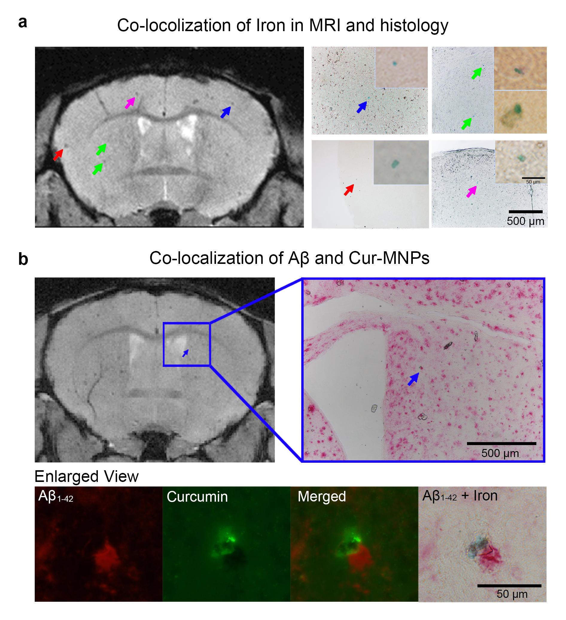
Figure 4 presents the histological validation of Cur-MNPs in targeting Aβ pathologies. Some hypointense spots induced by Cur-MNPs in T2*-weighted image (left, Figure 4a) were co-localized with iron (blue) in corresponding iron-stained 5xFAD mouse brain sections (right, Figure 4a). Furthermore, the co-localization of curcumin (green under fluorescent view) and iron (blue under bright-field view) were shown to bind to Aβ1-42 (red under fluorescent and bright-field view), revealing that Cur-MNPs targeted the Aβ pathologies (Figure 4b). These images demonstrate that Cur-MNPs can bind to Aβ pathologies and be visualized by MRI and immunohistochemistry.
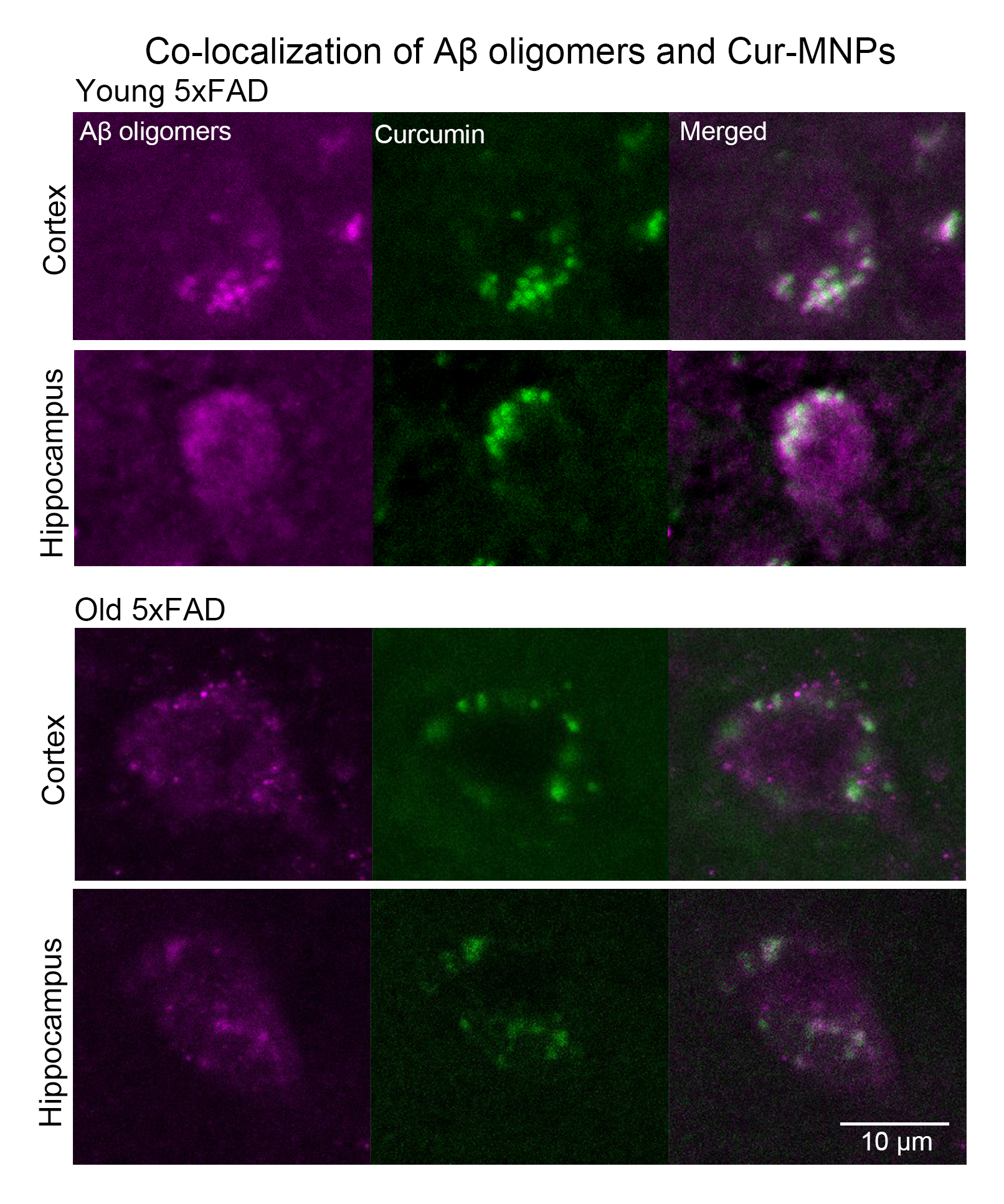
Figure 5 shows the co-localization of AβOs (imaged in magenta) and curcumin (green) in the cortex and hippocampus of young and old 5xFAD mice, demonstrating the ability of Cur-MNPs to target the AβOs at early and late stages of AD.
Discussion and Conclusion
The present study demonstrated that this novel MRI contrast agent, Cur-MNPs, could target the Aβ pathologies, including Aβ plaques and AβOs, at both late and early stages of AD by in vivo MRI measurements. Three months old 5xFAD mice are known to predominantly exhibit AβOs with little signs of cognitive decline10. It is likely that Cur-MNPs targeted more AβOs than Aβ plaques in young 5xFAD mice. In addition, our in vivo MRI findings indicated that old mice exhibit more Aβ plaques and AβOs than young mice, consistent with AD literature. Further, Cur-MNPs are mostly cleared from brain within one day after injection. Together, our MRI and histology findings strongly support that our proposed molecular MRI approach is capable of detecting and monitoring AD at both late and early stages. This approach will be valuable in preclinical AD research and drug development. More importantly, it holds potential in MRI detection and monitoring of AD progression in patient diagnosis or/and during therapeutic intervention.Acknowledgements
This work was supported by the Hong Kong Research Grant Council ( HKU17115116 to E.X.W.). We thank Dr. Kwok Kin Cheung for assistance in preparing the contrast agents.References
1. Association, A.s. 2018 Alzheimer's disease facts and figures. Alzheimer's & Dementia 14, 367-429 (2018).
2. Hardy, J.A. & Higgins, G.A. Alzheimer's disease: the amyloid cascade hypothesis. Science 256, 184 (1992).
3. Jack, C.R., Jr. & Holtzman, D.M. Biomarker modeling of Alzheimer's disease. Neuron 80, 1347-1358 (2013).
4. Wisniewski, T. & Goni, F. Immunotherapeutic approaches for Alzheimer's disease. Neuron 85, 1162-1176 (2015).
5. Sakono, M. & Zako, T. Amyloid oligomers: formation and toxicity of Aβ oligomers. The FEBS journal 277, 1348-1358 (2010).
6. Wadghiri, Y.Z., et al. Detection of Alzheimer's amyloid in transgenic mice using magnetic resonance microimaging. Magnetic resonance in medicine 50, 293-302 (2003).
7. Yang, J., et al. Detection of amyloid plaques targeted by USPIO-Aβ1–42 in Alzheimer's disease transgenic mice using magnetic resonance microimaging. Neuroimage 55, 1600-1609 (2011).
8. Sigurdsson, E.M., et al. A non-toxic ligand for voxel-based MRI analysis of plaques in AD transgenic mice. Neurobiology of aging 29, 836-847 (2008).
9. Cheng, K.K., et al. Curcumin-conjugated magnetic nanoparticles for detecting amyloid plaques in Alzheimer's disease mice using magnetic resonance imaging (MRI). Biomaterials 44, 155-172 (2015).
10. Oakley, H., et al. Intraneuronal beta-amyloid aggregates, neurodegeneration, and neuron loss in transgenic mice with five familial Alzheimer's disease mutations: potential factors in amyloid plaque formation. J Neurosci 26, 10129-10140 (2006).
Figures