0612
Whole brain depth-dependent task based connectivity with laminar fMRI1Donders Institute, Nijmegen, Netherlands, 2Department of Psychology, University of Birmingham, Birmingham, United Kingdom, 3Max Planck Institute for Psycholinguistics, Nijmegen, Netherlands, 4Erwin L. Hahn Institute for MRI, Universität Duisburg-Essen, Essen, Germany, 5Faculty of Science and Technology, University of Twente, Twente, Netherlands
Synopsis
Laminar resolution, functional magnetic resonance imaging (lfMRI) is a noninvasive technique with the potential to distinguish top-down and bottom-up signal contributions on the basis of depth-dependent interactions with distal regions. Hitherto, lfMRI has not been used to investigate whole-brain distributed networks nor complex cognitive tasks. We show here that lfMRI can reveal whole-brain directed networks during word reading. We identify language critical regions based on their association with the top-down signal stream and herewith establish lfMRI for the non-invasive assessment of directed connectivity.
Introduction
Interactions between top-down and bottom-up information streams are integral to brain function. It is difficult, however, to noninvasively measure these interactions. Laminar resolution functional magnetic resonance imaging (lfMRI) is potentially sensitive to information related to the interaction between brain regions.
A substantial body of work assessing the laminar sensitivity of the hemodynamic response (1,2,3,4), indicates that functional measures are reasonably well localized to the site of activation. Research on this topic has led to reports of task modulated effects at depth associated with the top-down and bottom-up termination sites of visual (5,6), auditory (7), motor (8), and hippocampal-entorhinal cortex (9). These findings notwithstanding, depth-dependent measurements have not yet been shown capable of identifying distributed networks unique to specific depths.
Here, we identified distinct distributed networks on the basis of directed signal flow through constituent regions. The networks corresponded to top-down and bottom-up signal pathways targeting the occipitotemporal sulcus (OTS) during word reading. We show that reading increased the top-down BOLD signal observed in the deep layers of the OTS. The depth-dependent signal could in turn be used to identify regions uniquely related to different depth compartments and thereby establish the directionality of signal flow within the reading network.
Methods
We extracted depth-dependent timecourses from 0.9mm3 GE-BOLD measurements (3DEPI). We additionally acquired Inversion recovery 3DEPI and MP2RAGE images. Gray matter volumes were parcellated into three equivolume depth-bins following (10), containing roughly the deep, middle and superficial layers. Depth-dependent signal was extracted using a spatial GLM (11). Generalized psychophysiological interaction analysis (gPPI) was used to assess depth-dependent connectivity. gPPI is often performed on fMRI data to model the interaction between stimuli and brain regions(12). With depth-dependent data, it is possible to leverage knowledge of laminae to inform directed interpretations of task-dependent interaction between regions. Thus, gPPI on depth-dependent data can be regarded as a directed measure. The successful application of gPPI to GE-lfMRI data is notable as it suggests that gPPI adequately modeled the main effect of task and returned a value related to the temporal correlation between regions. Thus, down-stream signal contamination effects endemic to GE-BOLD may be mitigated through the use of gPPI as downstream effects will be modeled as the main effect of task and thus eliminated by the gPPI.Manipulating top-down signal
We manipulated the top-down signal directed toward OTS by visually presenting real- and pseudo-words in Dutch (fig. 4). Pseudo-words are nonsense words that are orthographically and phonologically legal. As visual representations of real-words are related to information absent from the bottom-up stimulus, we expected to observe a relative increase in top-down signal during real-word reading(13,14). The contrast of real-words against pseudo-words was thus expected to target histological layers VI, V, or II/III --layers known to contain extrinsic feed-back terminations (see 16) -- subsumed within the deep (VI,V) or superficial (II/III) bins in our parcellation scheme(fig. 1).Results
A two-way ANOVA revealed significant main effects of condition and depth as well as a significant depth x condition interaction. T statistics for real- against pseudo-words showed higher values in the middle- and superficial-bins for the pseudo- relative to real-words. Despite the higher middle- and superficial-bin responses, response strength in the deep-bin was greater for the real-word condition (fig. 2).Connectivity
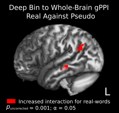
Our gPPI modeled the deep- and middle-bin task based interaction terms in OTS to predict activation in a standardized group brain. Regions predicted by the deep-bin were interpreted as primarily contributing top-down signal, whereas those predicted by the middle-bin were considered components of a forward directed network. Strikingly, the deep- and middle-bins did not show connectivity to similar brain regions. Following correction, the deep-bin exclusively targeted left temporal regions during real-word reading, regions associated with lexical retrieval. The middle-bin showed reduced whole-brain interaction generally, with no clusters surviving correction(fig. 3). A separate intraregional gPPI analysis showed that the deep- and middle bins have reduced interaction during pseudo-word reading, suggesting a suppressive effect of the deep-bin on the middle-bin.Conclusions
This work represents the first known use of lfMRI to assess directed connectivity within a distributed cortical network. We show that lfMRI can reveal whole-brain directed networks during word reading. In so doing, we identified language critical regions based on their association with the top-down signal stream and established lfMRI as a method for assessing directed connectivity. This also serves to demonstrate that the GE-BOLD contrast is capable of resolving spatially adjacent signal with sufficient precision to investigate the depth-dependent connectivity of distributed networks, and to observe interactions among bins within a region. The gPPI results suggest that the gPPI model can account for shared variance among the bins, reducing down-stream contamination effects arising from larger vessels and increasing the apparent resolution of the GE contrast.Acknowledgements
Funding from the Netherlands Organization for Scientific Research through the Language in Interaction consortium.References
1. T. Q. Duong, A. C. Silva, S.-P. Lee, S.-G. Kim, Functional mri of calcium-dependent synaptic activity: Cross correlation with cbf and bold measurements, Magnetic Resonance in Medicine 43, 383.
2. J. B. Goense, N. K. Logothetis, Laminar specificity in monkey v1 using high-resolution se-fmri, Magnetic Resonance Imaging 24, 381 (2006).
3. J. Goense, H. Merkle, N. K. Logothetis, High-resolution fmri reveals laminar differences in neurovascular coupling between positive and negative bold responses., Neuron 76, 629 (2012).
4. T. Jin, S. G. Kim, Cortical layer-dependent dynamic blood oxygenation, cerebral blood flow and cerebral blood volume responses during visual stimulation., Neuroimage 43, 1 (2008).
5. L. Muckli, et al., Contextual feedback to superficial layers of {V1}, Current Biology 25, 2690 (2015).
6. P. Kok, L. J. Bains, T. van Mourik, D. G. Norris, F. P. de Lange, Selective activation of the deep layers of the human primary visual cortex by top-down feedback., Curr Biol 26, 371 (2016).
7. F. De Martino, et al., Frequency preference and attention effects across cortical depths in the human primary auditory cortex., Proc Natl Acad Sci U S A 112, 16036 (2015).
8. L. Huber, et al., High-resolution cbv-fmri allows mapping of laminar activity and connectivity of cortical input and output in human m1., Neuron 96, 1253 (2017).
9. A. Maass, et al., Laminar activity in the hippocampus and entorhinal cortex related to novelty and episodic encoding., Nat Commun 5, 5547 (2014).
10. M. Waehnert, et al., Anatomically motivated modelingof cortical laminae, NeuroImage 93, Part 2, 210 (2014).
11. T. van Mourik, J. P. van der Eerden, P.-L. Bazin, D. G. Norris, Laminar signal extraction over extended cortical areas by means of a spatial glm, bioRxiv (2018).
12. D. G. McLaren, M. L. Ries, G. Xu, S. C. Johnson, A generalized form of context-dependent psychophysiological interactions (gppi): A comparison to standard approaches, NeuroImage 61, 1277 (2012).
13. R. Jackendoff, Storage and computation in the language faculty (Springer, 2002), pp. 23–58.
14. C. J. Price, J. T. Devlin, The interactive account of ventral occipitotemporal contributions to reading., Trends Cogn Sci 15, 246 (2011).
15. J. B. Goense, N. K. Logothetis, Laminar specificity in monkey v1 using high-resolution se-fmri, Magnetic Resonance Imaging 24, 381 (2006).
Figures