0610
A Novel Intravascular Contrast for Laminar Functional MRI1Section on Functional Imaging Methods, NIMH, Bethesda, MD, United States, 2Functional MRI Core, NIMH, Bethesda, MD, United States
Synopsis
In this work, we proposed a novel intravascular contrast for laminar specific fMRI in the human brain at 7T. This technique was shown to be sensitive to both cerebral blood volume (CBV) and flow (CBF) changes. We demonstrate that this new tool, with its highly specified functional layer profile, robust reproducibility and improved sensitivity, allows neuroscientific investigation of information flow across cortical microcircuits.
Introduction
Layer-dependent fMRI allows discrimination of activation that reflects cortical input and output1. Earlier research in cats has shown that both cerebral blood volume (CBV) and flow (CBF) could be used to visualize fMRI activation with superior spatial specificity to gradient-echo BOLD for layer-dependent research2. In humans at ultra-high field, CBF contrast of perfusion fMRI is technically challenging due to the intensive B1 inhomogeneity. CBV contrast for fMRI using vascular space occupancy (VASO) has been successfully used for laminar research. It is, however, compromised by interference of blood in-flow effect and a temporally limited acquisition window around the blood-nulling time point. Here we proposed to use DANTE3 (Delay Alternating with Nutation for Tailored Excitation) pulse trains combining 3D-EPI image technique5 to acquire an integrated VASO and perfusion (VAPER) contrast and demonstrated its predominant spatial specificity for laminar fMRI research in humans.Theory
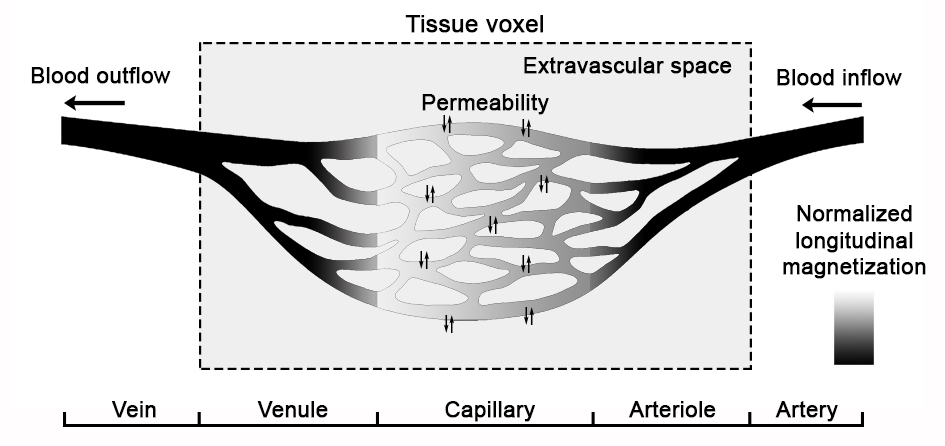
DANTE pulse trains3 can be used to suppress moving spins above a certain cutoff (2 mm/s) while retaining stationary tissue signal. In the human brain, blood in arteries, arterioles, venules and veins is travelling faster than the cutoff velocity4, thus the blood signal can be mostly diminished. Capillary blood may survive the DANTE suppression due to its low velocity (<1 mm/s)4. However, the suppressed blood in arteriole continuously flows into capillary, causing the blood signal in capillary partially attenuated. A four-compartment model with blood flow and permeability effect is used to describe the DANTE suppression effect (Fig. 1). Due to the limited B1 coverage of the head coil at 7T, this suppression process occurs only in subject’s head. During DANTE, the blood signal in human brain is nulled to achieve a VASO contrast. After DANTE, the fresh blood outside of the coil coverage flows into the image volume and replace the nulled blood, which generates a perfusion contrast.
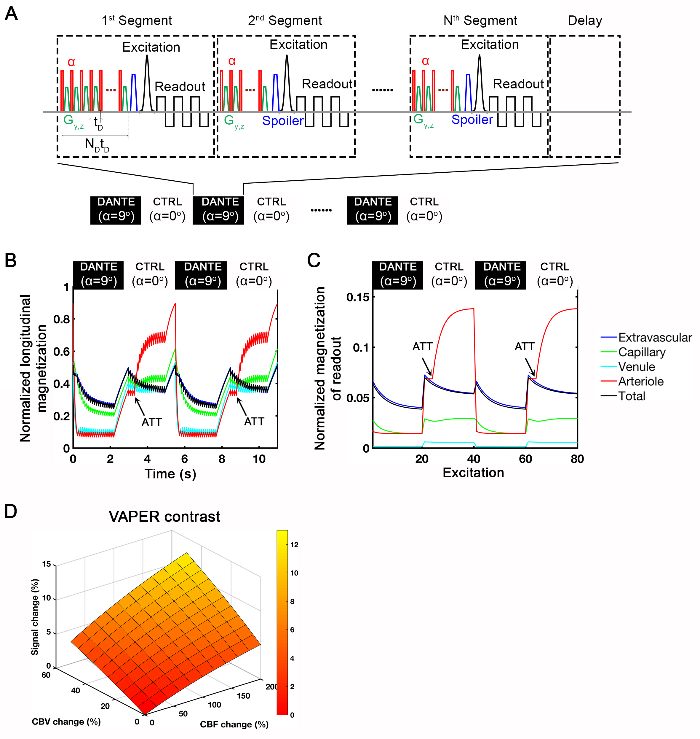
The sequence is implemented to acquire fMRI signal alternately between DANTE and CTRL conditions (Fig. 2A). In DANTE condition, 3D-EPI readout-modules5 are interleaved with DANTE pulses acquiring images sensitive to CBV changes known as VASO. The sequence scheme for CTRL condition is identical to DANTE condition, except that DANTE-RF pulses are switched off (α=0°). In CTRL condition, during the time between immediate after previous DANTE suppression and center k-space of current 3D-EPI acquisition, the blood signal recovers to a relatively high level due to T1-relaxaztion and further increases dramatically when fresh blood arrives (Fig. 2B-C; ATT, arterial transit time). Hence, the CTRL image is sensitive to CBF changes. The magnetization evolution of each compartment in longitudinal and readout directions is simulated based on Bloch equation of the four-compartment model (Fig. 2B-C).
The VAPER signal is generated as the difference between CTRL and DANTE conditions. It reflects an integrated effect of both CBV and CBF changes (Fig. 2D). Since the image intensity of DANTE condition is about 70-80% of the CTRL condition, the BOLD signal between these two conditions are quite similar. From the subtraction between DANTE and CTRL, BOLD contribution can be dramatically decreased while the intravascular contribution of CBV and CBF are boosted. To remove any remaining BOLD contribution from VAPER signal change, one may further divide the VAPER subtraction signal by CTRL signal to factor out the exp(TE/T2*) term.
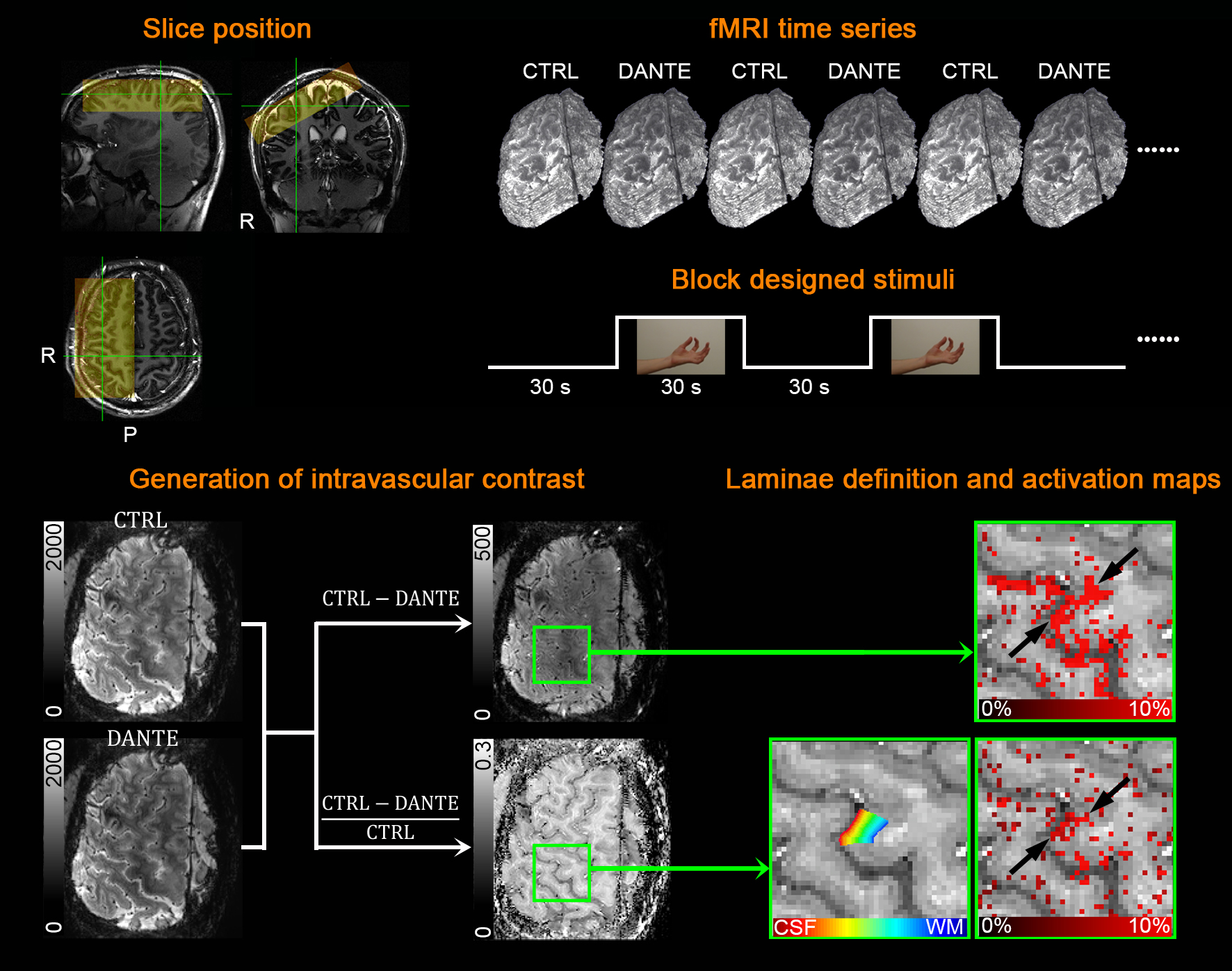
Experiments
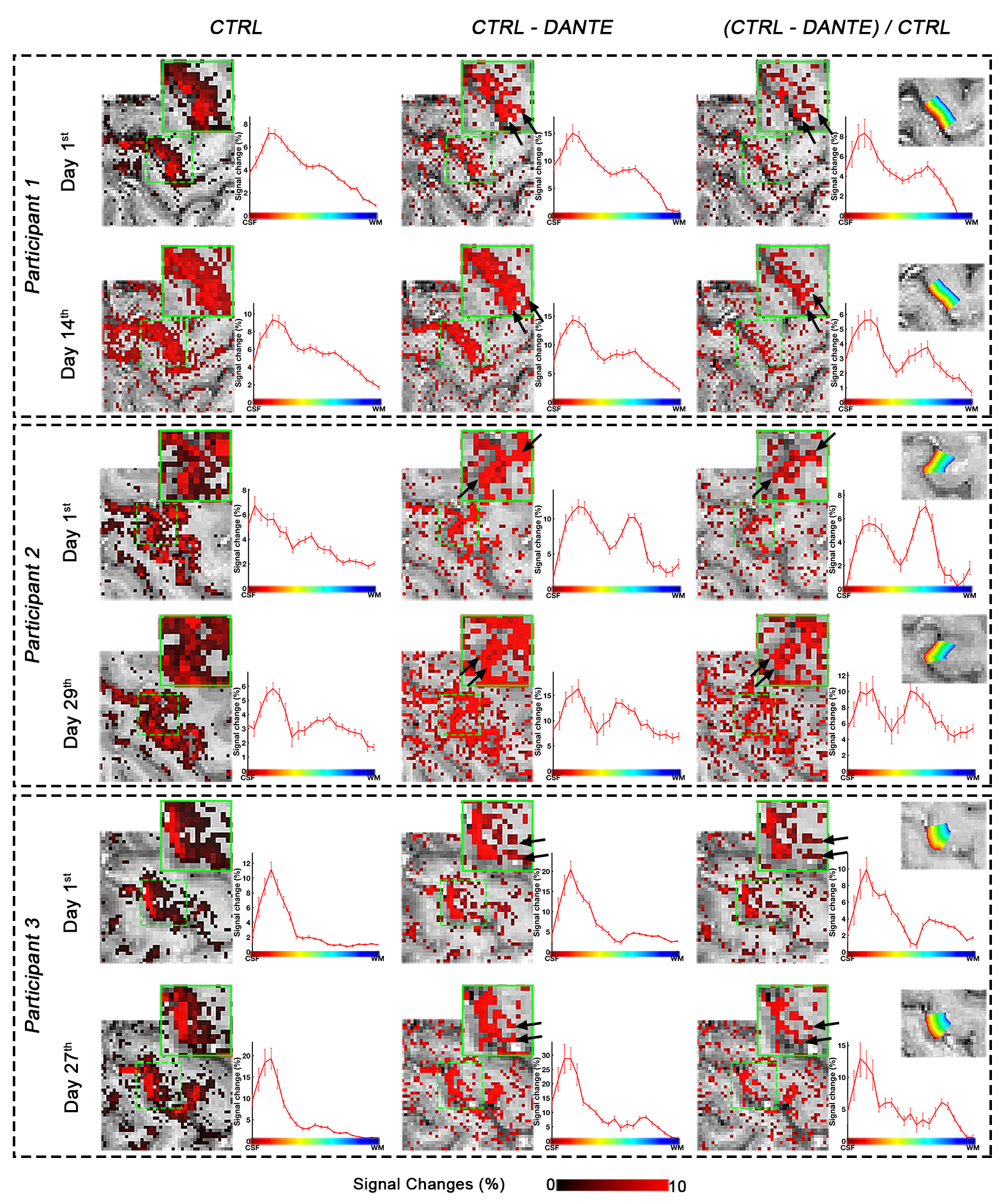
Six healthy volunteers (three females; aged 23-30) participated this study. Three participants were scanned a second time after two weeks. 3D-EPI VAPER was implemented on a Siemens MAGNETOM 7T scanner. fMRI data was acquired with TE/TR=30/3000ms, excitation FA=25°, 0.8mm in-plane resolution, 1.2-1.4mm slice thickness, 20 slices, no partial-Fourier, and GRAPPA 3. DANTE pulse number (ND) in 1st/later segment = 224/36,α/tD (interval) of DANTE RF pulse = 9°/1ms. Imaging slice position was adjusted individually for every subject to be perpendicular to the motor cortex area 4a (M1-4a) (Fig. 3). A 30-min left hand finger-tapping task paradigm (30s-rest/30s-tapping) was applied to induce a double-layer activation pattern1.Results and discussion
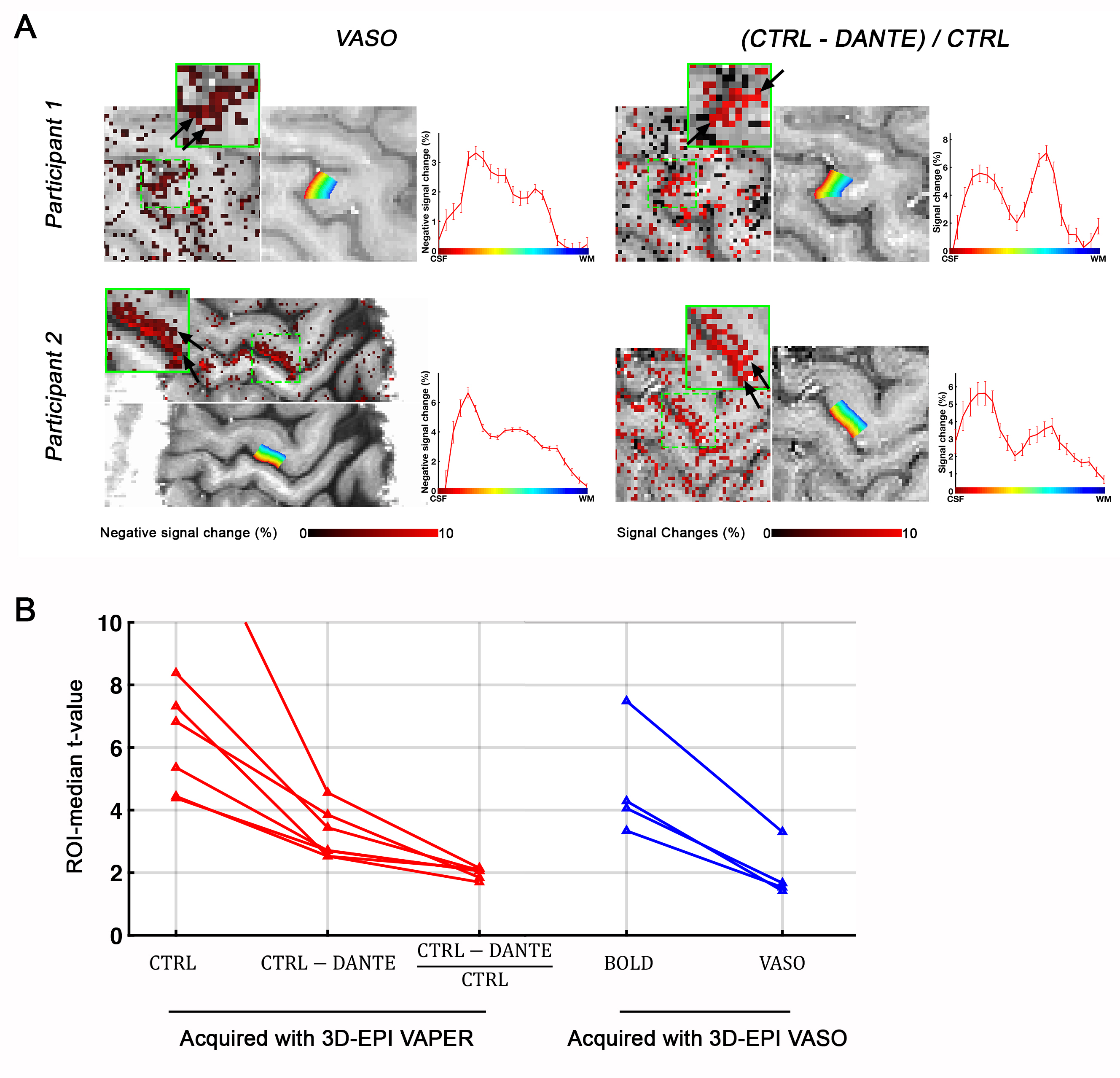
BOLD activity maps mainly show a monotonic decrease across cortical layers (Fig. 4). In contrast, VAPER signal changes, regardless of BOLD correction, show a clear feature of double response peak in different laminae: peak response in superficial layers reflect the input signal from other cortical areas while peak response in deep layers reflect the output signal to spinal cord. This double peak feature is highly reproducible across individuals and across days, and similar to the laminar response profile using VASO approach (Fig. 5A). Regarding to the sensitivity, BOLD corrected VAPER contrast is similar to VASO contrast in terms of the statistical t-value.Whereas, the BOLD uncorrected VAPER contrast has a significantly better sensitivity.Conclusion
VAPER technique with its highly microvessel-specific functional layer profile, robust reproducibility and improved sensitivity, allows neuroscientific assessment of information flow across cortical microcircuits.Acknowledgements
This work was supported by the NIMH Intramural Research Program. We acknowledge Benedikt A. Poser and Dimo Ivanov for contributions to the 3D-EPI sequence used here, and Arman Khojandi for help during scanning.References
1. Huber, L.et al.High-Resolution CBV-fMRI Allows Mapping of Laminar Activity and Connectivity of Cortical Input and Output in Human M1. Neuron96, 1253-1263 e1257, doi:10.1016/j.neuron.2017.11.005 (2017).
2. Jin, T. & Kim, S. G. Cortical layer-dependent dynamic blood oxygenation, cerebral blood flow and cerebral blood volume responses during visual stimulation. Neuroimage43, 1-9, doi:10.1016/j.neuroimage.2008.06.029 (2008).
3. Li, L. Q., Miller, K. L. & Jezzard, P. DANTE-prepared pulse trains: A novel approach to motion-sensitized and motion-suppressed quantitative magnetic resonance imaging. Magnetic Resonance in Medicine68, 1423-1438, doi:10.1002/mrm.24142 (2012).
4. Liu, D., Xu, F., Lin, D. D., van Zijl, P. C. M. & Qin, Q. Quantitative measurement of cerebral blood volume using velocity-selective pulse trains. Magnetic Resonance in Medicine77, 92-101, doi:10.1002/mrm.26515 (2017).
5. Poser, B. A., Koopmans, P. J., Witzel, T., Wald, L. L. & Barth, M. Three dimensional echo-planar imaging at 7 Tesla. Neuroimage51, 261-266, doi:10.1016/j.neuroimage.2010.01.108 (2010).
Figures