0589
Improvement and systematic analysis of the selective multiple quantum coherence 1H MRS lactate imaging pulse sequence for application to brain tumor and lymphoma patients1Department of Radiology, Perelman School of Medicine, University of Pennsylvania, Philadelphia, PA, United States, 2Division of Hematology Oncology, Perelman School of Medicine, University of Pennsylvania, Philadelphia, PA, United States
Synopsis
We have tested our revised Sel-MQC-CSI sequence for dependence of Lac/H2O on the B0 and B1 field inhomogeneity. The immunity of the Lac/H2O from our sequence on the above factors was demonstrated. We have improved lipid suppression from previously reported 100-fold to 22,000-fold while not losing the lactate signal. The revised sequence was applied to a glioma patient and a normal volunteer with a lactate phantom seated near the typical lymphoma occurring region of the body. In both human studies, satisfactory performance of the sequence was demonstrated.
Introduction
For application of the selective multiple quantum coherence chemical shift imaging (Sel-MQC CSI) of lactate into cancer patients,1-2 there are several issues that need to be carefully evaluated. One of them is the B0 and B1 dependence of the Lac/H2O ratio. Another is to fully suppress lipids in the tissue around the tumor nodes because remnant lipids may introduce artifact in the lactate signal of the tumor voxel of the CSI. These issues affect reliability of lactate imaging for routine clinical use for diagnosis or therapeutic response monitoring of cancer patients. In this study, we have evaluated the above mentioned issues in our revised Sel-MQC CSI pulse sequence for application to brain tumor and lymphoma patients.Methods
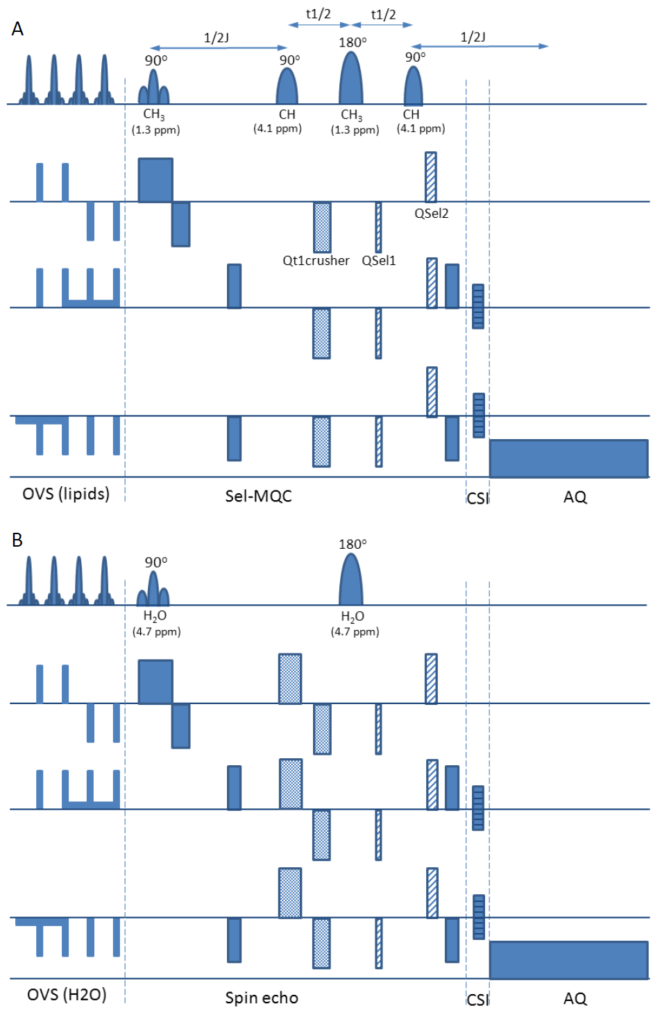
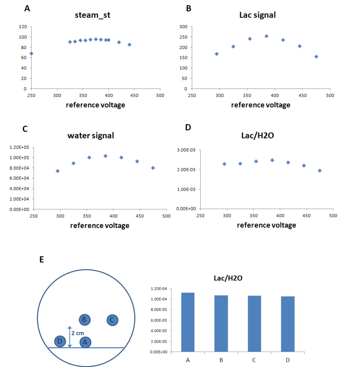
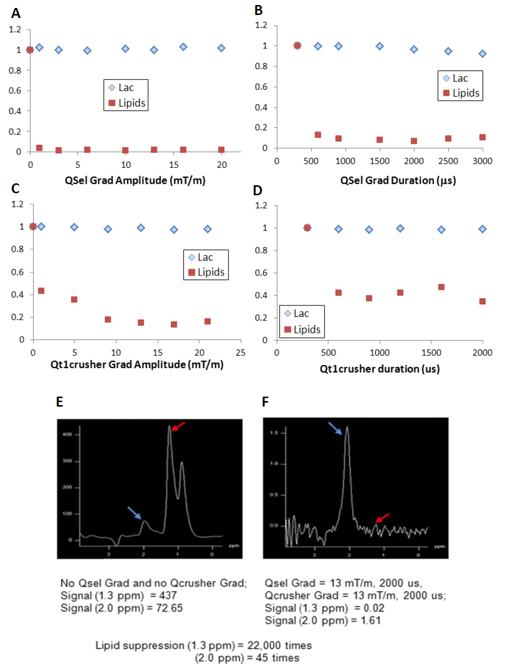
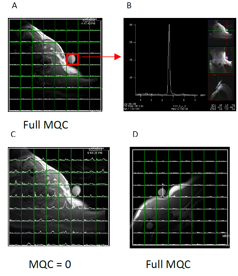
The study was performed in the 3T Tim Trio Siemens MRI systems. The 4-8 channel head coils were used for the phantom and the brain tumor patient study, and a home-built 7 cm two-channel surface coil was used for testing the sequence on phantoms located near the typical lymphoma occurring region of the body of a normal volunteer. Our previous sequence 2 has been modified in the following aspects: 1) Hadamard encoding inversion slice selection pulse was removed and a single slice selection was adopted for easy visualization of the CSI results in the scanner and to remove any possible motion artifact that may result when Hadamard transformation is performed. 2) The outer volume saturation slabs were added in case it is needed. 2) We used our own synthesized and tested Gaussian pulses to be sure of the flip bandwidth of the used RF pulses. The length of the 180o Gaussian pulse was reduced from 7800 μs to 5300 μs so that the 180o inversion width matches the 90o flip width of the 7800 μs Gaussian pulse. 3) The QSel gradients were adjusted to find maximal lipid suppression while retaining lactate signal. 4) The Qt1crusher gradients 3 were added and adjusted to achieve maximal lipid suppression while retaining lactate signal. 5) Water spin echo CSI sequence was built to have the same eddy current as in the Sel-MQC-CSI so that phase alignment and addition of the signals from multi-channel RF coils is possible. The pulse sequence is presented in Fig 1. We evaluated the coefficient of variation (CV) of the Lac/H2O at varying reference voltage and varying location of the lactate phantom inside a magnet. We evaluated the dependence of lactate and lipid signals on QSel and Qt1crusher gradient amplitude and duration in the revised sequence. We applied the sequence to the brain tumor patients and to a normal volunteer with lactate phantoms placed on top of the body regions where typical lymphoma lesions are observed.
Results
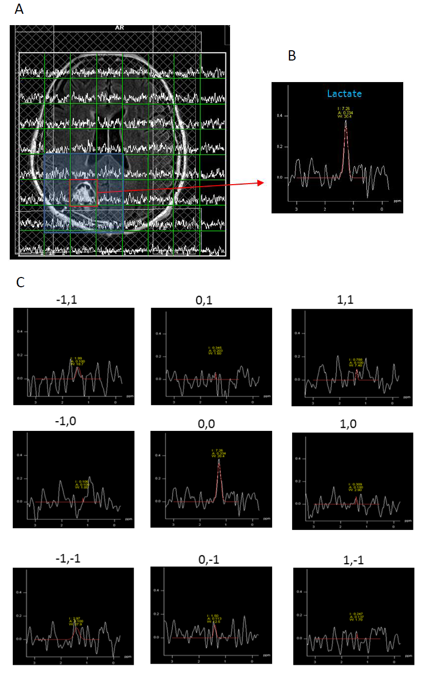
The CV of Lac/H2O was maintained within 4% while RF reference voltage is changed from 295V to 445V (Fig 2A-2D). With the phantom at various locations of the magnet (Fig 2E), the CV of Lac/H2O was maintained within 3%. With varying QSel and Qt1crusher gradients, the CV of lactate amplitude was preserved within 3%, while lipid signal showed abrupt dependence on these gradients (Fig 3A-3D). At the optimum combinations of those gradients, lipid signals dropped 22,000 times compared with signal at zero gradients of them. Application of the sequence into a recurrent glioma patient who was already treated with chemoradiotherapy resulted in clean lactate signal from the voxel of the tumor while only noise signals were observed in the surrounding voxels (Fig 4). Application of the sequence into a volunteer with 10 mM lactate phantom demonstrate that the sequence is well working in the human body circumstances and that lipid signals in the near skin are sufficiently suppressed leaving clean lactate signal from the phantom (Fig 5).Discussion
The immunity of the Lac/H2O on the RF reference voltage and on the location of the phantom inside the magnet demonstrated the robustness of Lac/H2O calculated from our sequence on B0 and B1 inhomogeneity. We have achieved a very high lipid suppression of 22,000 fold in this revised sequence. The previous lipid suppression by Sel-MQC in the clinical scanner has been ~100 fold.2 The increased lipid suppression has huge impact in applying the sequence when the tumor is surrounded by tissues with high amount of lipids.
Conclusion
We have demonstrated the improved characteristics of the revised Sel-MQC-CSI. The sequence is well suited for characterizing Lac/H2O of tumor from the brain tumor and lymphoma patients and possibly other types of cancer.Acknowledgements
R01-CA172820, R01-CA129544-03, ITMAT-TBIC, McCabe Foundation.References
1. Lee SC, Arias-Medoza F, Poptani H, et al. Prediction and Early Detection of Response by NMR Spectroscopy and Imaging. PET Clin. 2012;7(1):119-126.
2. Mellon EA, Lee SC, Pickup S, et al., Detection of lactate with a hadamard slice selected, selective multiple quantum coherence, chemical shift imaging sequence (HDMD-SelMQC-CSI) on a clinical MRI scanner: Application to tumors and muscle ischemia. Magn Reson Med. 2009;62(6):1404-1413.
3. He Q, Shungu DC, van Zijl PC, et al. Single-scan in vivo lactate editing with complete lipid and water suppression by selective multiple-quantum-coherence transfer (Sel-MQC) with application to tumors. J Magn Reson B. 1995;106(3):203-211.
Figures