0585
Magnetic resonance imaging for confirmation of catheter-selective tumor-targeting liposomes distribution1Medical College of Wisconsin, Milwaukee, WI, United States, 2Northwestern University, Chicago, IL, United States
Synopsis
Hepatocellular carcinoma is the most common form of primary liver cancer, and globally it is the sixth most common cancer. In this study, we use liposomes that can co-encapsulate the therapeutic agent (oxaliplatin or gemcitabine) in addition to iron oxide nanoparticles to enable MRI-monitored local delivery of the therapeutic agent to limit proangiogenic responses in non-resectable liver tumors following transcatheter embolotherapies. The results showed increased R2* values in the tumor regions at one week post-infusion, compared to the surrounding liver parenchyma and to same regions immediately after infusion, which allows for confirmation of procedural success and proper catheter-selective tumor targeting.
INTRODUCTION
Hepatocellular carcinoma is the most common form of primary liver cancer, and globally it is the sixth most common cancer. In patients with unresectable hepatocellular carcinoma, trans-arterial embolization or chemoembolization procedures can be conducted, where embolic and/or chemotherapeutic agents are co-delivered through the catheter locally to the tumors, which allows for specific targeting of tumors.
In this study, we use liposomes that can co-encapsulate the therapeutic agent (oxaliplatin or gemcitabine) in addition to iron oxide nanoparticles (IONP) to enable MRI-monitored local delivery of the therapeutic agent to limit proangiogenic responses in liver tumors following transcatheter embolotherapies.
This study includes a phantom experiment to investigate the relationship between IONP concentration and R2* relaxivity, followed by in vivo scans of a rat model with liver tumor to validate the capability of MRI-monitored tumor-targeted transcatheter delivery.
METHODS
Liposome nanoparticles were synthesized by preparing uniform iron oxide nanocubes using thermal decomposition. Then, the iron oxide nanocubes and oxaliplatin/gemcitabine were encapsulated in sterically stabilized polyethylene glycol (PEG)-coated liposomes, forming magnetic liposomes with a diameter of ~140 nm.
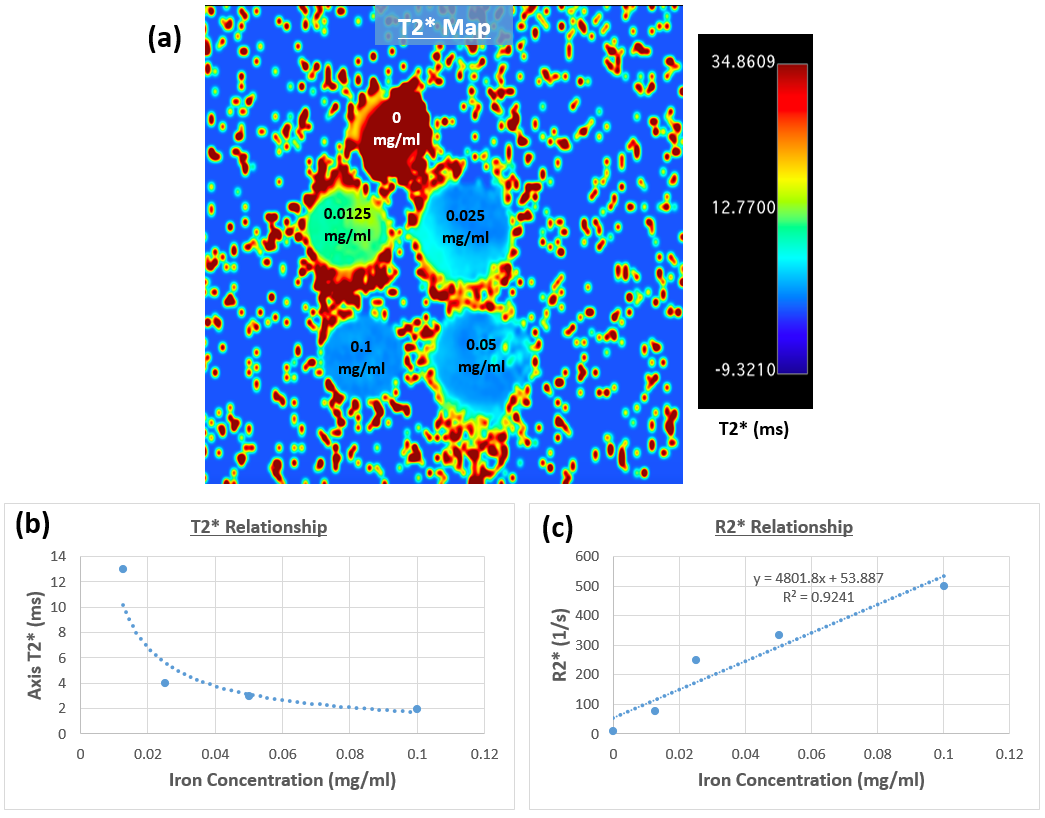
To characterize the T2* relaxivity properties of the liposomes, a phantom was created that includes five tubes filled with liposome nanoparticles, with iron-oxide concentrations ranging from 0 to 0.1 mg/ml, embedded in 1% agarose gels (Figure-1). The phantoms were imaged on a 9.4T Bruker MRI scanner. R2* relaxometry was assessed using a gradient-echo T2* mapping sequence. The imaging parameters were: repetition time (TR)=800ms, 9 echo times (TE) ranging from 4ms to 48ms in 5.5ms increments, flip angle=50⁰, matrix=128x128, field of view (FOV)= 45x45 mm2, slice thickness=1mm, acquisition bandwidth=586Hz/pixel, #averages=2, and scan time ~1 minute/slice. Signal intensities from the nine echoes were fitted to a mono-exponential decaying curve to derive T2* value, from which R2* was measured as the reciprocal of T2*. Least-square fitting was conducted between mean R2* and iron oxide concentration in the imaged phantoms.
Wistar rats (n=4), weighing 300-350g, were used for in-vivo scans. For the inoculation procedure, anesthesia was obtained using 2-3% isoflurane. A 3-cm midline incision was made and both the left and right hepatic lobes were mobilized to implant sub-capsular tumors. Approximately two weeks post-implantation, X-ray digital subtraction angiography was performed to selectively place a catheter into tumor feeding branches of the hepatic artery. After catheter placement, 500μL of liposomes were infused, after which the catheter was withdrawn.
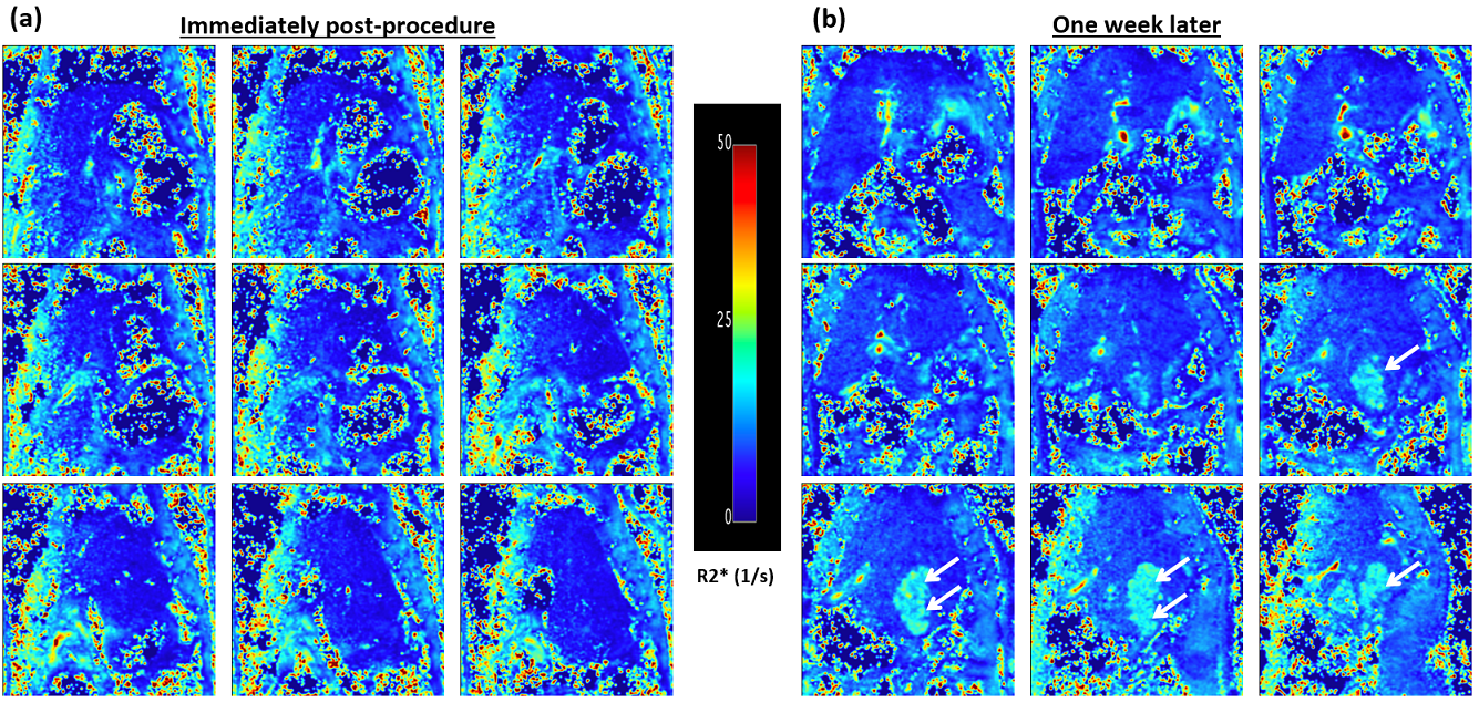
MRI was performed immediately post liposomes infusion and one week later on the same scanner and using the T2* imaging protocol as implemented in the phantom scan. Stacks of parallel axial or coronal slices covering the liver were acquired using respiratory gated acquisition to avoid breathing artifacts. Further, T1-weighted images were acquired before the procedure, which showed slightly hyperenhanced tumor regions. R2* maps were constructed from the T2* MRI images acquired immediately post-infusion and one week later. ROIs were drawn within the acquired maps to measure R2* in the tumor regions and surrounding tissues. Statistical t-test analysis was conducted to compare R2* measurements.
RESULTS
The phantom T2* map showed decreased signal intensity with increasing IONP concentration (Figure-1). T2* measurements showed decaying exponential relationship with IONP concentration, while R2* measurements showed linear relationship with IONP concentration (R2 = 0.924).
The R2* MRI maps (Figure-2) showed hyperintensity in the tumor regions at one week post-infusion (R2*=18±0.7 1/s), compared to the surrounding liver parenchyma (R2*=9.5±0.3 1/s; P<.001) and to the same regions immediately after infusion (R2*=7.2±0.9 1/s; P<.001), where the localized delivery of iron oxide resulted in low signal intensity immediately after infusion.
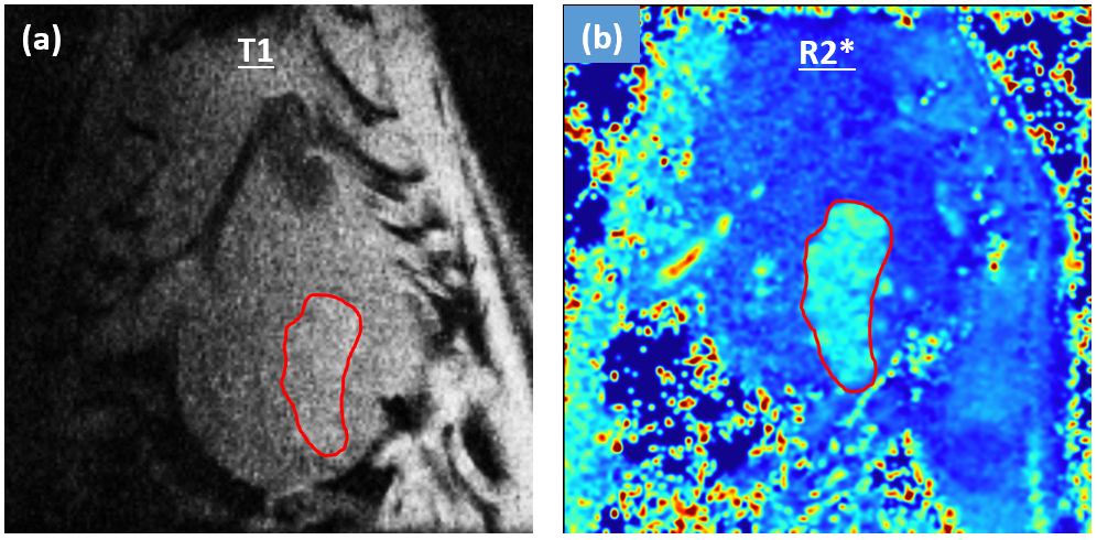
Figure-3 shows the appearance of hyperenhanced tumor region in the R2* map acquired one-week after infusion, matching the same region that appears slightly enhanced in the T1-weighted image acquired pre-infusion.
DISCUSSION and CONCLUSION
This study showed the capability of noninvasive assessment of liposome nanoparticles distribution to liver tumors using MRI imaging. Quantitative MRI enabled assessment of changes in tumor R2* values over time for confirmation of procedural success and proper catheter-selective tumor targeting.
The presence of iron oxide in the liposomes allows for triggered release of therapeutic agents by external triggers like alternating magnetic field or heat. Stimulated drug release reduces off-target effects and improves the efficiency of local high dose release. Further, the amount of therapeutic agent delivered to the targeted region can be quantitated and the outcome of the procedure can be assessed.
In conclusion, if translated, this technique would enable confirming the delivery of theranostic oxaliplatin or gemcitabine/iron oxide nanoparticles to the tumors, which is of importance for the combination therapy with embolic platforms currently in use.
Acknowledgements
No acknowledgement found.References
1. Wahidiyat et al, Hematology; 22: 501-507.
2. Chen et al, Biomaterials; 61: 299-306.
3. White et al, Radiology; 285: 809-819.
4. White et al, PLOS ONE; 11(5): e0155334.
Figures