0570
Performance of a novel cryogen-free cryostat with an automated temperature control for fine tuning of High Temperature Superconducting RF coils: high-resolution MR imaging at 1.5 T1IR4M, UMR8081, Université Paris-Sud/CNRS, Université Paris-Saclay, F-91405 Orsay, France, Orsay, France, 2Irfu, CEA Paris-Saclay, Université Paris-Saclay, F-91191 Gif-sur-Yvette, France, Gif-sur-Yvette, France
Synopsis
We present the performance of an MR-compatible cryogenic system dedicated to cool High Temperature Superconducting (HTS) radiofrequency coils for micro-MRI at 1.5 T. A real time control and regulation of the temperature were performed to finely tune the HTS coil to the Larmor resonance frequency with a precision of 1 Hz. Firstly, we demonstrated that this cryostat does not cause any electromagnetic disturbance. Secondly, MR images of a 1 mm-cubic liquid phantom were acquired using the HTS coil as a transceiver with a spatial resolution down to (100 μm)3 under real clinical research experimental conditions.
Purpose
Small-sized high Temperature Superconducting (HTS) radiofrequency (RF) coils have been successfully involved in several micro-MRI applications and have achieved a substantial improvement in Signal-to-Noise Ratio (SNR)1,2. The main issue with HTS coils is the degradation of the electrical properties such as the resonance frequency (f0) and the quality factor (Q) in the presence of B0 because of the vortices formation containing normal carriers. The frequency shift in presence of B0 depends on both the coil plane orientation relative to B03 and the cooling temperature4. HTS coils present a high sensitivity of detection and exhibit a narrow bandwidth (BWRFcoil) around tens of kHz, imposing to perform a very fine tuning of the coil to the Larmor frequency. The previously reported tuning method for monolithic HTS coil is based on inductively coupling a closed copper loop, but this was technically complex and has degraded the overall electrical properties of the coil1. For these reasons, we have developed a novel MR compatible cryogen-free cryostat to control the operating temperature of an HTS coil with a sufficient stability to ensure real-time tuning of the coil during MRI exam5. In the following, the performances of this cryostat and high-resolution images acquired in a 1.5T clinical MR are presented.Methods
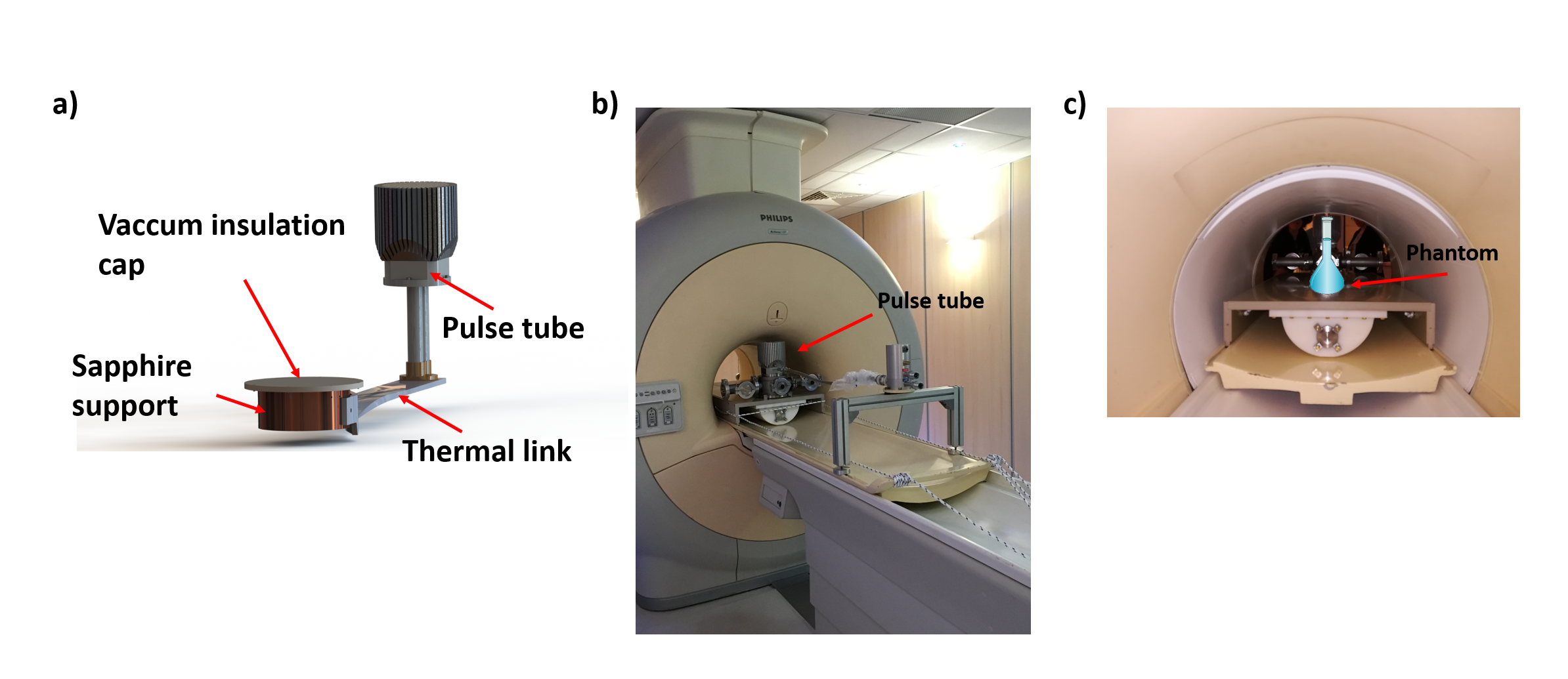
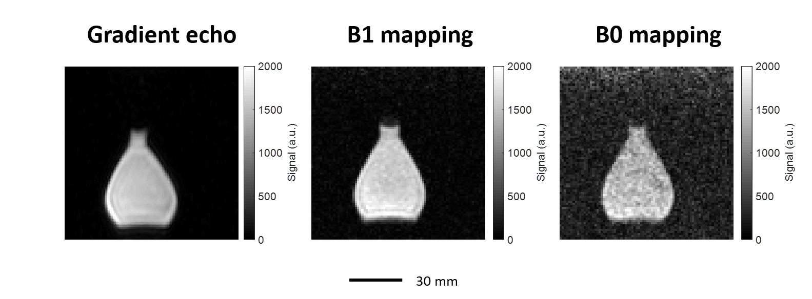
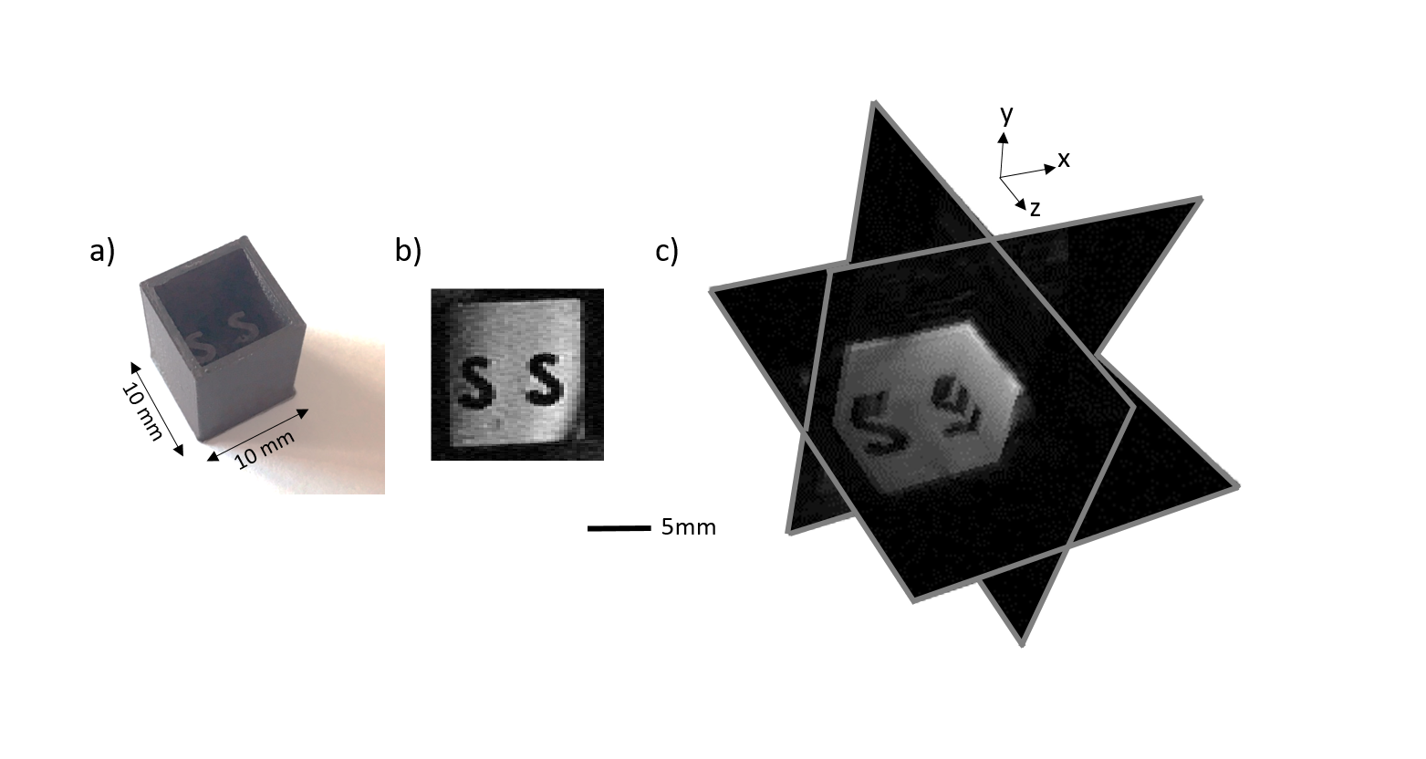
The cryostat is made of polymers designed to avoid any kind of static or RF field perturbation. It is based on a single stage pulse tube cryocooler linked to a support (figure 1a). The coil to be cooled is installed on a circular platform made of sapphire enclosed between its support and a vacuum insulation removable cap5. RTD sensors and heaters are placed at different locations along the cryostat for a real-time control and regulation of the temperature using a temperature controller (CTC100, SRS). The used RF coil is a 12 mm-diameter HTS coil made of YBa2Cu3O7 thin film superconductor etched on both sides of a 500μm-thick lanthanum aluminate substrate. Matching the coil to 50 Ω and the RF signal pickup were done using inductive technique6. The resonance frequency was finely adjusted through controlling the temperature of the coil7. MR imaging experiments were performed on a 1.5T clinical MRI (Achieva, Philips). First, we performed a preliminary study without the HTS coil to assess the influence of the cryostat, at ambient temperature, on the homogeneity of the B0 and B1 fields. 3D-gradient-echo MR images of a flask of water were acquired using the whole body RF coil of the scanner (figure 1b-c). A second study was carried out with the HTS coil as a transceiver using a 3D-gradient-echo sequence. The flip angle calibration was performed with a constant B1emission magnitude fixed to 1μT and by varying the application RF time to avoid any distortion due to the non-linear behavior of superconducting materials regarding the transmitted RF power. A plastic cubic-phantom containing water doped with gadolinium (C=0,00125mmol/mL, T1=0.2sec) was placed at the isocenter of the imager (figure 3a).Results
Figure 2 shows the homogeneous MR image, B1 and B0 maps of the tested water flask without any deformation. At Earth field and 60K, the quality factor of the HTS coil was around 5500 at f0=63.636250MHz (BWRFcoil=1Hz). The Q-factor is decreased to 3000 and f0=63.899313MHz at B0=1.5 T. The operating temperature was changed to 61K, to compensate the f0 shift due to the B0-field and finally retune the HTS coil to the Larmor frequency. MR images of the phantom along the three planes were obtained with a spatial resolution of (100μm)3 in 20 minutes as shown in figure 3. The measured SNR was around 60.Discussion and conclusions
MR images (figure 2) have shown that the cryostat does not cause any electromagnetic perturbation regarding the magnetic fields. We demonstrated that the cryostat, associated to a temperature controller, allows a regulation of the temperature and provides an efficient tool for a fine tuning of the HTS coil to the Larmor frequency, without degradation of its electrical properties. A vacuum of around 10-6mbar, a temperature of 61K, and a f0 of 63.899313MHz were kept stable during all the experiments in the MRI (more than twelve hours). Besides, an excellent image quality was achieved with the HTS coil, proving that, this cryostat would be a suitable platform to accommodate small animals for future experiments and to use multiple HTS or array coils placed on the sapphire support (150mm-diameter). For now, the acquisitions were performed using one HTS coil as an RF transceiver. The next step will be to evaluate a promising detuning approach9,10 allowing the use of the highly sensitive HTS coils in receive mode only.Acknowledgements
This work was performed on a platform of France Life Imaging network partly funded by the grant ANR-11-INBS-0006 and has been supported by the French Agence Nationale de la Recherche (ANR), under grant ANR-14-CE17-0003 (SupraSense project).References
1. Poirier-Quinot M., Ginefri J.-C., et al. Performance of a miniature high-temperature superconducting (HTS) surface coil for in vivo microimaging of the mouse in a standard 1.5T clinical whole-body scanner. Magn. Reson. Med. 2008;60:917–927.
2. Laistler E. et al. In vivo MR imaging of the human skin at subnanoliter resolution using a superconducting surface coil at 1.5 tesla. J. Magn. Reson. Imaging 2015;41:496–504.
3. Ginefri J.-C., Darrasse L. et al. High-temperature superconducting surface coil for in vivo microimaging of the human skin. Magn. Reson. Med.2001;45:376–38
4. Black R. D., Early T. A. & Johnson, G. A. Performance of a High-Temperature Superconducting Resonator for High-Field Imaging. Journal of Magnetic Resonance, Series A 1995;113:74–80.
5. Authelet G., Poirier-Quinot M., et al. Conceptual design of a cryogen-free μMRI device. IOP Conf. Ser.: Mater. Sci. Eng. 2017;278:1-7.
6. Ginefri J.-C. Poirier-Quinot, M., et al. Technical aspects: Development, manufacture and installation of a cryo-cooled HTS coil system for high-resolution in-vivo imaging of the mouse at 1.5T. Methods 2007;43:54–67.
7. Lambert S., Ginefri J.-C., et al. High-temperature superconducting radiofrequency probe for magnetic resonance imaging applications operated below ambient pressure in a simple liquid-nitrogen cryostat. Review of Scientific Instruments 2013;84:1-7.
8. ANR funded project. ANR Available at: http://www.agence-nationale-recherche.fr/en/anr-funded-project/?tx_lwmsuivibilan_pi2[CODE]=ANR-14-CE17-0003.
9. Geahel et al, Proc. ISMRM 2017, 2704.
10. Geahel et al, Proc. ESMRMB 2017, 62.
Figures